Characteristics of the main protein fractions
Albumins . Albumin accounts for more than half (55–60%) of human plasma proteins. Mol. the mass of albumin is about 70,000. Serum albumins are renewed relatively quickly (the half-life of human albumins is 7 days).
Due to their high hydrophilicity, especially due to the relatively small molecular size and significant concentration in serum, albumins play an important role in maintaining blood oncotic pressure. It is known that serum albumin concentrations below 30 g/l cause significant changes in blood oncotic pressure, which leads to edema. Albumins perform the important function of transporting many biologically active substances (in particular, hormones). They are able to bind to cholesterol and bile pigments. A significant portion of serum calcium is also bound to albumin.
During electrophoresis in starch gel, the albumin fraction in some people is sometimes divided into two (albumin A and albumin B), i.e. such people have two independent genetic loci that control albumin synthesis. The additional fraction (albumin B) differs from regular serum albumin in that the molecules of this protein contain two or more dicarboxylic amino acid residues that replace tyrosine or cysteine residues in the polypeptide chain of regular albumin. There are other rare variants of albumin (Reading albumin, Gent albumin, Maki albumin). Inheritance of albumin polymorphism occurs in an autosomal codominant manner and is observed over several generations.
In addition to hereditary albumin polymorphism, transient bisalbuminemia occurs, which is sometimes mistaken for congenital. The appearance of a fast component of albumin in patients receiving large doses of penicillin has been described. After discontinuation of penicillin, this component soon disappeared from the blood. There is an assumption that the increase in the electrophoretic mobility of the albumin–antibiotic fraction is associated with an increase in the negative charge due to the COOH groups of penicillin.
Rice. 17.2. The structure of the immunoglobulin molecule (scheme). Explanations in the text.
Globulins . When salted out with neutral salts, serum globulins can be divided into 2 fractions - euglobulins and pseudoglobulins. The euglobulin fraction mainly consists of γ-globulins, and the pseudoglobulin fraction includes α-, β- and γ-globulins, which during electrophoresis, especially in starch or polyacrylamide gel, can be separated into a number of subfractions. The α- and β-globulin fractions contain lipoproteins as well as metal-bound proteins. Most of the antibodies contained in serum are in the γ-globulin fraction. When the level of proteins in this fraction decreases, the body's defenses sharply decrease.
Immunoglobulins, or antibodies, are synthesized by B lymphocytes or plasma cells formed from them. There are 5 known classes of immunoglobulins: IgG, IgA, IgM, IgD and IgE, with IgG, IgA and IgM being the main classes; IgD and IgE are minor classes of human plasma immunoglobulins. The immunoglobulin molecule consists of two identical pairs of polypeptide chains. Each pair in turn consists of two different chains: light (L) and heavy (H). In other words, the immunoglobulin molecule consists of two light (L) chains (molecular weight 23000) and two heavy (H) chains (molecular weight 53000–75000), forming a tetramer (L2H2) using disulfide bonds (Fig. 17.2). Each chain is divided (perhaps somewhat arbitrarily) into specific domains, or regions, that have a specific structural and functional significance. The half of the light chain, including the carboxyl terminus, is called the constant region (CL), and the N-terminal half of the light chain is called the variable region (VL).
About a quarter of the heavy chain, including the N-terminus, is classified as the variable region of the H chain (VH), the rest of it is the constant region (CH1, CH2, CH3). The immunoglobulin region that binds to a specific antigen is formed by the N-terminal variable regions of the light and heavy chains, i.e. VH and UL domains. Higher vertebrates have all 5 classes of antibodies (IgA, IgD, IgE, IgG and IgM), each with its own class of H chains: α, δ, ε, γ and μ, respectively. IgA molecules contain α-chains, IgG molecules contain γ-chains, etc. In addition, there are a number of immunoglobulin subclasses IgG and IgA. For example, in humans there are 4 subclasses of IgG: IgG1, IgG2, IgG3, and IgG4, containing heavy chains γ1, γ2, γ3, and γ4, respectively. Different H chains give the hinge regions and tail regions of antibodies different conformations and determine the characteristic properties of each class and subclass (for more information, see immunology manuals).
In clinical practice, there are conditions characterized by changes in both the total amount of blood plasma proteins and the percentage of individual protein fractions.
Hyperproteinemia is an increase in the total content of plasma proteins. Diarrhea in children, vomiting due to obstruction of the upper small intestine, and extensive burns can increase the concentration of proteins in the blood plasma. In other words, the loss of water by the body, and therefore the plasma, leads to an increase in protein concentration in the blood (relative hyperproteinemia).
In a number of pathological conditions, absolute hyperproteinemia may be observed due to an increase in the level of γ-globulins: for example, hyperproteinemia as a result of infectious or toxic irritation of the macrophage system; hyperproteinemia in myeloma. Specific “myeloma” proteins are found in the blood serum of patients with myeloma. The appearance in the blood plasma of proteins that do not exist under normal conditions is usually called paraproteinemia. Often with this disease, the protein content in plasma reaches 100–160 g/l.
Sometimes in multiple myeloma, abnormal plasma proteins cross the kidney barrier and appear in the urine. These proteins, which are light chains of immunoglobulins, are called Bence Jones proteins. The phenomena of paraproteinemia can also be observed with Waldenström macroglobulinemia. Waldenström's disease is characterized by the appearance in the blood plasma of proteins with a large molecular weight (1,000,000–1,600,000); the content of macroglobulins can reach 80% of the total amount of protein, which in this case is 150–160 g/l.
Hypoproteinemia, or a decrease in the total amount of protein in the blood plasma, is observed mainly with a decrease in albumin levels. Severe hypoproteinemia is a constant and pathogenetically important symptom of nephrotic syndrome. The total protein content decreases to 30–40 g/l. Hypoproteinemia is also observed when liver cells are damaged (acute liver atrophy, toxic hepatitis, etc.). In addition, hypoproteinemia can occur with sharply increased permeability of capillary walls, with protein deficiency (damage to the digestive tract, carcinoma, etc.). Therefore, we can assume that hyperproteinemia is usually associated with hyperglobulinemia, and hypoproteinemia with hypoalbuminemia.
In many diseases, the percentage of individual protein fractions often changes, although the total protein content in the blood serum remains within normal limits. This condition is called “dysproteinemia”. In Fig. Figure 17.3 schematically shows the nature of changes in protein fractions of blood serum in a number of diseases without taking into account the form and stage of the disease.
Rice. 17.3. Changes in the electropherogram of serum proteins in certain diseases (according to Emmrich).
During many diseases associated with general inflammation (infectious diseases, rheumatism, etc.), several stages are observed, which undoubtedly affects the protein spectrum of the blood.
As noted, α- and β-globulin fractions of serum proteins contain lipoproteins and glycoproteins. The carbohydrate part of blood glycoproteins mainly includes the following monosaccharides and their derivatives: galactose, mannose, rhamnose, glucosamine, galactosamine, neuraminic acid and its derivatives (sialic acids). The ratio of these carbohydrate components in individual serum glycoproteins is different. Most often, aspartic acid (its carboxyl) and glucosamine take part in the connection between the protein and carbohydrate parts of the glycoprotein molecule. Somewhat less common is the connection between the hydroxyl of threonine or serine and hexosamines or hexoses.
Neuramic acid and its derivatives (sialic acids) are the most labile and active components of glycoproteins. They occupy the final position in the carbohydrate chain of the glycoprotein molecule and largely determine the properties of a given glycoprotein.
Glycoproteins are present in almost all protein fractions of blood serum. When electrophoresis on paper, glycoproteins are detected in large quantities in the α1- and α2-fractions of globulins. Glycoproteins associated with α-globulin fractions contain small amounts of fructose, and glycoproteins found in β- and especially γ-globulin fractions contain significant amounts of fructose.
An increased content of glycoproteins in plasma or serum is observed in tuberculosis, pleurisy, pneumonia, acute rheumatism, glomerulonephritis, nephrotic syndrome, diabetes, myocardial infarction, gout, as well as in acute and chronic leukemia, myeloma, lymphosarcoma and some other diseases. In a patient with rheumatism, the increase in the content of glycoproteins in the serum corresponds to the severity of the disease. This is explained, according to some researchers, by depolymerization of the main substance of connective tissue, which leads to the entry of glycoproteins into the blood.
Previous page | Next page
CONTENT
Work No. 1. Separation and quantification of proteins
Blood serum fractions by electrophoresis on paper.
Principle of the method. Electrophoresis is the movement of charged particles in a field of direct electric current. The speed of movement of protein molecules in an electric field depends on the charge, molecular weight, pH, and ionic strength of the solution.
Serum proteins are placed on a strip of paper moistened with a buffer solution, through which a direct electric current is passed. At pH 8.6, serum proteins are negatively charged and, under the influence of an electric field, move to the anode.
Human blood serum contains various proteins. Using electrophoresis, 5 fractions are isolated on paper - albumin, α1-, α2-, β-, γ-globulins.
Clinical and diagnostic significance. Many pathological conditions are accompanied by quantitative changes in the ratio of protein fractions in the blood - dysproteinemia. A decrease in the content of the albumin fraction is characteristic of liver diseases due to a decrease in the protein-synthesizing function of hepatocytes. Hypoalbuminemia also accompanies kidney disease due to protein loss in the urine. An increase in the content of α1- and α2-globulin fractions is observed under stress, the presence of inflammatory processes due to “acute phase” proteins, collagenosis and metastasis of malignant neoplasms. The β-globulin fraction increases with hyperlipoproteinemia. The fraction of γ-globulins increases during immune reactions caused by viral and bacterial infections. A decrease in the γ-globulin fraction can occur in primary and secondary immunodeficiency.
Work order
1. Device design for electrophoresis. The device consists of a rectifier that supplies direct current at the required voltage, and a chamber for electrophoresis. The chamber itself consists of 2 baths; one of them has a fixed partition where a platinum electrode (+ anode) is placed, and the other contains a stainless steel electrode (- cathode). Between the baths filled with the appropriate buffer, there is a connecting bridge on which strips of special filter paper are placed.
2. Carrying out electrophoresis. Fill both chamber baths with a solution of veronal buffer with pH 8.6. There should be enough buffer solution in the baths so that it covers the fixed partition, but is below the movable partitions.
Insert electrodes into the baths. Cut strips of filter paper of the required size depending on the size of the chamber (usually 4-6 cm wide) and with a simple pencil mark the place where the serum will subsequently be applied (start). Soak these strips in Veronal buffer. Insert the connecting bridge into the bath chambers. Place strips of paper on dry plates with forceps, immersing the ends of the strips in baths with buffer, and apply 0.025-0.005 ml of serum to pre-marked areas of the paper at a distance of 5-6 cm from the edge of the bridge. Serum is applied from the cathode side.
Figure 1. Diagram of a chamber for electrophoresis of proteins on paper:
1-stabilizer; 2-chamber for electrophoresis; 3-buffer solution; 4-supporting connecting bridge-electrode; 5-filter paper for electrophoresis.
After applying the serum to the paper strips, the chamber is hermetically sealed with a lid. There is a locking clamp on the camera cover that is used to turn on the camera. The attached rectifier supplies the camera with a constant current of 2 to 4 mA at a constant voltage of 110-160V. Electrophoresis is carried out at a potential gradient of 3 to 8 V per 1 cm of strip at room temperature. Good separation occurs in 18-20 hours.
3. Turn off the device and identify protein fractions. Turn off the device. Remove the cameras and remove the paper strips from the device. Then each strip is placed in a drying oven for 20 minutes at a temperature of 1050C. In this case, the protein fractions are fixed on paper. Proteins are stained with a bromophenol blue solution for 30 minutes, then the electropherograms are washed with a 2% acetic acid solution. The resulting electropherograms are dried in air. Protein fractions turn blue-green.
4. Quantitative determination of protein fractions. The colored protein spots are cut out, the dye is eluted with a 0.01 N alkali solution. The color intensity of each fraction is determined colorimetrically using FEC.
Quantitative determination of protein fractions on an electropherogram can be determined in two ways: by elution of the dye and photocolorimetry and by the densitometric method.
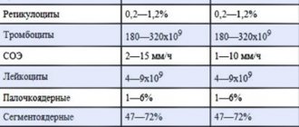
The content of protein fractions of blood serum, obtained using paper electrophoresis, on average for an adult is:
albumins 55.4-65.9%
α1-globulins 3.4-4.7%
α2-globulins 5.5-9.5%
β-gdobulins 8.9-12.6%
γ-globulins 13-22.2%
Densitometric method. In a special apparatus (densitometer), a beam of light is passed through the electropherogram, the absorption of which depends on the optical density of the colored protein spots. The light passing through the electropherogram is captured by a photocell and converted into an electric current, the vibrations of which are recorded on a paper sheet in the form of a curve, each peak of the curve corresponds to a certain protein fraction.
Figure 2. Electropherogram of human serum.