A person who does not have the necessary amount of medical knowledge may be somewhat confused when distinguishing between professional terms and trying to understand why the fetal week differs from the obstetric one. In approximately the same way, some women consider the pregnancy period to be 9 months, wondering why 9 months is not 36 weeks, but 40. All differentiation in matters of gestation and conception stem from the fact that the third obstetric week of pregnancy coincides with the first embryonic week. At the moment when conception occurs, the doctor already calculates, for convenience, the gestational age in relation to the last menstruation.
Why is the study done and to whom is it recommended?
The three-week period refers to the early stage of fetal development, which lasts about 10 weeks. Next, the embryo becomes an embryo and takes on human shape.
At week 3, a woman often doesn’t even know that a baby is developing inside.
Hormonal changes and biochemical processes in her body have already started, but visually nothing has changed yet.
Important! It is at the 3rd week from conception that it is determined whether the pregnancy continues to develop, or whether the egg, not implanted correctly, is removed from the body through the bloodstream.
So, why do you need an ultrasound examination at 3 weeks?
- In order to find out the cause of the delay (pregnancy or inflammation).
- To start treatment on time, if necessary.
- To exclude ectopic pregnancy, which has become more common in recent years.
- To confirm or refute the results of a pharmacy pregnancy test.
Two views on the deadline
For convenience, any doctor will count from the first day of a woman’s last menstruation. The concept of obstetric term is based on this system. According to another system (embryonic), the period is calculated from the middle of the cycle. It is at this time that ovulation and conception most often occur.
In obstetric practice, the third week is the period of fertilization . The egg has matured and is moving towards the uterine cavity. At some point, a sperm penetrates into it, and then the zygote will continue to move. This new cell has a full set of chromosomes and is capable of dividing very quickly.
According to the embryonic method, once a single cell is already developing vigorous activity and actively living its special, hidden life. What about the expectant mother? If she doesn’t yet know exactly about her pregnancy, she probably guesses. After all, her period is already about a week late.
In order not to get confused in the lines , read a detailed article about this (terms of pregnancy: obstetric and embryonic).
From guesswork to certainty
Three weeks after conception, a pharmacy test will show those same two stripes. At the same time, the level of the hCG hormone in the woman’s body will increase. An appropriate blood test will confirm pregnancy. But if we talk about the obstetric third week, pregnancy has barely begun and is not yet determined. Moreover, a woman can still get her period.
According to medical research, 70-75% of fertilized eggs are rejected by the female body even before they are fixed in the uterine cavity. This happens for many reasons:
- initial cellular defects;
- hormonal disorders;
- infectious diseases;
- stress.
Sometimes rejection occurs without any prerequisites at all. Since the embryo has not actually begun to develop, this phenomenon is not considered a miscarriage. The woman may, in principle, not know what happened. For her, one day the next menstruation will simply come.
What will the examination show?
On average, ovulation occurs on the 14th day of the cycle, then the egg moves through the fallopian tube into the uterine cavity. If, during the short period of its existence, a sperm manages to fertilize this egg, then a delay in menstruation will begin. According to obstetric calculations, this will be the third week of pregnancy.
Of course, it is still impossible to see the baby at this stage, simply because it is still a cluster of cells - a blastocyst, which is constantly “busy” with further division. During the division process, the formation of the baby's future organs occurs. The number of cells in the blastocyst at the end of 3 weeks is approximately 250.
So what does an ultrasound show at 3 weeks of pregnancy:
- hyperplasia (thickening) of the endometrium;
- the presence of the corpus luteum and the functioning of its blood flow;
- the place of thickening of the mucosa, which determines the place of attachment of the fetus. Its location indicates whether there is an intrauterine pregnancy or not;
- the shape and condition of the appendages and uterus, the presence or absence of pathologies in this area;
- if a fertilized egg is present, measurements are taken and the cavity is examined. At 3 weeks, the fertilized egg looks like a tiny speck on the monitor screen.
The implantation period has already been completed by this time, and each woman can feel it differently: some will complain of toxicosis, while others will not feel anything. At this time, there is a high risk of the formation of various pathologies and developmental anomalies, in other words, deformities.
Reference! It is from the third week of pregnancy that all the organs and systems of the unborn child are formed, so the mother needs to think now about her lifestyle, nutrition, emotional and physical state.
If the birth of a baby is planned, then this is not difficult to do.
Feelings at 3 weeks of pregnancy
As mentioned above, the third week of pregnancy is often perceived by a woman as a period of PMS. The engorged mammary glands and the associated increased breast sensitivity are misleading. Hormonal changes in the body are also accompanied by changes in mood: moodiness, irritability, emotional instability, nervousness. All these symptoms are characteristic both of the time “shortly before menstruation” and the period “the first weeks of pregnancy”.
Among the not very pleasant sensations for early pregnancy, one can recall the phenomenon of increased fatigue: many accomplished mothers tell how in the early stages of pregnancy they “didn’t have enough strength for anything and constantly wanted to sleep.”
It should be remembered that there are not and cannot be any special, universal signs and sensations of the third week of pregnancy: each woman experiences it individually. Moreover, often the third week of pregnancy passes “secretly” for the expectant mother: if the couple did not plan the pregnancy, what happened can well be explained by the woman as the next period is approaching.
Mother's condition and fetal development
The first signs of pregnancy are:
- increased urge to urinate;
- breast swelling;
- increased basal temperature;
- change in food habits, worsening or increased appetite;
- nausea;
- aversion to certain odors;
- discharge, not abundant and homogeneous, possibly bloody;
- possible pain in the lower abdomen;
- rapid fatigue and severe drowsiness during the day.
Each woman handles pregnancy differently, and a lot depends on how you set yourself up.
If the baby is long-awaited, love and understanding reign in the family, and the woman is not prone to impulsiveness and soul-searching, the pregnancy will pass without unnecessary drama.
And if some medical difficulties arise, a calm and confident mother will endure them more easily, at the same time saving the child from stressful situations.
How do they do it?
At 3 weeks of pregnancy, an ultrasound will help determine the exact period of fetal development. If there is no menstruation, but pregnancy is not detected by ultrasound, with the help of an examination you can find out exactly what diseases caused this.
Ultrasound examination in early pregnancy is performed with a transvaginal sensor. This method has almost universally replaced the outdated transabdominal ultrasound; it is considered safer and more informative.
The procedure goes like this: the doctor asks the patient to take off her underwear and, lying on the couch, spread her knees. The doctor puts a special disposable condom on the vaginal sensor for ultrasound (it happens that you have to bring it with you, this needs to be clarified in advance).
The thickness of the sensor is no more than a couple of centimeters, it is about 20 cm long, so there should be no pain or discomfort during the examination.
Who is recommended for early stage ultrasound?
At 3 months of pregnancy, experts recommend performing an ultrasound examination for women with complaints of pain and discomfort in the abdomen and its lower part.
Such symptoms may occur with an ectopic pregnancy or threatened miscarriage. Bloody discharge is also a reason to prescribe an ultrasound examination.
Sometimes a woman has all the signs of pregnancy, but there is no fetus in the uterus. Instead, bubbles appear, their number is constantly growing. This disease can be detected by ultrasound.
Photo:
If there are any negative manifestations of pregnancy, ultrasound makes it possible to identify all problems at an early stage and carry out the necessary treatment.
If the study is carried out at an early stage, the gestational age can be more accurately determined. At later stages, information about the time of embryo development will be less accurate.
A missed period does not always mean pregnancy. Sometimes it is the result of a disease. This is why an ultrasound is recommended.
A disease detected in its early stages can be cured faster than an advanced disease.
If there are no negative signs of early stage pregnancy, then specialists most often will not prescribe an ultrasound examination.
There is an opinion that ultrasound has a bad effect on the fetus and its development, on the structure of DNA. But this statement has not been proven.
There is also no evidence that ultrasound is completely harmless to the unborn baby. All theories about the dangers of ultrasound are just hypotheses.
Ultrasound diagnostics is the most accurate way to determine the presence of pregnancy. An ultrasound will show an “interesting” position already in the third week after fertilization has occurred.
Thus, pregnancy can be determined 4-5 days after a delay in menstruation occurs.
READ Features of fetal development on ultrasound at 16 weeks of pregnancy
Video:
Sometimes a woman feels changes in her body, but during the research procedure the result is negative.
In this case, the specialist may refer the woman for a repeat ultrasound. If the fertilized egg is not detected, it is worth conducting more thorough studies to exclude or confirm an ectopic pregnancy.
How to prepare?
For periods of 3 weeks, abdominal examination (that is, through the anterior wall of the abdomen) is rarely performed due to its low information content. Most likely, the doctor will suggest examination using a vaginal sensor. Preparation for such a procedure is to maintain hygiene: before the examination, you should take a shower and empty your bladder.
If the ultrasound is performed in a budget clinic, prepare:
- condom for ultrasound;
- a towel or sheet for covering;
- a towel or napkins if you need to remove any remaining gel.
If the study is planned in a private clinic, then all the necessary details are usually already included in the cost of the procedure.
Often the diagnostic room has a pre-registration. It is better to find out about this in advance from the medical registrar, so as not to waste time, but to meet the appointed time.
Preparing for diagnosis
In the 3rd week, preparation before diagnosis is required. Without certain measures, the survey will not be effective. For a better examination, you need to reconsider your diet. Excluded from the menu:
- cabbage;
- corn;
- legumes;
- carbonated drinks;
- radish;
- radish;
- beets;
- sauces;
- marinades;
- fried foods;
- fatty foods;
- fast food;
- peaches;
- pears;
- apples.
Fried and spicy foods should be excluded from the diet
It is better to drink 1 liter of water. Meals should be fractional and balanced.
You should not overeat or fast on the day of the examination. You should give preference to a light breakfast.
In the 3rd week, most women experience toxicosis. That is why it is recommended to take a bottle of water and an apple with you for a snack. Another important condition is not to worry. Otherwise, the pregnant woman may become very ill during the examination.
During a transvaginal ultrasound, a woman should take a condom with her. In the morning you need to take a shower and put on clean underwear. During transabdominal diagnostics, you should first drink a large amount of fluid. This is required in order to raise the uterus. There is not enough amniotic fluid for examination.
Read also: how to choose a condom for ultrasound.
If you are having a transvaginal ultrasound, you should take a condom with you.
Regardless of the diagnostic method you choose, you should take with you:
- replacement shoes;
- towel;
- napkins;
- exchange card.
All data obtained during the examination are entered into a protocol.
In what cases is it prescribed?
The attending physician may recommend an ultrasound even in such a non-standard period for examination as 3 weeks from conception, if a woman:
- experiences pain of any kind in the lower abdomen;
- sees bloody discharge when pregnancy is established;
- She thinks she is not pregnant, but there is a delay.
In other cases, the doctor will not prescribe an ultrasound at 3 weeks of gestation; it can only be performed by the woman herself if she feels the need for the procedure.
Pathological signs
It happens that in the third week of pregnancy, the expectant mother feels a tug in the lower abdomen, and the pain radiates to the area where the pelvic bones join the spine or to the area of soft formations located in front, between the pubic bones. To relieve these symptoms, you should lie down and rest for a while.
From time to time, doctors advise taking medications and vitamins that reduce the tone of skeletal muscles. They prevent painful contractions of the uterine muscles. But in cases where the pain does not stop, and the number of spasms increases, an ambulance should be called. Such symptoms may indicate the development of a spontaneous miscarriage.
Ectopic pregnancy
There are often cases when the fetus is attached outside the uterine space. However, qualified gynecologists, together with new diagnostic methods, are able to recognize a bad situation in the early stages and prevent risks.
Spontaneous termination of pregnancy
When the fetus is already attached to the uterine cavity, but suddenly it is rejected, a miscarriage results. The most common symptoms of spontaneous miscarriage are severe pain accompanied by spotting or even bleeding.
If a woman reacts to dangerous signs in time and also pays attention to the pulling sensations in the lumbar region and lower abdomen, there is a chance of saving the pregnancy.
When expectant mothers are admitted to the clinic “for safekeeping,” gynecologists-obstetricians most often put the threat of unauthorized termination of pregnancy as the reason. The most dangerous period is the first 12 weeks. Sometimes women are unaware of their condition when they arrive at the gynecological department.
Frozen pregnancy
Another dangerous situation for women's health is frozen pregnancy. Often signs of fetal developmental arrest are invisible. The situation can be diagnosed in a timely manner in medical institutions using laboratory tests.
hCG
On days 6-8 after fertilization, a special hormone begins to be produced in the pregnant woman’s body. The outer shell of the embryo has tissue called chorion, which produces the hormone human chorionic gonadotropin.
The content of this protein constantly increases in the first months of pregnancy, which indicates that the fetus is developing normally.
Ultrasound
Ultrasound examination also helps to ensure the harmonious development of the embryo. It confirms the results of blood tests for hCG. Moreover, ultrasonic waves are so gentle that they cannot harm a fetus at 3-5 weeks of development. If there are symptoms of a threatened miscarriage, an ultrasound is always prescribed to monitor the situation.
It is very difficult for a woman to lose a child and often after the incident she becomes despondent. However, you should get through the difficult period as quickly and easily as possible, because the next pregnancy may come very soon, and you need to meet it without stress.
What can be seen in the photo of the fetus?
Three weeks is such a short period of time that it’s premature to talk about the baby’s first photo..
It is still an egg, about 4 mm in diameter. In the picture it looks like a small dark spot in the uterine cavity.
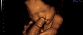
3D ultrasound is usually not performed at this stage, as it is not informative. It is recommended to conduct this study starting from week 16.
Is it harmful to have an ultrasound examination?
Neither safety nor obvious harm has yet been proven by ultrasound. However, there are scientific studies whose results are alarming. Since the advent of medical ultrasound diagnostics, many experiments have been carried out, the purpose of which was to identify the possible harm of the procedure on the human body.
One of them concerned the functioning of DNA: it is known that DNA molecules have their own acoustic spectrum, that is, a sound that differs in different tissues and organs.
Domestic biologists isolated cellular DNA, placing it in a special solution, and established the acoustic spectrum of the sound of molecules, which turned out to be very wide - from a tenth of a hertz to several hundred hertz. The DNA was then treated with ultrasonic waves at the frequency of a medical device.
A control measurement showed that the sound produced by the molecules became uniform and amounted to 10 Hz for DNA of any type. This effect lasted for several weeks and can hardly be called a positive effect of ultrasound on human DNA.
Scientists have come to the conclusion that a failure in the acoustic properties of DNA increases the risk of developing pathologies of internal organs and plays the role of a carcinogen - a substance that provokes the development of oncology.
Similar experiments were carried out by the Americans, who officially do not recommend that pregnant women undergo ultrasound without strict indications, especially 3D and 4D ultrasound, the radiation of which is stronger.
Where to do it and how much does it cost?
But if the harm of ultrasound is only a matter of debate in scientific circles, then the benefits of the procedure are more obvious.
In some cases, medical diagnostics are vitally necessary to identify dangerous pathologies such as hydatidiform mole or ectopic pregnancy .
An ultrasound examination can be performed at the 3rd (5th obstetric) week in a clinic at your place of residence or, if the decision about the need for an ultrasound is made independently, in any private clinic.