The level of modern medicine allows expectant mothers to trust doctors and be calm about the health of their unborn baby. After all, ultrasound diagnostics, as one of the mandatory studies, makes it possible to monitor the state of fetal development and, if necessary, prevent force majeure circumstances. Ultrasound at 32 weeks is the third planned ultrasound examination, which is performed by a doctor to identify delayed development of the baby and examine the condition of the placenta. Let's talk about this stage of pregnancy and the study itself in more detail.
A little about ultrasound as a diagnostic method
Ultrasound has been used in medicine for more than half a century. It’s hard to say whether it’s a lot or a little, because the speed of scientific and technological progress is very high. During this time, ultrasound research methods have developed and improved, making them one of the most widespread throughout the world.
- The reasons why this method does not stand still are very significant. Ultrasound is one of the safest diagnostic methods. Ultrasound radiation does not cause any harm to the body of the fetus or mother. This radiation itself is a very high-frequency sound that is inaudible to the human ear.
- Also, one more, almost the most important point is the quality and accuracy of the results. Ultrasound diagnostic methods fully satisfy all the requirements of doctors. And more developed techniques, for example 3D ultrasound, allow you to create a full-fledged three-dimensional model. The 3D ultrasound video obtained as a result of the study at 32 weeks is transmitted to parents for their personal viewing. Many families celebrate this as a pleasant and unforgettable moment.
In addition to ultrasound, magnetic resonance imaging (MRI) can be cited as an example of safe diagnostic methods, but this method is much less accessible and some structures of the fetal body may not be visible.
As for the question: “Is ultrasound wrong at 32 weeks?”, then everything is very ambiguous. If the diagnosis is carried out by a qualified specialist using good equipment, then the likelihood of an error is extremely low. The issue of error is closely related to the choice of institution where the diagnosis will be performed. Also, in case of suspected defect, the study is not carried out once.
How many times is ultrasound performed during pregnancy?
Normally, if a woman is registered at the antenatal clinic from the moment pregnancy was established, she is prescribed 3 planned tests.
- The first is at 10-14 weeks. The goal is to identify gross malformations of the fetus, size, and also to identify genetic pathology.
- The second ultrasound is performed at 20-24 weeks. There is a spectrum of vices that can be defined more broadly. The development of internal organs, limbs, etc. is assessed. This will allow us to identify diseases that threaten not only the fetus, but also the mother, as early as possible. This is due to the fact that sometimes an artificial termination of pregnancy is necessary, which should be performed as early as possible, because it is safer and will entail much fewer consequences. It is for this reason that, in the case of a pathological decrease in hCG, an ultrasound is immediately performed, and then a therapeutic tactic is developed.
- Ultrasound at 32 weeks and above, up to 36 weeks, is the last. The condition of the placenta, blood circulation, as well as other indicators that were assessed at previous stages are assessed. The peculiarity of this ultrasound is that it most often determines intrauterine developmental delays, which are often associated with circulatory failure. But in no way should the last ultrasound be singled out more than the previous ones, because problems with blood circulation, including heart defects, can be detected at an earlier date.
Sensations signaling danger
Complications leading to gestosis, placental abruption, oligohydramnios, polyhydramnios, and the appearance of discharge are among the main ones. Considerable danger can come from various unpleasant sensations:
- The appearance of heartburn associated with a compressed stomach and acid entering the digestive system;
- Presence of baby kicks in the upper abdomen. At 32 weeks, these tremors may indicate improper placement of the fetus;
- The presence of swelling in the face, arms and legs, pronounced veins, accompanied by pain symptoms, acute pain in the back, neck and lower back.
What do they look for on an ultrasound at 32 weeks of pregnancy?
As mentioned above, an ultrasound at 32 weeks of pregnancy allows one to assess the condition of the placenta very well. This is very important in the management of childbirth. The placental insertion site is not significant for the fetus, but is extremely important for planning a cesarean section. The relationship between the location of the lower edge of the placenta and the internal opening of the cervix is important. Too close a position (less than 4 cm) can lead to severe bleeding during childbirth.
In addition to the location, the thickness of the placenta is also assessed. If the placenta is too thick or, conversely, small, this indicates its insufficiency. Thickening may also indicate the presence of an inflammatory process.
- At 32 weeks of pregnancy, an ultrasound scan also evaluates the internal structure of the placenta. It is called maturity and varies in degrees 0, I, II and III. Doctors widely use the expression “aging” or “maturing” of the placenta. Today, premature aging of the placenta is quite common. This represents premature onset of grades II, III, or both. This is not a terrible pathology and there is no need to panic. But in this case, treatment is prescribed that is aimed at preventing fetal hypoxia, that is, at improving blood flow in the uteroplacental and fetoplacental systems.
- These are not all the pathologies that can be detected. A very common disease is placental calcification. In most cases, it occurs in women who smoke. In this case, a spasm of blood vessels occurs in the placenta, due to which, due to oxygen starvation of the tissues, they die. Then, in place of the dead tissue, calcifications are formed, which represent an accumulation of calcium, in other words, “stones” are formed. These areas lose their functions. But, the placenta is a very “strong” organ and can compensate for this deficiency with the help of other, healthy vessels, if, of course, they are still able to do so and the process has not started. But even in this case, one cannot refuse therapy aimed at improving blood flow.
The indicators that ultrasound shows at 32 weeks are very important data. Their number is much greater than it might seem at first glance, and the range of pathologies is much wider than that listed above.
It is also important to understand that if, before the 32nd week of pregnancy, ultrasound indicators were normal, then this does not provide great guarantees, the absence of defects and pathologies, which means it is worth conducting this study, because it is safe and will not harm in any way, and monitoring the child’s health is much more important. more important than anything else.
What happens to a baby at 32 weeks?
At the 32nd week, fetal development proceeds at a rapid pace. The baby already weighs 1.7-1.9 kg and is 43-45 cm long. The formation of the brain is almost complete, the head becomes proportional. The baby’s layer of subcutaneous fatty tissue increases, causing the body to smooth out, wrinkles disappear, and facial features are more clearly formed. The central nervous system is working better and better, and the child is no longer so afraid of the bright light penetrating inside the mother’s tummy.
The baby’s connection with his mother also strengthens: he feels how all her internal organs work, hears his mother’s voice, and instantly responds to changes in her mood. After birth, when the baby is placed in the woman’s arms, he will immediately recognize the closest person in his life.
After an ultrasound at the 32nd week of pregnancy, the position of the fetus in the uterus may still change, but usually at this time the baby is already completely turning head down. In this case, his legs may rest against his mother’s ribs, and this causes discomfort. To alleviate the condition, doctors advise pregnant women to try to support their back.
How an ultrasound is performed at 32 weeks of pregnancy and preparation for it
- No preparation is necessary for this fetal examination. There are also no contraindications to the procedure. The only reason why an ultrasound at 31-33 weeks may not be performed is the human factor and the prejudices of parents. Ultrasound examination is performed only using the transabdominal method. At this stage, there is enough amniotic fluid, which is a good conductor for ultrasonic waves.
- The pregnant woman arrives at the appointed time and lies down on the couch. The doctor then applies a special gel to the area to be examined. It is necessary to improve the conductivity of ultrasound; more precisely, it removes excess air between the sensor and the skin, which prevents the conduction of ultrasound waves, as it leads to distortion and a significant decrease in the reliability of the results.
The ultrasound will take 10-30 minutes, after which there will be a short wait and the woman will receive the results of the study, with which she should go to her obstetrician-gynecologist.
What can be seen in the photo of the fetus?
In addition to anatomical indicators, an ultrasound examination is a chance to “get to know” the child in absentia.
In a budget clinic they do not print out ultrasound images, but in a private medical center this is easy to do.
Today, many pregnant women begin their baby’s photo album with ultrasound photographs, in which you can see the baby’s profile, the outlines of his arms and legs (and if the photo is high-quality, you can count the fingers on your hands).
In girls, with sufficient magnification, you can clearly see the labia, in boys - the penis. However, in an ultrasound photo without a doctor’s advice and without searching for the right angle, this can be problematic.
Photo of a girl
Photo of a boy
Photo 3D
Interpretation of ultrasound results and norms at 32 weeks of pregnancy
There are many indicators that may differ between different devices, below are some of them :
- Biparietal size. An indicator that evaluates the width of the head between the temples using the minor axis.
- Head circumference. Actually the circumference of the entire head.
- Fronto-occipital size. Another head size. It is calculated by measuring the segment drawn between the most distant points of the forehead and the back of the head.
- Abdominal girth. An indicator from which the volume of the abdominal cavity can be calculated.
- The dimensions of the shoulders, shin bones, hips, forearms, and necessarily height and weight are also assessed.
When assessing indicators, you can highlight ultrasound norms at 32 weeks of pregnancy, but this will not be one specific number, but a range, because all people are unique and this uniqueness is reflected even before birth. Below is a table with normal ultrasound results from 31 to 33 weeks of pregnancy.
Preparation and conduct of the study
No special preparation is required for screening in the third trimester (diet or drinking water before the procedure). A woman needs to sign up for an examination and take a shower before going to the ultrasound. You need to have a towel for the couch, disposable wipes for removing gel and shoe covers.
If a woman smokes at 32 weeks of pregnancy, she needs to refrain from smoking for 2 hours before the procedure.
A typical two-dimensional ultrasound lasts no more than 20 minutes. It is carried out as follows:
- The doctor invites the patient into the office and invites her to lie down on a couch covered with a towel. You need to put a roller or fist under your side. The belly is exposed.
- A layer of special gel is applied to the patient’s stomach. It is cold, so it often causes discomfort.
- The doctor moves the sensor over the abdomen and examines the image that appears on the screen. In the process, he tells the expectant mother the most important facts about her fetus.
- After the procedure, the woman gets up from the couch, removes the remaining gel, and gets dressed. After 5–10 minutes, she receives a description of the examination and, if desired, a photo of an ultrasound scan of the fetus.
Three-dimensional and four-dimensional ultrasounds take longer (40–60 minutes). But the expectant mother gets the opportunity to watch a real color film about her baby.
Interpretation of ultrasound from 31 to 33 weeks of pregnancy
| A week | 31 | 32 | 33 |
| Height, cm | 39-42 | 40-43 | 43-44 |
| Weight, g | 1300-1500 | 1600-1700 | Around 2000 |
| BDP (Biparetal size) mm. | 80-87 | 82-89 | 84-91 |
| Fronto-occipital size, mm. | 101-109 | 104-113 | 107-116 |
| Head circumference, mm. | 294-315 | 304-325 | 311-333 |
| Abdominal girth, mm | 274-301 | 286-314 | 296-325 |
| Femur length, mm. | 61-66 | 63-68 | 65-70 |
| Length of shin bones, mm. | 56-61 | 58-63 | 60-65 |
| Length of humerus, mm. | 56-60 | 58-62 | 59-63 |
| Length of forearm bones, mm. | 49-53 | 50-54 | 52-56 |
The results are assessed by an obstetrician-gynecologist who “knows” this pregnant woman from the very beginning of pregnancy, and also has all the necessary data, especially if there is a risk of congenital pathologies, such as heart defects.
Based on this, when he evaluates the results of an ultrasound at 32 weeks of pregnancy and the normal indicators, he can evaluate certain deviations from the norm in dynamics, because he knows the entire course of pregnancy and can assume some characteristics of the fetus, because those same deviations can be and the norm. Therefore, you should not get too attached to numbers, especially without a medical education.
Decoding
An ultrasound scan of the fetus at 32 weeks shows the level of its development. It allows you to carefully look at the brain, analyze the cerebellum, hemispheres, and lateral ventricles. Their width varies around 10 ml. If this indicator is more than average, then this may indicate the presence of hydrocephalus. Most often, this pathology is caused by the pressure exerted by amniotic fluid on the baby's head.
Next, they look at the ultrasound of the face, which should include proportionally located and formed eye sockets. During examinations using 3D equipment, pathologies may be detected that are invisible during two-dimensional ultrasound. If diagnostics are done using modern equipment, then hernias and spina bifida can be examined. Ultrasound waves allow you to assess the condition of the skin. Now it is distinguished by density and a flesh-colored tint. The baby seems to be smoothing out as a result of the accumulation of the subcutaneous layer.
More to read:
How to cure cough during pregnancy
Decoding the results has the following parameters:
- BPR – within 85 mm;
- LZR – 102 mm;
- Exhaust gas – 311 mm;
- Coolant – 272 mm;
- DB – 62 mm;
- height – about 43 cm;
- weight – within 2000 g.
The gynecologist can compare the size of the fetus with the pelvis of the expectant mother. Particular attention is paid to the level of lung development; it is assessed whether the baby will be able to breathe after birth.
- the period from 27 to 34 weeks corresponds to the first degree of maturity;
- from 35 to 39 weeks - second degree;
- from 37 to 40 weeks - third maturity.
The following indicators are also assessed. Location of the placenta. Its lower edge should be within 7 cm of the internal os, at first maturity, about 32.2 mm thick. The volume of amniotic fluid is from 77 to 269 mm. The umbilical cord consists of 1 or 2 veins, without entwining the fetal neck, the length of the cervix is within 30 mm, the uterus should be without increased tone.
Carrying out diagnostics with Doppler has the following standards:
- SDO of the umbilical artery – from 2.48 to 2.52;
- SS – from 32 to 39;
- IR of the umbilical cord arterial vessel from 0.52 to 0.75;
- PI of the umbilical artery – from 0.64 to 0.89;
- IR of the uterine artery – from 0 to 0.65;
- SDO VSA – from 4 to 6.5;
- IR of the ICA – from 0.79 to 0.81;
- Aortic IR – 0.83;
- SDO – from 3.9 to 8.
What other methods are used
In addition to conventional ultrasound, ultrasound and 3D ultrasound are used:
- Doppler ultrasound, also known as Doppler ultrasound, is a method used to assess blood flow. Using the Doppler effect, you can determine the speed, direction and possible problems in the blood flow. When there is fetal growth restriction, an ultrasound scan is almost always performed, because the most common cause is problems with blood flow. They are especially often observed in pregnant women who smoke, because in addition to placental calcification, there are other pathologies that lead to fetal hypoxia.
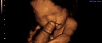
- But three-dimensional ultrasound diagnostics (3D ultrasound) is one of the most modern methods. It allows you to create a full-fledged three-dimensional model of the fetus. Previously, it was believed that this method was harmful to the fetus and should be used only when necessary, but today some parents are increasingly turning to this method. After all, it’s not every day that you can look at your child even before his birth, and even more so get a photo that is a good memory and improves contact between mother and child, even before his birth.
3D ultrasound at 32 weeks of pregnancy photo
Diagnosed pathologies
The third screening can diagnose the presence of the following pathologies:
- fetoplacental insufficiency, which occurs when placental maturity does not correspond to the term;
- umbilical cord entanglement. This condition does not pose a threat to the fetus if it does not cause disruption of blood flow;
- discrepancy in the size of the baby, breech presentation;
- predisposition to premature birth, which manifests itself in a shortened cervix;
- altered volume of amniotic fluid, which can manifest itself as both polyhydramnios and oligohydramnios;
- if there is a scar, identify its failure;
- oxygen starvation of the fetus.
An ultrasound performed at 32 weeks allows us to assess the condition of the baby and the woman. This diagnosis is important because it makes it possible to improve the baby’s condition.
Conclusion
Ultrasound diagnostics is an integral part of modern obstetrics and gynecology. Ultrasound during pregnancy has good reviews from both doctors and parents. Thanks to her, it became possible to prevent very serious and life-threatening diseases for the child and mother. And this is not some local data - this is information from all over the world and it’s hard to imagine how many parents and their children this method has helped.
However, it is worth understanding that you cannot limit yourself to ultrasound alone. It is also worth using other diagnostic methods, both instrumental and laboratory. Also, you cannot do without consulting a doctor, since without a correct assessment, the cost of this study is too low.