Impaired functioning of the gallbladder (hereinafter referred to as the gallbladder) affects not only the functioning of the entire digestive system, but also the health of the body as a whole. Any tiny pathologies can develop to extreme stages, and if they are not diagnosed and treated, they can even cause death. Ultrasound of the gallbladder has long been recognized as one of the simplest and most accessible methods for examination, allowing one to assess the functional state of the organ itself, as well as the bile ducts. The procedure, despite its simplicity, requires careful preparation of the patient and is distinguished by a special technique.
Indications for ultrasound
This study is prescribed by a gastroenterologist or therapist in the following cases:
- dull or paroxysmal pain in the right hypochondrium;
- suspicion of cancer;
- bitterness and dry mouth in the morning;
- cholelithiasis;
- biliary dyskinesia;
- icteric staining of the sclera and skin;
- dynamic monitoring of chronic diseases;
- changes in the level of bilirubin in the blood;
- obesity;
- chronic alcohol intoxication;
- abdominal injury;
- abuse of fatty foods;
- depleting diets;
- eating disorder.
Cholecystitis in women is a contraindication to taking hormonal drugs. When choosing contraceptives, an ultrasound of the abdominal cavity is performed.
Problem 2
An 8-year-old boy complains of abdominal cramps after eating.
Gallbladder on an empty stomach:
The topography has not been changed. Shape – S-bend. The walls are not changed. The contents are homogeneous, there are no stones. Perivesicular tissues are not changed. Choleretic “breakfast” – Chofitol 20% 8 ml. After taking the medicine, nausea and abdominal pain appeared.
| Time, minutes | 0 | 10 | 20 | 30 | 40 | 50 | 60 | 70 |
| Volume, cm3 | 36 | 21 | 18 | 27 | 33 | |||
| FV, ml | 18 | |||||||
| EF, % | -50% | |||||||
| Reduction | + | + | ||||||
| Relaxation | + | + | ||||||
| OZhP, mm | 2 | 2 | 2 | 2 | 2 |
Time of maximum contraction - from taking a choleretic drug to the minimum volume of the gallbladder (N 20-40 minutes): 20 minutes.
Latent period - from the moment of taking the choleretic drug to the beginning of contraction of the gallbladder (N up to 5 minutes): up to 5 minutes.
The primary reaction is an increase in the volume of the gallbladder during the latent period: absent.
Duration of contraction - from the beginning of contraction to the minimum volume of the gallbladder (N 15-30 minutes): 15 minutes.
Ejection fraction – EF(%)=(1-Vmin/Vmax)*100%, where Vmin is the minimum and Vmax is the maximum volume (N 40-70%): 50%.
Conclusion: Accelerated emptying of the gallbladder due to strong contraction of the muscle wall.
How to prepare for research
Preparing for an ultrasound of the gallbladder involves following a diet and taking medications. It is recommended to follow a diet for 2 or 3 days before the session.
The recommended diet includes foods:
- buckwheat, oatmeal with water;
- low fat cottage cheese 2.5%;
- soft-boiled egg;
- a piece of boiled chicken or beef.
Foods that cause flatulence are excluded from the menu:
- yeast baked goods;
- legumes – lentils, beans, peas;
- fruits, vegetable salads, greens without heat treatment;
- coffee, alcohol, carbonated drinks, milk.
Classic patient pose for pain in the gallbladder area
Medication preparation for the procedure for 2–3 days:
- It is recommended to take enzymes three times a day with meals - Festal, Creon, Pancreatin, Panzinorm;
- sorbents Enterosgel or Activated carbon 3 times a day between meals;
- Taking the carminative drug Espumisan three times eliminates flatulence.
Necessary actions before the ultrasound:
A light dinner at 7 p.m. is recommended. Before going to bed, you need to empty your bowels naturally. If there was no stool, put a glycerin suppository or make a Microlax microenema.
Actions on the morning of the examination:
- You only need to come to the procedure on an empty stomach. Can I drink liquid before the procedure? Drinking water is not recommended. Otherwise, a reflex release of bile will occur. A shortened gallbladder will give false results.
- What to do if the ultrasound is scheduled in the afternoon? In the morning, have a snack with crackers and tea. There should be 6 hours between breakfast and the session. If necessary, you can drink water 2-3 hours before the scan.
- Infants under 1 year of age are not given food and water 3–3.5 hours before the ultrasound.
- A child under 3 years of age is not given food or water 4 hours before the procedure. For children over 8 years old, the interval is 6 hours.
- Before the procedure, you should not smoke or use chewing gum.
Problem 1
A 10-year-old girl complains of periodic abdominal pain, unrelated to food intake.
Gallbladder on an empty stomach:
The topography has not been changed. The shape is a bend in a funnel. The walls are not changed. The contents are homogeneous, there are no stones. Perivesicular tissues are not changed. Choleretic “breakfast” – Chofitol 20% 10 ml.
| Time, minutes | 0 | 15 | 25 | 35 | 40 | 50 | 60 | 70 |
| Volume, cm3 | 18 | 12 | 10 | 13 | ||||
| FV, ml | 8 | |||||||
| EF, % | -44% | |||||||
| Reduction | + | + | ||||||
| Relaxation | + | |||||||
| CBD, cm | 0,17 | 0,3 | 0,3 | 0,2 |
Time of maximum contraction - from taking a choleretic drug to the minimum volume of the gallbladder (N 20-40 minutes): 25 minutes.
Latent period - from the moment of taking the choleretic drug to the beginning of contraction of the gallbladder (N up to 5 minutes): up to 5 minutes.
The primary reaction is an increase in the volume of the gallbladder during the latent period: absent.
Duration of contraction - from the beginning of contraction to the minimum volume of the gallbladder (N 15-30 minutes): 20 minutes.
Ejection fraction – EF(%)=(1-Vmin/Vmax)*100%, where Vmin is the minimum and Vmax is the maximum volume (N 40-70%): 44%.
Conclusion: Timely emptying of the gallbladder. At the time of the study, gallbladder dysfunction was not determined.
Ultrasound technique
You must enter the scanning room without metal objects on your clothes or head. The person is asked to lie on his back and free the abdominal area from the shirt. The doctor applies a gel to the transducer to eliminate the air cushion between the body and the sensor upon contact. If the gallbladder is not visualized, the patient, at the doctor’s request, inhales deeply, holds his breath, or turns on his left side. To identify stones, a person must bend forward several times.
Preparing for Function Scanning
An ultrasound of the gallbladder with determination of function is performed by a functional diagnostics doctor. The method reveals changes in the organ and its ducts after a choleretic breakfast. To determine contractility, monitoring is carried out with a functional test. But you won’t be able to do the research quickly. Preparation for the procedure is the same as for scanning the abdominal organs.
Diet before ultrasound a week before diagnosis:
- refusal to drink alcoholic beverages;
- exclude from the diet foods that cause flatulence - vegetables and fruits, whole milk, legumes and black bread;
- You are allowed to eat boiled fish and lean meat, water-based porridge, steamed cutlets, and dried bread.
Medication preparation 3 days earlier (you must coordinate the appointment with your doctor):
- Enzymatic agents - Pancreatin 10,000 units with each meal, washed down with a glass of water.
- For chronic constipation, it is recommended to take Lactulose daily at night.
- Drugs that stimulate the action of the intestines - Domperidone, Simethicone.
Actions the day before the procedure:
- Dinner no later than 8 pm consists of porridge with a minimum amount of sugar.
- In the evening, before going to bed, you should empty your bowels. If there was no stool, use a glycerin suppository. Attention! You can't do an enema.
Procedure on the day of the examination:
- Separate the yolks from the boiled eggs and bring them to the procedure. Instead, you can use 200 g of 20% sour cream as a choleretic agent. An alternative is also 20 g of sorbitol per 1 glass of warm water.
- If an ultrasound examination is performed in the middle of the day, you can have unsalted cheese, dried bread, and tea for breakfast at 7 a.m.
- You should not drink water before the session. Otherwise, the bile will be released before scanning. The results will be wrong.
When preparing a child, follow the same recommendations, only without the use of medications. Before the session, children under 3 years of age are not given food for 3 hours. The same interval before ultrasound of the gallbladder of a child up to one year old. Older children are not fed for 6 hours.
Research methodology
Ultrasound of the gallbladder with determination of function takes place in 4 successive stages.
First stage
Similar to conventional ultrasound examination. The procedure is carried out before taking a choleretic breakfast and is performed using a special sensor using a gel. The patient lies on his back.
Second phase
The patient takes breakfast prepared the day before. The ultrasound examination is repeated, the results are compared with those before the load.
Third stage
Wait 15-20 minutes and repeat the ultrasound examination.
Fourth stage
A repeat study is done 40-45 minutes after taking a special breakfast.
The difference between this echography and an ordinary ultrasound is that during a stress test the functional state of the gallbladder is assessed.
Interpretation of results
The interpretation of the ultrasound of the gallbladder is done 45 minutes from the start of the scan. During the scan, the doctor analyzes the following indicators: organ localization, bladder parameters, contractility, wall thickness, presence of sand or stones, diameter and patency of the ducts, whether there are neoplasms or polyps. The analysis takes into account the norms of organ parameters.
Dimensions of the gallbladder when examined by ultrasound:
- width 2–4 cm;
- length from isthmus to bottom from 4 to 10 cm;
- wall thickness does not exceed 3–4 mm;
- diameter of the common duct 6–8 mm;
- the internal diameter of the lobar ducts does not exceed 3 mm;
- The volume of the bladder in an adult is 35–70 cm3.
Qualified doctor's opinion
The normal size of this organ in children depends on height and body weight. The capacity of the bubble is calculated using the formula 0.5 x A x B x C. The value of A, B, C is length, width, thickness.
If the volume of the gallbladder is 60–80%, they speak of unimpaired function of the organ. A level above 80% indicates an increase in the contractility of the organ. In this case, based on ultrasound, a diagnosis of gallbladder dyskinesia of the hypertensive type is made. Volume less than 60% indicates dyskinesia with reduced motor function.
The results of the study are influenced by preparation for ultrasound of the gallbladder. Therefore, you need to strictly follow the doctor’s recommendations. Normally, an organ without pathology is pear-shaped, the cavity does not contain sand and stones. The walls are of normal thickness and contract after breakfast.
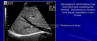
How to decipher an ultrasound photo of the liver
The results will be reliable if the preparation for ultrasound of the liver and the procedure itself were performed correctly. After diagnosis, an assessment of the liver condition is required. Many patients have questions not only about how to prepare for an ultrasound of the liver, whether they can eat before the examination and what foods, but also about the possibility of deciphering the ultrasound photo themselves. However, it is difficult to do this without the help of a specialist, since many important signs need to be taken into account.
| Name of the indicator under study | Standard parameter | Deviations from the norm |
| The degree of echogenicity of the parenchyma | It must be homogeneous, that is, ensure uniform transmission of ultrasonic waves. The value of this indicator is lower compared to | If the ultrasound picture is heterogeneous, the doctor may assume that the liver tissue has changes. This may be a sign of such diseases |
| Organ length value | Right lobe – up to 15 cm, left – up to 12 cm | An enlarged liver may indicate the presence of hepatitis, cirrhosis, etc. If the liver is reduced, the doctor will diagnose the last stage |
| Condition of the fabric in terms of grain | Fine-grained appearance | If the surface is lumpy or has tissue nodules, the doctor may diagnose cirrhosis. |
| Volumetric formations | No | If changes in the form of space-occupying formations are detected, then this is a sure sign of the presence of cysts or an abscess. |
When interpreting the results of an ultrasound examination of the liver, the doctor also establishes the presence or absence of pathological changes in the vessels through which blood circulates.
The presence of pathological changes in the portal vein will be indicated by a deviation from the norm in the transverse size (no more than 13 mm). This portal vein indicator is influenced by the patient’s breathing state at the time of diagnosis. Also, the movement of blood through the portal vein should be towards the liver. The opposite direction is a sign of the development of a disease such as portal hypertension. To measure the speed of blood flow, the breath-holding method is used. At the same time, the norm for this indicator is within 24 cm/sec.
If there are no pathological changes in the structure of the hepatic artery, then it is a branched vessel, characterized by a normal size of no more than 6 mm and a blood flow speed of 80 cm/second. The structure of the hepatic veins can be presented in two variants: with numerous or three branches. In the absence of pathological changes, their size is within the normal range from 0.6 to 1 cm. When assessing the inferior vena cava, the doctor focuses on its diameter, which according to the norm should be 2-2.5 cm.
To get a more complete picture of the nature of blood flow, a medical specialist will complement the vascular ultrasound with an additional procedure - Dopplerography.
What diseases does ultrasound detect?
What does the examination show? Ultrasound detects diseases:
- The most common pathology is cholecystitis. The scan reveals an enlarged bladder with thickened walls. The cavity contains bubble inclusions and partitions. The contours of the light-colored walls are not clearly visible on the monitor screen. In a chronic process, the organ decreases in size and becomes deformed.
- Cholelithiasis is a gallstone disease. Ultrasound identifies stones in the bladder and ducts. When the body position changes, they shift. The walls of the organ are thickened with uneven edges. A sign of small stones on ultrasound is an expansion of the duct above the blockage. Stones are more often found in women than in men.
Cholelithiasis
- Dyskinesia of the biliary tract is manifested during scanning by increased tone and thickening of the walls of the bladder. A bend in the neck is detected.
- The tumor is visualized as a mass. The walls of the deformed bladder are thickened.
- Polyps appear on the monitor as round formations. A size greater than 1 cm requires dynamic monitoring, as there is a risk of a malignant process.
- Congenital pathology – double gallbladder or diverticulum.
The study of this part of the liver is carried out at the gastroenterology center. It is better to combine the procedure in combination with all digestive organs.
What can a gallbladder ultrasound reveal?
Ultrasound diagnostics shows all abnormalities in the functioning of the organ, including at the initial stage, when it is possible to effectively treat them. It can reveal:
- inflammation (cholecystitis) in both acute and chronic forms;
- the presence of sand and hard stones in the gallbladder and ducts;
- tumors, polyps and other neoplasms;
- cessation of organ function as a result of blockage of the bile ducts and other reasons (disconnected gallbladder);
- congenital abnormal location and size of the organ;
- violation of contractile function - during dynamic studies.
The most frequently detected are cholecystitis and cholelithiasis. These diseases usually arise due to poor nutrition and accompanying digestive disorders.