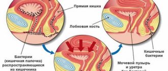
The bladder is a very vulnerable organ, especially in women. Due to the short urethral canal, infection from the genital tract can enter it, causing inflammatory processes of various types. During the last trimester of pregnancy, when the baby becomes quite large, the bladder is often subject to compression, which can also lead to pathologies.
To examine this organ of the urinary system, many diagnostic techniques are used, which in most cases are non-invasive and cannot always provide a complete picture of its condition. Whereas cystoscopy of the bladder in women allows you to thoroughly examine the entire internal surface, without missing a single area.
What is cystoscopy
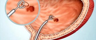
Cystoscopy is an instrumental examination of the internal structure of the bladder and urethra. At the same time, the condition of its walls, ureteral orifices, contractility, capacity, and the presence of pathological foci (inflammation or suspicion of cancer) is assessed. During cystoscopy, it is possible to take biopsy material (a piece of tissue from the most suspicious area of the mucosa) in order to clarify the diagnosis, as well as many therapeutic measures.
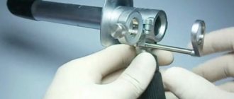
The cystoscopy procedure is performed using a cystoscope; this is a device made of a long tube (tube), a lighting device and auxiliary parts.
Cystoscopes come in standard rigid (stiff) and flexible types.
The essence of the examination
The term cystoscopy is formed by two ancient Greek words - kystis, which means “bladder”, and σκοπέω - I look, observe, examine. That is, the procedure involves examining the bladder using special instruments. The instruments for this study are called cystoscopes, and they come in two types - hard (metal tube) and soft (flexible fiber optic cord).
A special lighting system is built into the end of the devices, allowing the doctor to examine the inner surface of the urethral canal and bladder. It should be noted that recently, diagnosis is most often performed with a soft cystoscope, as this significantly reduces the risk of injury to the areas being examined.
Some women, having received a referral for cystoscopy and learned the essence of the procedure, begin to worry that such an examination will most likely be painful. This is wrong. Yes, of course, the procedure is not particularly comfortable, but, firstly, a soft cystoscope can easily penetrate the bladder, and secondly, anesthetics are almost always used to reduce sensitivity. Therefore, it may be unpleasant, but not painful!
Does it hurt
Most patients (especially women) tolerate cystoscopy well. The procedure is unpleasant, but most often it is painless. Discomfort is noted when the cystoscope is inserted and rotated. During the collection of biological material, patients complain of a burning sensation.
Types of cystoscopes
Hard or traditional
A rigid cystoscope is a metal structure with a long thin tube and a magnifying device with a lens.
A tube (tube) is inserted into the urethra and the doctor, gradually moving the device into the cavity of the bladder, examines all areas of the mucous membrane step by step.
The device then reaches the bladder cavity. For a full examination, about 200 ml of a sterile isotonic solution (usually saline) is injected into the bladder.
During the examination, indications for intervention are determined. In this case, cystoscopy becomes not only a diagnostic measure, but also a therapeutic procedure.
Flexible cystoscope or fibrocystoscope
A flexible cystoscope has a movable tube that can be given any bend, unlike the metal tube of a rigid cystoscope. Flexible cystoscopes have a mini camera at the end. The image is transferred to the computer screen.
The image is transferred to the computer screen.
The advantage of flexible cystoscopy is that images can be saved to digital media and then the dynamics of the disease can be monitored.
The procedure itself is the same in both cases, but the procedure with a flexible cystoscope is much more comfortable for patients.
Examination with a flexible cystoscope also has its disadvantages. It is often less informative than inspection with a rigid instrument.
If necessary, additional instruments are inserted into the cystoscope tube to cauterize ulcers, remove polyps and stones, and dissect adhesions.
Advantages and disadvantages of the cystoscopy method
The undoubted advantage of this technique over many other studies in urological practice is its high information content. In particular, during the procedure, malignant tumors can be detected in the bladder in the early stages, thanks to which the patient is successfully treated. In addition, cystoscopy is used to perform a wide range of therapeutic procedures, from restoring the patency of the urinary tract to removing tumors.
The disadvantage of the study is that for its successful implementation a number of conditions must be met:
- Normal urethral patency.
- The bladder must have sufficient capacity.
- The fluid used to fill the bladder during the procedure must be clear (heavy bleeding or large amounts of pus will interfere with the examination).
- The patient’s readiness for the study, absence of acute painful sensations during the procedure. Severe discomfort or the appearance of an uncontrollable urge to urinate are indications for forced termination of cystoscopy of the bladder.
The disadvantage of cystoscopy is its technical complexity. To perform the procedure efficiently, accuracy and certain experience are required, since unprofessional actions can lead to injuries to the organs of the lower parts of the urinary system.
Book a consultation 24 hours a day
+7+7+78
How to prepare for cystoscopy
Conversation with your doctor
The issue of pain relief is discussed. Most patients tolerate the procedure using local anesthetics well. Other types of pain relief are rarely used. A date and time for cystoscopy is set.
Arrive on an empty stomach
Firstly, everyone has a different reaction to the feeling of pain and bloating, which often occurs. If you do not arrive on an empty stomach, you may experience nausea and weakness.
Secondly, sometimes the procedure is started under local anesthesia, and then a short-term general anesthesia is necessary. This is necessary if during the examination there is a need for cauterization or removal of a polyp, removal of stones, and so on. The procedure is lengthened and the local anesthetic may not be enough.
During general anesthesia, all the muscles in a person are greatly relaxed, food from the stomach is thrown into the esophagus and can enter the lungs through the larynx. Food in the stomach is mixed with acidic gastric juice, and if it enters the bronchi and lungs, a real acid burn occurs. This is a dangerous condition that requires hospitalization and long-term treatment and observation.
To prevent this, you need to have dinner the night before no later than 20.00 (light salads, dairy products, cereals) and not eat in the morning.
Get tested
- CBC (complete blood count: hemoglobin, signs of inflammation)
- UAM (general urine analysis)
- coagulogram or it is also called hemostasiogram (blood clotting test).
Sometimes the patient is examined at the clinic and brings the tests with him, less often the examination is carried out upon admission.
Hygiene preparation should be carried out.
Shared showers the night before and in the morning are recommended for everyone. Shaving the perineum is recommended for those with active hair.
Preparation for the procedure
Before agreeing to the examination, be sure to inform your doctor about all the factors that concern you, find out the details of the procedure, and inquire about the composition of the medications used to exclude the possibility of allergic reactions or complications. Tell the specialist in detail about all medications, nutritional supplements or biologically active substances that you have taken recently. If no contraindications are identified, then you will need to give your written consent to conduct this type of research.
Preparation for cystography involves following a special diet prescribed by your doctor. You will also have to stop drinking carbonated drinks with dyes. On the eve of the procedure, measures to cleanse the gastrointestinal tract may be indicated, namely the use of a laxative and a cleansing enema.
How is cystoscopy performed?
The patient, after preparation and on an empty stomach, arrives at the clinic.
The duration of the cystoscopy procedure is from 10 to 60 minutes. The time depends on the complexity of the examination, the technical capabilities of the equipment and the diseases identified during the examination.
The examination procedure itself takes place either in a chair resembling a gynecological one or on a couch.
If you lie down in a chair, your legs rest on special supports with fasteners, this helps create good access for inserting the device.
If the diagnosis is carried out on a couch, then you lie on your back, bend your legs and spread your knees to the sides.
Before inserting the cystoscope, the external genitalia are treated with an antiseptic solution. This is necessary to prevent the introduction of pathogenic flora from the skin.
After anesthesia, a cystoscope tube pre-lubricated with sterile glycerin is inserted into the urethra. Glycerin improves glide and helps avoid friction and injury to the mucous membrane. Moreover, it is transparent and does not interfere with the clarity of the picture.
After inserting the device tube, urine (even its residual amounts) is removed from the bladder. To create a transparent, uniform environment, about 200 ml of saline solution is injected into the bladder. If these conditions are met, the walls of the bladder are clearly visible, their color and folding can be assessed, and areas of inflammation, ulcers, polyps and neoplasms (tumors) can be identified. At this point, the patient may experience moderate bursting or pulling pain in the suprapubic region.
The bladder cavity is examined according to a certain algorithm: the front wall, the left side wall, then the right side wall, then the bottom of the bladder, the so-called Lieto triangle, and the orifices of the ureters. Pathologies (polyps and tumors, including cancer) are most often localized in the bottom of the bladder, so this area is examined very carefully. As for the orifices of the ureter, their location, patency, symmetry, and even presence and quantity are assessed (anomalies of the urinary tract are not as rare as it seems).
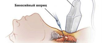
Further, if necessary, a piece of tissue is taken for examination (biopsy) and therapeutic measures (cauterization, removal of polyps, foreign bodies and stones).
For therapeutic manipulations, special instruments are inserted into the tube: to remove stones and foreign bodies - a Dormi cystoscopic basket, to cauterize papillomas - an electrocoagulator, and so on.
To take a biopsy, I use special forceps, which are also inserted into the cystoscope tube and bite off an area from a suspicious location. This tissue is then placed in a container with formaldehyde and sent for histological examination.
After the procedure is completed, the cystoscope is removed and sent for sterilization.
If local anesthesia was used, the patient is sent home. If you had general anesthesia, you will need some time of observation in the hospital, and then a refusal to drive.
After the procedure, discomfort may persist for some time:
- itching and burning in the urethra
- nagging pain above the womb
- a small amount of blood in the urine
- painful urination
This is a consequence of the introduction of a foreign object into such a delicate organ as the bladder. As a rule, these complaints can last for about a day, then they go away. If it hurts to urinate, and the urine has an unusual color, even after 3-5 days. You should immediately consult a urologist to rule out complications.
Cystoscopy in men
In men, the length of the urethra averages from 15 to 22 cm. In this case, the curvature of the urethra is observed. Therefore, cystoscopy in men is a more technically complex procedure for the doctor and more painful for the patient. Local anesthesia is used in most cases of the procedure. A flexible cystoscope is most often used.
Cystoscopy in women
In women, the length of the urethra ranges from 2.5 to 5 cm, while it is wider than in men. This facilitates the process of inserting the cystoscope. Diagnostics takes less time and complications occur much less frequently. Cystoscopy is sometimes recommended for urinary incontinence of unknown origin. This helps to distinguish whether the patient has a urological problem or a gynecological one. This means which specialist should be treated and observed.
Cystoscopy in children
The procedure is rarely performed in children. Only in cases where all types of ultrasound, radiography and MRI give an unclear result. For children, a special flexible cystoscope of smaller size (length and diameter) is used. In boys at birth, the length of the urethra is about 5 cm, in girls it is 1.5 cm. As the child grows, the approximate length of the urethra is calculated. In children, cystoscopy is always performed under anesthesia, usually with local anesthetics. If there is an allergy to novocaine/lidocaine or the child is still very small, then short-term general anesthesia is performed.
Cystoscopy in pregnant women
In pregnant women, cystoscopy is rarely performed, in exceptional cases. Preference is given to ultrasound diagnostic methods.
For therapeutic purposes, catheterization of the bladder can be performed.
Pain in the urethra and lower abdomen, bursting sensations due to the filling of the bladder can provoke hypertonicity of the uterus, which threatens miscarriage. At longer stages of pregnancy, the procedure is not so dangerous, but it becomes technically more difficult due to the enlarged uterus.
Thus, in terms of examining pregnant women, cystoscopy is not a common method.
Features of cystography in childhood
How is cystography done in children? Before carrying out this diagnostic method, the baby’s parents must be instructed about the rules for its implementation and other important nuances.
During the examination, children may be frightened by extraneous sounds or actions of health workers; therefore, first of all, the child must be psychologically adjusted correctly.
Preparation for cystography in children includes bowel cleansing. For 2-3 days before the manipulation, the baby should eat only dietary products.
Cabbage, beans and soda are excluded from his diet. If we are talking about an infant or newborn child, then 1-2 days before the test he should be given dill water to reduce gas formation in the intestines.
Immediately before the procedure, the baby is given a cleansing enema. Also, on the eve of the examination, parents should monitor the child’s hygiene and wash him thoroughly.
This condition must be observed for all types of diagnostics of diseases of the genitourinary organs. In this situation, this is very important, since the presence of bacteria on the skin of the genitals can cause infection of the urinary tract when a catheter is installed.
What else should parents know? Doctors warn adults in advance about the following nuances of performing cystography in children:
- Before the procedure, the child must empty his bladder.
- The baby must be calm; he is not allowed to move during the examination. Adults explain this to the little patient on the eve of the diagnosis.
- X-ray equipment makes quite loud and unpleasant sounds - you should also tell your child (especially a small one) about this.
- During the examination, boys' genital glands are covered with a special screen. This helps prevent radiation exposure to the testicles.
If urinary functions are impaired, voiding cystography is indicated in children and adults. At the end of the manipulation, the doctor checks the condition of the little patient, and his well-being is monitored throughout the day.
Cystography is a diagnostic method used in urology; such an examination is carried out in patients of any gender and age. The purpose of the procedure is to evaluate the condition of the bladder after the administration of a contrast agent.
The cost of diagnostics is determined based on the type of technique and the contrast used. The total amount also changes when additional examination of the kidneys, ureters, and urethra is performed.
The term “cystography” consists of 2 words: “kystis” (bladder), “graphō” (draw). During the diagnostic procedure, contrast is injected into a child's vein or through the urethra.
The essence of using contrast is that the substance becomes visible under X-ray radiation. Since the product easily penetrates into all folds of the bladder, the intracavitary contents and the line of the walls are clearly visible in the image. This makes it possible to assess the anatomical features and size of the organ.
Thanks to the X-ray contrast agent, the following pathologies can be detected:
- place of breakthrough (in case of injury);
- strictures (narrowing of the cavity);
- diverticula;
- urinary fistula;
- ureteral artesia (absence of opening);
- tumor;
- stones;
- blockage of the ureter/neck with an intravesical cyst;
- duplication and other birth defect.
For cystography, liquid or gas contrasts are now used. The first are prepared from polyhydric iodine preparations, mixing them with infusion solutions.
The concentration is selected in accordance with the instructions for use of the product. The second type includes carbon dioxide, air and nitrous oxide. They are administered only through the urethra. Paramagnetic substances are used in contrast cystography with MRI.
The essence of using x-rays lies in highlighting different tissue densities in black and white. After turning on the device, radioisotope particles saturate the soft and hard structures of the body to varying degrees.
Diagnostics are prescribed for newborns for urination disorders in order to exclude anatomical defects. For infants and older children, cystography is performed if malformations of organs or the appearance of neoplasms are suspected.
Indications for examination are enuresis, symptoms of urolithiasis, vesicoureteral reflux, trauma and pathological damage to the walls of the urinary tract.
There are four types of cystography. They differ in the method of administering the contrast agent. The appropriateness of using each method is assessed by the doctor.
| Method name | Brief description of the technique | Advantages and disadvantages of the procedure |
| Retrograde cystography (direct) | The bladder is completely filled with a contrast solution through a urethral catheter. The substance will rise upward during administration, but will not penetrate the ureters and kidneys. | The procedure does not cause nephrological complications. There is a risk of mechanical damage to the urethra and transmission of a sexually transmitted infection to the bladder. |
| Descending cystography (indirect) | Preparation and initiation of the procedure are the same as for excretory urography. Cystograms begin to be taken after the bladder is filled with contrast fluid. | Allows you to assess the condition, functions and evacuation capacity of the kidneys, ureters, and bladder. The drugs are nephrotoxic and allergenic. They can provoke acute kidney failure, anaphylactic shock or Quincke's edema. |
| Pneumocystography | The bladder is filled with gas through the urethra. | Allows you to identify non-contrast neoplasms or stones. |
| Cystography lacunar (double contrast) | A liquid and then a gaseous substance is injected into the bladder through a urethral catheter. | It has the advantages of the ascending technique and pneumocystography. |
| Vaccine cystography | A contrast agent is injected, then images are taken while urinating | Allows you to evaluate the evacuation function of the bladder, the amount of residual urine, and the condition of the urethra. It is difficult for young children to make such a diagnosis. |
It is important to know! The bladder and kidneys can be examined using MSCT cystourethrography without the use of contrast. After 14 years, it is allowed to do PET-CT with the introduction of radionuclide agents.
Cystography of the child’s bladder is performed in three directions. This is psychological preparation, prevention of flatulence and cleansing of the intestines.
The need for cystography must be explained to the child. On the recommendation of a doctor, sedatives can be given. Regardless of the research methodology used, children over 12 months of age need to follow a 2-day diet.
Exclude from the diet:
- bread;
- raw vegetables;
- fresh fruits;
- bean dishes.
During the preparatory period, they drink little, consume semi-liquid cereals, boiled meat, and unsweetened tea. Infants are fed as usual, only the last meal is given 6-8 hours before.
The doctor may recommend that the child drink Espumisan, Simethicone and similar carminatives. They are given at the same time as the diet to prevent flatulence.
A small child should be given a cleansing enema with chamomile decoction the evening before the procedure and in the morning. Older children can take castor oil 2 hours after dinner, and in the morning after bowel movements they cleanse the intestines with an Esmarch mug.
Before performing cystography using the descending method, you need to re-donate urine with blood for general and biochemical examination. Liver tests are also prescribed to study the functional capacity of the kidneys.
https://www.youtube.com/watch?v=ys0m3YBF7qI
Attention! Lactating women also need to follow a diet. This will reduce the likelihood of flatulence in breastfed babies.
Progress of the examination
Let's look at how cystography is done in children. The beginning of the diagnosis will be the same for any child, regardless of gender. In medical centers they allow you to change into a loose shirt for procedures.
With the retrograde method, you need to remove your underwear to open access to the genitals. In the case of using the descending method, the injection can be given to the child under the skin, into a vein, or into the muscle under the shoulder blade. Algorithm for performing ascending cystography:
- The child is given an antihistamine.
- The patient lies on a table under an X-ray device.
- Anesthesia is used according to indications. The boys are given local anesthesia.
- The scrotal area is covered with a protective plate.
- A survey urogram is performed before cystography.
- Contrast is injected into the bladder until the organ is filled.
- An X-ray is started and pictures of the bladder area are taken with the patient positioned on the back, side, or stomach.
- They take out a catheter, ask the child to pee, or use a Janet syringe to remove the liquid.
- The last photo is taken after urination.
- Upon completion of the procedure, the child remains in the hospital as an outpatient for the purpose of medical monitoring of his condition.
The technique differs in different methods. With voiding cystography, organs are photographed at the time of bowel movement and after urination. With the descending method, after infusion of the drug, wait until contrast begins to be released through the kidneys and the liquid completely fills the bladder.
Anesthesia
Cystoscopy with a rigid (standard) cystoscope
This is a rather painful procedure, it all depends on individual sensitivity, but the use of anesthesia greatly facilitates the process.
local anesthesia
Local anesthetic drugs are injected into the urethra immediately before insertion of the device. A solution of novocaine 2%, about 10 ml, or more modern gels based on lidocaine (catedgel, instillagel) are used.
A few minutes are allotted for “freezing”, and then the doctor begins the procedure.
clamping
If we are talking about cystoscopy of men, then sometimes a gentle clamp is used just below the head of the penis. The duration of applying the clamp is no more than 10 minutes.
spinal anesthesia
Spinal (spinal, epidural) anesthesia is used in patients with high sensitivity, in the presence of somatic diseases (for example, arterial hypertension with crises) or when a therapeutic and diagnostic procedure is planned (removal of a stone or polyp).
general anesthesia
Given the short duration of the procedure, general anesthesia is performed very rarely: in children, psychiatric patients and in other rare individual cases.
As a rule, local anesthesia is quite sufficient. This is the most common type of pain relief during cystoscopy.
Cystoscopy with a flexible device
The flexible device brings less discomfort, so cystoscopy can be performed without anesthesia.
If a decision has been made about anesthesia, then the same drugs for local anesthesia are used as in the case of a rigid cystoscope.
In children
Cystoscopy of the bladder in children is rarely done. It is prescribed only if the child’s problem cannot be identified after ultrasound, radiography and MRI. For children, a special small flexible cystoscope is used. In this case, anesthesia is always used.
If the child behaves restlessly, general anesthesia is used. In modern urology, there are even cystoscopes for performing the procedure on six-month-old infants.
The examination of the child is carried out only with the consent of the parents, given in writing.
Indications
Diagnostics:
- interstitial cystitis (disease of the bladder with deep damage to its walls and difficulty urinating)
- chronic cystitis (to clarify the cause and depth of damage to the bladder walls)
- bladder tumors (benign and malignant)
- diverticula and polyps (pathological growths and protrusions of the inner wall of the bladder)
- stones of the urethra, bladder and stones blocking the mouths of the ureters (in the place where the ureter “flows” into the bladder, small stones often become lodged)
- foreign bodies. Foreign bodies in the bladder are not such a rare situation. The literature often describes cases of small objects and parts getting into the bladder through the urethra. This is often due to the desire to diversify your sex life. A long-term stay of an ordinary mercury thermometer in the cavity of the bladder was once described.
- injuries and consequences of old injuries. The consequences of old injuries and damage to the bladder can be very different. Fistula (pathological opening) between the bladder and vagina or rectum, in which case urine involuntarily flows out of the vagina or anus.
- structures or pathological narrowings along the urethra can be the result of injury, inflammation or tumor.
- hematuria (blood in urine) of unknown origin
- enuresis in adults and children
- diagnostic search for identifying atypical (suspicious of cancer) cells in a general urine test
- frequent urination and chronic pelvic pain that could not be treated conservatively, and a more accurate diagnosis is needed.
- benign prostatic hyperplasia (BPH) in men
Reviews
Nelly, 36 years old: I received a referral for cystoscopy because I was very often bothered by cystitis. I was afraid at first, of course, but then I somehow relaxed or the doctor was friendly. When I inserted the device, I felt almost nothing, then I carefully looked into it for about 10 minutes and turned it as it was comfortable. It was a little unpleasant when I got it, but I got through this even without painkillers. So I think that the procedure is not scary.
Svetlana, 49 years old: I’m undergoing cystoscopy for the third time. The first time was a little scary, but I found one of the best specialists in our city, and everything went with a bang. The doctor is an expert in his field, so I only go to him. Spray a little lidocaine and off you go. 15 minutes and you're done. The last time there was not even any discomfort.
Irina, 38 years old: After suspensions were detected in urine tests, I was sent for cystoscopy. I was terribly afraid - I even took a sedative. But to my surprise, everything went very quickly and did not hurt at all. It stung a little when the doctor took out the tube, and then all day it was a little uncomfortable when urinating. But my bladder was completely examined, and the frightening diagnoses were not confirmed.
Treatment
Often the diagnostic procedure turns into a therapeutic one. If the doctor has the technical capabilities and sufficient experience, the patient can avoid repeating such an unpleasant procedure and solve two problems in one visit.
- removal of foreign bodies and stones from the bladder and urethra. If stones or foreign bodies are small, accessible to grip, have rounded edges and are not welded to the inner wall, then there is a high probability of their safe removal. If there are large stones, you can try to split the stone and remove it piece by piece.
- removal of a small tumor or polyp
- taking a piece of tissue for histological examination. Any removed formation and biopsy material are in any case sent for histological examination to a pathologist. This allows you to diagnose cancer, determine its prevalence and type, or, conversely, confirm that the tumor/polyp is benign and there is nothing to worry about.
- dissection of strictures. When the urethra or openings are narrowed, urine does not pass properly, is retained and stretches the urethra. This is manifested by pain, difficulty urinating and requires treatment. In these cases, cystoscopy is a very gentle and effective method; the narrowing areas are stretched or incised and urine flow is restored.
- installation of stents. Stents are cylindrical devices that are inserted to widen a specific area. Stenting may be used if cutting the adhesions helps for a short time and narrowing and difficulty in passing urine occurs again.
What diseases are diagnosed and treated using cystoscopy?
Cystoscopy is prescribed when ultrasound, despite the symptoms, fails to detect neoplasms, stones and sand, modifications or injuries, as well as traces of inflammation in the bladder.
What symptoms may be a reason to undergo cystoscopy:
- Damage to the mucous membrane of the bladder due to a weakened immune system (interstitial cystitis).
- Frequent urination.
- Difficult or intermittent flow of urine.
- The appearance of blood in urine.
- Involuntary urination.
- Chronic pain in the pelvic area.
- The presence of atypical cells in the urine, detected during laboratory tests.
The question often arises: is it possible to do cystoscopy for cystitis? Infectious cystitis and inflammation of the bladder mucosa in the active phase are contraindications to cystoscopy. However, this procedure is prescribed if relapses of this disease become frequent.
Modern medicine has great potential for solving problems of varying complexity.
So, when other methods for diagnosing diseases of the urinary system have not helped to establish an accurate diagnosis or develop an effective treatment regimen, cystoscopy is performed. It can help identify diseases such as:
- chronic cystitis;
- congenital anomalies and injuries of the genitourinary system;
- urolithiasis disease;
- benign tumors;
- leukoplakia (formation of plaques on the mucous membrane of the bladder);
- antonia (impaired control over the accumulation and excretion of urine);
- hematuria (blood in urine);
- polyps;
- tuberculosis;
- ulcer;
- blockage or narrowing of the ureters;
- bladder cancer.
Doctors of the American Urological Association concluded that bladder cancer is a disease of men and older people. The likelihood of developing this disease in the stronger sex is almost three times higher than in women, and the average age of patients is 73 years.
In most cases, a tumor is diagnosed at the stage of damage to only internal tissues. In almost 35% of patients, the cancer does not spread beyond the organ, and in 65% of patients the disease affects the tissue adjacent to it. This type of cancer rarely affects other organs by metastases.
This diagnostic method is also used for medicinal purposes. It helps solve the following bladder problems:
- remove the tumor;
- stop internal bleeding;
- conduct electrocoagulation of a benign tumor;
- crush and remove stones;
- eliminate blockage and narrowing of the urinary tract;
- eliminate congenital anomalies.
And also for other medical procedures using cystoscopy, you can install a catheter in the ureter and perform a biopsy of tissue of the urinary tract.
Cystoscopy as an additional method is often used in medical fields such as proctology and gynecology. In this case, the pelvic organs are subject to examination. This procedure allows proctologists and gynecologists to diagnose:
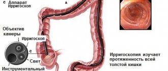
- malignant tumor of the sigmoid and rectum;
- cancer of the uterus and appendages in women;
- prostate cancer, hypertrophy, hyperplasia and prostate adenoma in men.
There are also contraindications for cystoscopy:
- cystitis and urethritis in the active stage of the disease;
- renal failure;
- liver failure;
- post-infarction state;
- angina pectoris;
- heart defects;
- pregnancy.
Contraindications
- acute inflammatory diseases of the genitourinary system (cystitis, urethritis, prostatitis, orchitis and others). During the period of acute inflammation, the tissues are loose, painful, swollen and very easily damaged.
- severe somatic diseases. Patients with renal, hepatic and pulmonary-cardiac insufficiency are at risk of worsening their condition during cystoscopy. If the procedure is necessary, you should first achieve a stable state for cardiac or other disease, and then begin diagnostics.
- blood clotting disorders (high risk of bleeding even from minor damage to the mucosa)
- early pregnancy. The uterus and bladder are located close. Stretching the bladder with a solution, movement of the cystoscope tube, and pain can provoke uterine hypertonicity and even lead to miscarriage.
Among women
The length of the urethra in women is 2.5–5 cm. It is wider than in men. So, there are often no problems with the introduction of the device. Diagnostics takes less time and does not cause unpleasant consequences.
Cystoscopy is often prescribed to patients with urinary incontinence of unknown origin. In this case, it is possible to determine what type of problem: gynecological or urological. An examination may also be prescribed for pain in the bladder area, poor urine tests, etc.
Although it is generally accepted that cystoscopy of the bladder in women is easier, many patients are afraid of having it performed, especially after viewing photographs of the device. Young girls are particularly sensitive people, so they often require general anesthesia.
In pregnant women
This procedure is rarely performed on pregnant women. To perform it there must be indications. Generally, doctors prefer to use ultrasound as a diagnostic tool. For treatment purposes, bladder catheterization may be performed.
Pain can provoke hypertonicity of the uterus, and this can lead to miscarriage. In the last months of pregnancy, the procedure becomes safer, but diagnosis is complicated due to the enlarged uterus.
Complications of cystoscopy
- urethral trauma
The urethral mucosa is very delicate and thin, so it can be damaged, especially when using a rigid cystoscope. The risk increases if the patient is restless, moves or tries to interfere with the process (pulls out the device, tries to get up).
- false move
This is a very serious variant of traumatic injury to the urethra. In this case, the cystoscope creates a hole in the wall of the urethra and goes into the surrounding tissue. If the cystoscope penetrates the prostate tissue, threatening bleeding is possible.
- cystitis
If the hygiene of the external genitalia was insufficient and irregular, then during the procedure, an infection gets inside the skin and mucous membranes. One treatment with an antiseptic solution cannot remove all types of bacteria.
If left untreated, cystitis can develop into pyelonephritis, which is a much more serious disease that affects the renal pelvis. With pyelonephritis, lower back pain, high fever with chills develop, and blood and urine counts worsen.
- bladder perforation
A puncture (perforation) of the bladder can occur due to insufficient experience of the doctor, an abnormal structure of the bladder, or a change in the location of organs due to adhesions.
Features of the course of different categories
Differences in the anatomical structure of the urethra, age and purpose of the study reveal the peculiarities of the operation in different categories of citizens. In women, the urethra is 3-5 centimeters, which allows infection to easily pass directly to the bladder, ureters, and kidneys. Therefore, cystoscopy in women is performed more often; it makes it possible to identify cystitis, nephrolithiasis or tumors. The examinations cause virtually no discomfort and in most cases are performed without anesthesia. The male urethra is 15-18 centimeters, so the procedure is painful and the use of anesthesia is necessary. Cystoscopy of the bladder in men is more difficult because the path of the cystoscope passes through the prostate. If the examination is carried out incorrectly, there is a risk of injury to the mucous membranes, inflammation of the seminal tubercle, and impaired potency. This procedure allows you to identify inflammatory processes, tumors, stones, and enlarged prostate. Cystoscopy of the bladder in children is carried out using a flexible cystoscope, which is smaller in size than a regular one; written consent of the parents is required. Cystoscopy during pregnancy is possible, but it is recommended to wait until the baby is born. The bladder is located next to the uterus, so there is a risk of damage to the wall of the reproductive organ, which can lead to premature birth or miscarriage.
Sources
- https://MyCistit.ru/diagnostika/cistoskopiya
- https://kdc.clinic/urologiya/operatsii/tsistoskopiya-mochevogo-puzyrya/
- https://apkhleb.ru/endoskopiya/cistoskopiya-mochevogo-puzyrya-zhenshchin
- https://diagnostica.DocDoc.ru/articles/endoskopicheskie-issledovaniya/cistoskopia/kak-delaut-cistoskopiu/
- https://apkhleb.ru/endoskopiya/chto-takoe-cistoskopiya
- https://MyCistit.ru/diagnostika/cistoskopiya-u-muzhchin-zhenshchin-detej
- https://urolog.klinika-abc.ru/tsistoskopiya.html
- https://zdravotvet.ru/cistoskopiya-mochevogo-puzyrya-pokazaniya-kak-delayut-u-zhenshhin-muzhchin-podgotovka-rezultaty-stoimost/
- https://www.euroonco.ru/glossary-az/tsistoskopiya-mochevogo-puzyirya
- https://www.smclinic.ru/professional-services/diagnostika-v-urologii/tsistoskopiya/
- https://www.wp-german-med.ru/sovremennaia-endoskopia/tsistoskopiya-kak-provoditsya.html
- https://mypochka.ru/lechenie/13-cystoscopy.html
Cost of bladder cystoscopy procedure
In government institutions, the procedure is performed free of charge under compulsory medical insurance upon the direction of the attending physician, or through paid services. In private clinics, the patient pays all expenses independently. Prices in different regions vary greatly, the price range is from 800 rubles to 25,000 rubles.
The price of the issue also depends on whether it is a primary or secondary appointment, whether diagnostics are needed or whether treatment measures are already being planned. The cost increases in accordance with the technical complexity and the need to use additional tools in the process. The price of the study also includes a histological examination of a tissue biopsy of the bladder or urethra.
Locations of the procedure
Cystoscopy is an informative procedure. But it is difficult to carry out. Therefore, it is performed by experienced urologists. It is offered both in public clinics and in private specialized medical institutions.
When visiting public hospitals, the examination can be carried out free of charge. Private clinics offer paid diagnostics. However, its price will depend on each specific institution.
Medical office for the procedure.