X-ray of the hand and wrist is a way to study their internal structure without invasive intervention. The hand and fingers are complex anatomical parts of the body, consisting of many small bones and performing important tasks in human functioning. This is the ideal and precise tool we use. Every day they are exposed to mechanical stress, their condition is affected by diet, quality of metabolism and other life processes. With the help of radiography, diseases in this area, cracks, dislocations and fractures are detected. Timely diagnosis prevents the development of severe diseases of the hands and wrists.
What does a hand x-ray show?
In everyday life, the human skeleton is subject to constant loads, including the hands. Excessive physical activity, infectious diseases, and mechanical damage change the structure of this part of the body and can sometimes lead to partial or complete loss of functionality.
To diagnose the disease and its cause in a timely manner, radiography is used. This method has been known for a long time and is still popular among traumatologists, surgeons, and therapists.
Content:
- What does a hand x-ray show?
- Indications for radiography
- Features of the study
- Research results
- Contraindications for hand radiography
- Alternative to radiography
- Where to take the study
- The myth about the dangers of research
The technique works on the principle of x-ray radiation. Beams of rays pass through the object and leave a contour mark on the film. Depending on the density of the tissue, the beam passes through it differently. Thus, bone tissue is reflected best, while soft tissue is reflected poorly.
However, based on the shadow in the image, a radiologist or other doctor can see changes in the lymph nodes, tendons, and muscles. The x-ray image shows the entire internal structure of the wrist and hand.
In the picture you can see all parts of the hand: wrist, phalanges of fingers, metacarpus. In turn, each of these sections contains several subgroups of bones, ligaments, joints, and muscles. Damage to even one unit of this system leads to pain, swelling, and deformation of the entire arm or its parts.
An x-ray clearly shows any damage to the bone tissue: thinning, cracks and microcracks, dislocations, inflammatory processes. To accurately diagnose problems with soft tissue, the doctor may prescribe a more informative computed tomography scan, or angiography to examine blood vessels.
Today medicine uses several options for examining the hands and wrist; they are more modern, more accurate, and faster. These include MRI, CT, MSCT and others. However, conventional X-rays continue to be used because they are simpler and more accessible.
Every clinic has such equipment; it costs much less or is completely free. In addition, the radiation dose for all progressive options and radiography is almost the same.
X-ray of the wrist joint: indications and contraindications
X-ray is contraindicated:
- During pregnancy and lactation. During this period, radiation can provoke hormonal imbalance, which will lead to serious consequences. It is better not to prescribe an x-ray examination if it is possible to replace it with a more gentle procedure.
- In critical and serious condition of a person. Such an event can greatly worsen your health. If the patient does not feel well, this is reported to the doctor before the procedure.
- X-rays should be prescribed with caution in patients with schizophrenia. It is recommended to first consult with the patient's physician.
You need to prepare for the event in advance.
To do this you should:
- get rid of metal objects and remove jewelry;
- remove plasters and oil dressings;
- if necessary, cover the area to be examined with an aseptic bandage;
- remove any remaining sticky patches, iodine, brilliant green, or creams from the desired area.
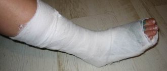
If the patient has a hand in a cast, a doctor should be consulted before the procedure to make sure that an x-ray can be taken without removing the cast.
Features of the procedure:
- A special protective uniform made of dense material (lead) is put on. Such clothing will help reduce the harm from radiation to those parts of the body that do not need x-rays.
- For children, only their limbs are left exposed.
- The arm, bent at the elbow, is lowered onto a special surface.
A series of radiographs is taken for about three minutes in different projections:
- Straight. Projection involves scanning both the back and palm of the hand. In medical practice, direct projection is most often used. The X-ray beam passes through the middle of the hand.
- Lateral. The hand is positioned edgewise, and the thumb is moved to the side. The position of the palm during the procedure makes it possible to determine the state of the contour of the hand and palm and identify various types of displacements.
- Palmar oblique. The pose involves placing the hand with the palm up at an angle of 45 degrees. The projection is used to examine the trapezius or scaphoid bone.
- Dorsal oblique. The palm is positioned with the back side up at an angle of 45 degrees. The images will show bones: pisiform, hamate and triquetrum.
The main characteristics of the method are speed and reliability, allowing the specialist to assess the integrity of the bones and their anatomical details in the area of the wrist joint and the part of the forearm articulated with it.
Sometimes swelling or pain in the palm and wrist can occur without prior injury, but the limitation of motor activity and reduction in range of motion forces patients to react to such manifestations and look for ways to quickly diagnose.
The pain of this joint can be caused by two categories of reasons, which are characterized by the intensity of their occurrence:
- Acute course. Such disorders include injuries of varying complexity, dislocations and subluxations, complete or partial ruptures of the ligamentous-articular apparatus. The increased risk of injury to the wrist and palms is due to the fact that during a fall a person reflexively puts both hands in front of him, and the force of the impact falls on the palms and their complex articulations with the forearm - the wrist joints. At this moment, their fragile structure is subjected to strong mechanical stress, which exceeds the safety margin of the joint. Therefore, during accidents, falls and other incidents, a person often receives similar injuries of varying degrees of severity.
- Chronic course. Such pathologies do not develop so quickly, but the severity of pain can be in no way inferior to acute diseases. Such cases include problems with slow progression - carpal syndrome, osteomyelitis, carpal tunnel syndrome, etc. This category also includes pathologies caused by constant excessive physical activity, exhaustion of the body and insufficiently balanced nutrition, wear and tear of cartilage structures, age-related changes in the body, and poor lifestyle with low physical activity or constant stress on the joint when sitting for many hours at the computer.
To identify pathological changes in the wrist joint, an x-ray examination is most often prescribed, as one of the most informative and necessary in this situation. In the vast majority of cases, this single examination is sufficient to make a final diagnosis.
After detecting symptoms of an acute or chronic nature, the patient is sent for an initial examination at a clinic or paid medical institution.
The initial examination is carried out by a therapist or family doctor, who draws up a description and gives a referral to a specialist based on the existing clinical picture.
The general practitioner determines which specialist needs to be contacted based on a survey of the patient, examination and palpation of the lesion.
Most often, with such complaints, the patient is redirected to undergo further detailed diagnostics to a traumatologist with a mandatory X-ray examination. Sometimes X-rays are supplemented with studies using magnetic resonance imaging or computed tomography.
Based on the data obtained, the therapist can refer the patient to clarify the diagnosis to the following specialists, who will refer the patient for an x-ray:
- Rheumatologist - in case of various types of arthritis and other inflammatory processes.
- An arthrologist is a specialist in pathological changes in joints of various origins.
- Orthopedist - if a dislocation, fracture or sprain is suspected.
- Neurologist - in the case of a painful syndrome in the wrist, presumably associated with a degenerative condition of the nerve bundles, pinched or damaged nerve fibers.
- Occupational pathologist - in case of possible pain due to the constant action of harmful factors present at work.
- Endocrinologist - if the disease is associated with abnormal functioning of the endocrine system.
All of the above specialists can refer the patient for an x-ray of the wrist joint to confirm or refute their suspected diagnoses associated with a narrow specification.
Indications
The causes of degenerative-destructive changes in the wrist joint have different origins. But most of them are accompanied by the following manifestations that require examination with x-rays:
- fractures of varying degrees in any of the eight bones of the wrist;
- bruises;
- dislocations;
- cracks;
- the presence of acute or chronic pain syndrome;
- swelling in the palm or joint area;
- subcutaneous hematomas;
- inflammatory or neoplastic changes;
- numbness in fingers;
- abnormalities in the visual contours of the carpal region.
In addition to the above symptoms, this diagnostic test is prescribed when crunching or clicking, painful or any discomfort is heard when moving the hand. X-rays are also prescribed in case of severe limitation of the motor ability of the wrist joints.
Sometimes, to assess the effectiveness of therapy, X-rays are taken and the feasibility of the chosen type of therapy is weighed.
The range of possibilities of X-ray examination is quite wide and allows you to determine the presence of various kinds of changes in hard, soft and cartilaginous tissues. This study helps evaluate:
- ligament damage;
- abnormal condition of the tendon flexors and extensors of the phalanges of the fingers and hand;
- pinched ganglia or nerve fibers;
- fractures of all bone structures in the visualization area;
- condition of the synovial membrane;
- degree of damage to bone tissue;
- presence of fibrosis;
- the degree and presence of arthritis, tensinovitis, arthrosis, osteoporosis, bursitis;
- the presence of fragments and solid foreign inclusions in the muscles;
- effusions and fluids inside joints;
- scleroderma;
- condition of articular cartilage;
- presence of neoplasms;
- osteophytes and any bone growths;
- various pathologies of the blood vessels of the circulatory system.
All data obtained using digital radiology does not require a long time for processing, which distinguishes the method.
Methodology
X-ray examination is based on the ability to record projections of x-rays onto film or a monitor (in the case of a digital device). The high resolution of the obtained images makes it possible to examine in detail even thin areas of the ligamentous-tendon apparatus of the hand.
X-ray diagnostics does not require any special preparation. It is necessary to remove clothing from the area being examined, remove bracelets, watches and other jewelry from your hands. If other examinations have been previously performed, their results must be provided to the radiologist before the examination for review.
Choosing the correct projection angle for x-rays is the key to obtaining good results. The image should clearly show all the structures of the wrist and ligamentous-articular apparatus.
The research is carried out according to the following algorithm:
- During the procedure, the person being tested can stand or lie down, depending on the condition.
- The hand is placed in a palm down position, with the elbow in a bent, relaxed state.
- It is important to keep your palm stationary - the quality of the pictures directly depends on this. The palm should be pressed as tightly as possible to the surface.
- If the radiologist decides to take an image in an oblique projection, then the hand should be positioned at 45° in relation to the surface. In order to achieve immobility in this position, a special hard lining in the form of a pillow is placed under the palm.
- During the image, the patient is required to hold his breath briefly, as this reduces the number of involuntary movements that degrade the quality of the image.
- If images need to be taken in different positions, the radiologist will take a series of images while advising the patient on how to position the arm correctly.
- If necessary, the same steps are repeated on the other hand.
The diagnostic time does not exceed 10 minutes for both hands.
The main inconveniences during the manipulation may be the relatively low air temperature in the office and the forced fixation of the hand in a certain position if this is associated with pain. To reduce the radiation dose, the rest of the hand is covered with a special protective apron made of materials that do not transmit rays.
Indications for radiography
Like every part of the body, the hands and wrists are subject to illness and injury, and the latter happens to them most often.
To accurately establish the diagnosis and future treatment plan, the doctor prescribes an x-ray of the hand and wrist. The images also help to clearly establish the location of the injury, determine its severity and recovery methods.
X-rays of the wrist and hand are prescribed for:
- mechanical damage: bruises, fractures, dislocations;
- arthritis and arthrosis;
- rheumatism;
- swelling and hyperemia of the joints;
- osteomyelitis;
- congenital anomalies in the structure of the hands;
- neoplasms: tumors, cysts, papillomas;
- systemic lupus erythematosus;
- scleroderma.
It is impossible to accurately determine the cause of the disease and its stage by visual examination, so the doctor first prescribes a diagnosis. Any changes in the structure of the hand and wrist skeleton will be visible in the photographs. In addition, radiography is prescribed for children if a lag in physical development and too rapid development is noticed. The patient can undergo this procedure if he experiences pain in any part of the hand, notices curvature of the fingers, numbness, and swelling.
X-ray examination
This type of examination is called radiography of the hands. This is a procedure that can detect damage in the bone skeleton. X-rays are used to detect changes in joints, for example, in the elbow joints, and damage affecting muscle and bone tissue.
Diagnosis of diseases is possible by assessing the condition in which the joints of the hands are. Pathologies, the nature of their manifestation, the affected area and its size can greatly facilitate the diagnosis and determination of the stage of the disease. To accurately determine the disease, it is important in which joints pathological processes are detected.
The joints of the hands are primarily affected by changes in rheumatic diseases. An X-ray of the hand is necessary to check soft tissues and bone structures in order to determine the extent of changes that have occurred. This is most important when there is calcification of the tissues that make up the joint.
If, as a result of the development of the disease, there is no damage to the soft tissues, then the image will show the presence of changes in the joint, which take the form of compactions and thickenings. When obtaining such a result, it is impossible to determine exactly which structures were subject to pathological changes.
Rheumatoid diseases are accompanied by the following pathologies, discovered as a result of radiography:
- thickening of the wrist joint;
- calcification affecting soft tissues;
- osteolysis;
- necrosis to which bone tissue is exposed.
Features of the study
Pictures of the hands are taken quickly and painlessly. No preparation is required for the test; you need to take with you to the session directions from the doctor who prescribed it. X-rays are carried out in a separate room equipped with equipment. Before diagnosis, the patient removes jewelry so that it does not shade the image.
It is worth warning the doctor if there are metal inserts in the palms and wrists. To protect the rest of the body, the technician provides a lead membrane that blocks X-rays.
The installation consists of an X-ray table and a chair. The patient sits at the table and bends his limbs at the elbows, placing his hands on the table. Depending on the desired area of study, the projection of the images is changed. The patient's hands can be placed in different positions to obtain the most informative data.
For wrist x-rays, use:
- Direct projection. A cassette is placed under the palms, on which the photograph will then be reflected. Pictures can be taken from the back or from the palm, sometimes from two projections at once. The patient takes a stationary position, the laboratory assistant takes a photo using a remote button. This way the doctor will be able to examine all segments of the wrist: scaphoid, triquetrum, capitate and others. The phalanges and metacarpal regions are also visible. However, there are several segments that are not visible with this installation.
- Lateral projection. The hand is placed sideways on the cassette, with the thumb protruding slightly forward. This arrangement allows you to view the phalanges of the fingers, carpal and metacarpal bones from a different angle.
- I slant my palm. For this projection, the patient's hand is positioned so that the palm with the cassette creates an angle of 45 degrees. It is important that during the examination the hand does not move or shift; the laboratory assistant will monitor the correct positioning. Images from this position show the trapezium, scaphoid, and trapezius bones.
- I slant the back. In this case, the back of the hand creates a 45-degree angle with the cassette. In this way, the pisiform, hamate and triquetrum bones are best visible.
In addition to these methods, other types of styling can be used to examine the wrists. The radiologist can take targeted pictures of each individual bone. I also have my own radiography methods for reflecting fingers.
Each phalanx is placed in a direct and lateral perspective; the doctor can assess the condition of each of them separately. The accuracy of the results depends on the experience and competence of the doctor; he must set the minimum level of radiation and ensure the correct position of the patient.
For the patient, this procedure takes place without discomfort and lasts 5-15 minutes. If necessary, the doctor can take pictures in all projections for a detailed and complete study of the hands; for example, in case of infectious diseases, it is necessary to establish the entire affected area.
Also, in case of injuries or impaired development, a set of images is needed. No matter how many images need to be taken, the process will be quick and painless.
Photo gallery
The photo shows x-rays of the hands.
X-ray of a healthy hand in direct projection
X-ray of a broken arm
Arthrosis on x-ray
Fractured finger on x-ray
Wrist fracture on x-ray
X-ray of the right hand
Research results
After the images are taken, they must be interpreted and described by a radiologist. This will take 15-40 minutes, depending on the severity of the condition. Based on these data, the attending physician will determine the treatment regimen.
For hand and wrist injuries, therapy is carried out and monitored by a traumatologist; for other diseases, this can be a surgeon or therapist. The patient must take the results of the x-ray to the doctor who referred him for research. The images may reveal mechanical damage or rheumatic diseases.
Rheumatic diseases include scleroderma, joint inflammation, rheumatoid arthritis and other diseases. X-rays can also reveal dangerous joint psoriasis, bone calcification, and cysts. Advanced types of such inflammation lead to erosion of bone tissue and loss of functionality. In severe cases, X-rays indicate surgery, but systematic treatment is usually sufficient.
When is a hand x-ray prescribed?
X-rays of the hands are prescribed to clarify the preliminary diagnosis made by a surgeon or therapist.
Indications for the procedure:
- control of bone tissue growth;
- checking synovial fluid;
- diagnosis of painful symptoms;
- swelling of the hands;
- redness and calcification of joints;
- diagnosis of fractures of fingers, wrist bones;
- identification of bruises, dislocations, ligament tears;
- monitoring bone condition in chronic diseases and inflammation;
- checking for foreign bodies.
Contraindications for hand radiography
Radiation examination carries a dose of radiation to the patient, so patients are often afraid of this procedure. However, when examining the hands and wrists, the dose is very small, so there are no categorical contraindications. This procedure is carried out even for small children; for maximum protection, they are completely covered with a lead apron, freeing only the hand.
Pregnant women are still not taken pictures unless absolutely necessary, but in case of fractures, the patient is also covered with a membrane. Breastfeeding mothers also undergo x-rays if necessary.
The session may be complicated if the patient has serious mental problems. To obtain clear images, the patient requires complete temporary immobility, which is not always possible in case of mental disorders.
Also, in a critically ill patient, such a study cannot be performed. If x-rays cannot be taken for some reason, alternative methods of examination are prescribed.
The process of preparing for the study
X-ray, like most diagnostic methods, has a number of preparatory measures:
- Before the procedure, you will need to remove all metal and jewelry items, as they distort the image and, as a result, an incorrect diagnosis may be made;
- during an x-ray examination of the hand, iodine must be removed from the skin, large oil dressings, which can be replaced with simpler aseptic ones, sticky residues of the plaster must be removed
- the doctor must be warned if there is any suspicion of a possible pregnancy, to protect the child from harmful radiation rays;
- If you have a plaster bandage on your arm, you should consult your doctor about the need to remove it. If it is necessary to remove the plaster, you should contact a specialist and follow all his further recommendations.
Preparing for an X-ray
You should not move during the procedure. If a child needs an x-ray, a relative can help record it. To take an X-ray, you must have an outpatient card and a referral for examination. If an x-ray is performed on a child without the presence of relatives, the doctor must obtain written permission from the parents to perform the procedure.
During the research process, it is necessary to connect the fingers of the hand and place them flat on the surface of the cassette. The hand should be positioned so that its axis coincides with the axis passing through the wrists and forearms. A standard shot of a hand is taken in straight and side projections. Other hand placement options can also be used for more detailed information.
To take an x-ray of the right and left hands, no preliminary preparation is needed. Before the procedure, you need to remove rings, bracelets, watches and place your hands (or arm) on the work table of the X-ray machine as directed by a radiologist.
To protect from the influence of ionizing radiation those parts of the patient’s body that are not involved in the study, special lead aprons or vests are used.
Most often, radiography is a norm included in the diagnostic standard and is mandatory for any bone injuries. It allows you to determine the severity of damage to bone and muscle tissue, regardless of which area is damaged, including the right or left hand, foot, knee or elbow joint.
Before performing the examination, preliminary preparation of the patient is necessary:
- Before starting the procedure, it is necessary to remove all jewelry (watches, bracelets, rings), the presence of which negatively affects the quality of the image and the determination of the subsequent result;
- you need to remove the bandage and iodine residues from the area being examined, as well as traces of the adhesive plaster;
- the question of the need to remove the plaster before X-ray diagnostics is decided by the attending physician, who will give all the necessary consultations on further immobilization of the limb.
You need to prepare for the event in advance.
To do this you should:
- get rid of metal objects and remove jewelry;
- remove plasters and oil dressings;
- if necessary, cover the area to be examined with an aseptic bandage;
- remove any remaining sticky patches, iodine, brilliant green, or creams from the desired area.
If the patient has a hand in a cast, a doctor should be consulted before the procedure to make sure that an x-ray can be taken without removing the cast.
Alternative to radiography
Radiological diagnostic results can be obtained in other ways. The main competitor to radiography is computed tomography. It is based on the same radiation, but at a lower dose.
The CT scan process is faster and the results are more comprehensive. Thus, with a regular x-ray, soft tissues are poorly distinguished, although they are also often susceptible to disease. CT scan reflects vessels, muscles, and ligaments much better. However, the conventional technique is better suited for diagnosing bones.
Also, sometimes the X-ray method is replaced by MRI. This option is considered, like computed tomography, to be very informative. It provides comprehensive information about the condition of bone tissue and its surrounding elements. In this case, the patient does not receive radiation exposure at all, since the process is based on electromagnetic resonance. For a detailed examination of individual systems, such as blood or lymphatic vessels, lymphangiography or angiography is prescribed.
Limitations and benefits of hand X-ray therapy
The main advantage of X-ray therapy of the hand is the ability to study the internal state of bone and altered soft tissues. Most often, radiography is used to study mechanical damage and diagnose chronic pathologies, such as arthrosis.
The procedure is used to a limited extent, as it can have severe side effects on health. After frequent use of X-rays, leukopenia and the risk of developing leukemia may occur in the human body. This diagnostic method cannot be used more than once within a month.
X-ray of the hands is prescribed quite often. This type of examination is popular and informative. The procedure allows you to identify a wide range of pathologies in the shortest possible time period. Where can I get this examination? In any district clinic that has the necessary equipment installed.
The main indications that indicate the need for fluoroscopy of the hand are possible injuries and pain caused by inflammation.
The doctor will give the patient a referral for this type of examination, subject to the following complaints:
- Pain in the hands during periods of rest and movement.
- The presence of swelling and the appearance of an enhanced pattern of blood vessels.
- For joint deformation.
- After injury.
- If you suspect the development of osteomyelitis.
- With congenital abnormal developments.
An absolute indication for x-rays of the hands is the development of rheumatoid diseases.
Where to take the study
If a simple x-ray is prescribed, it can be done at any emergency room, clinic, or hospital. The procedure requires conventional equipment, which is available in private and public medical institutions. More advanced techniques such as MRI or CT will have to be sought in private clinics.
The price for a regular photo of one brush in one projection will be 5-10 dollars. The cost may vary depending on the reputation and financial policies of the institution.
Two projections will cost twice as much. The price includes the examination and photographs; interpretation of the results in a public clinic should be free, in a private clinic you will need to pay an additional 5-8 dollars.
Tactics for performing the examination
Normally, an X-ray of the arm can be taken on any generally healthy adult patient. But if this type of diagnosis does not bring the expected results, other options for examining the hands are prescribed. For example, CT is used to detail the nature of the damage and identify the source of pathology.
To take an x-ray, the patient is seated on a chair next to the x-ray machine table. The arm should be bent at the elbow and the hand should be placed on the table. The information content of the resulting image depends on how correctly the brush is placed. In practice, several installation options are used:
- Direct projection. For such shooting, the brush is placed horizontally, on the table - with the back side or palm. The X-ray beams will pass through the brush perpendicular to the film cassette. This image will reveal all the bones of the wrist, with the exception of the pisiform. The wrist joint, metacarpal bones, carpometacarpal joints, intercarpal joint, and phalanges will also be visible.
- Lateral projection. To shoot this way, you need to place your palm with the ulnar edge on the cassette. The thumb should be slightly moved forward. In such a picture, the contours of the carpal bones, metacarpal bones, and phalanges will be clearly visible. This projection turns out to be most useful in traumatology, when it is necessary to identify displacements of the metacarpal bones.
- Oblique dorsal projection. The brush must be positioned so that its back surface forms a 45° angle with the cassette. Such an image will show the condition of the triquetral bone, pisiform bone, hamate bone, as well as the first and fifth metacarpal bones.
- Oblique palmar projection. The hand is positioned so that the palm surface forms an angle of 45° with the cassette. This image will show the scaphoid, trapezius, and trapezium bones.
In all cases of X-ray examination, the patient is put on a special apron with lead coating, which reduces ionizing radiation.
Advantages:
- diagnosing mechanical damage;
- detailed study of pathologies;
- timely detection of diseases;
- relieving anti-inflammatory method for purulent infections;
- extinguishes inflammatory processes without the presence of suppuration.
The disadvantages of the method include:
- the possibility of developing leukemia;
- the occurrence of leukopenia (decrease in the number of red blood cells in the blood).
Pros and cons of x-ray examination. The author of the video is Doctor Komarovsky.


