Three times ultrasound examination is mandatory for pregnant women. However, it is carried out in two-dimensional mode, and in such an image it is difficult to make out anything without a doctor’s prompting. This is why three-dimensional ultrasound is gaining popularity - during such an examination the child is clearly visible on the screen and you can see the features of his appearance.
And for the most curious mothers, medical centers offer 4D ultrasound, where a fourth dimension – time – is added to the three spatial dimensions. Such research provides a unique opportunity to look into the intrauterine everyday life of your baby and watch a short film about him.
Why do they do it and who do they recommend it to?
Computer technologies are developing dynamically, bringing new opportunities to all areas of our lives. Medicine was no exception - four-dimensional computer ultrasound began to be performed relatively recently, but it is already clear how much more informative this method is than the usual ultrasound examination.
A more detailed study, three or four-dimensional, may be prescribed to a woman if there is a suspicion of abnormal development of the baby. This method allows you to much better see small details of the baby’s appearance, for example, the bones of the nose or the features of the skull and the structure of the limbs.
Thanks to a detailed image, it is possible to more accurately determine whether a child has problems with the spine or diagnose incipient disorders in the formation of organs.
Important! Early diagnosis plays a significant role in the correction of intrauterine pathologies of child development, and this role is increasing. And, of course, four-dimensional ultrasound is an excellent way to get a complete picture of the unborn child before his birth.
3D and 4D ultrasound during pregnancy
May 30, 2014022653 Category: Pregnancy
Indications for 3D ultrasound during pregnancy
A 3D ultrasound during pregnancy is done either on the recommendation of the attending physician, if there is a risk of abnormal development of the fetus, or if the appearance of various types of neoplasms is suspected. This study can also be completed without a referral, but at will.
3D ultrasound makes it possible to see the unborn child even before his birth as realistically as possible, take a photo of the fetus, capture it while moving, examine separately all the organs and parts of the body (arms and legs: check for the presence of five fingers on each limb, see the face, etc.). d.), determine which parent the child resembles, find out the gender of the baby.
3D ultrasound at 16 weeks of pregnancy
During the study, the first psycho-emotional connection between the future parents and the child is established.
This type of ultrasound in gynecology is a fairly new diagnostic method, through which the condition of the fetus is determined, anomalies in the normal formation and development of organs and systems are identified, and it becomes possible to analyze the parameters of the fetus in a three-dimensional projection (length, height, depth). The result of the study can be obtained immediately after this procedure. Three-dimensional ultrasound is safe and does not harm either the expectant mother or the fetus.
At what stage of pregnancy is it best to do 3D and 4D ultrasounds?
3D and 4D ultrasounds take at least an hour. During it, the first contact of parents with their unborn child is directly carried out. There are often cases when it is quite difficult for a woman to get close to her child after he is born. This happens due to insufficient visual contact with the baby during pregnancy, since feeling the presence of a living being inside is something different than seeing it, already born, next to you and starting to take the first care of it.
Therefore, thanks to the opportunity to get to know the unborn child while he is still in the womb, it is much easier for parents to contact him after birth. Moreover, in addition to awakening the maternal instinct, 3D and 4D ultrasound is very useful for expectant fathers, since the birth of a child in their life is the same exciting and responsible moment as for the expectant mother.
It is advisable to undergo these types of studies in the second half of pregnancy. It will be even better if you combine 2D research with 3D. 3D ultrasound provides additional information to 2D ultrasound results. In this case, the power of the ultrasonic wave is the same. The optimal time for an ultrasound is 20-28 weeks of pregnancy.
If the doctor suspects a defect that was not detected in a timely manner, then the woman is sent for a 3D or 4D ultrasound at 13-18 weeks of pregnancy. Until the 16th week of pregnancy, the entire fetus can be seen; at the 18-24th week, the gender of the child can be clearly distinguished; from the 22-24th week, facial features are clearly visible.
Conducting expert 3d and 4d ultrasound
Ultrasound of pregnancy in 3d and 4d format is a diagnosis of pregnancy using ultrasound therapy based on echolocation. The essence of the method: reflection of an ultrasonic wave from tissues and organs, captured by a sensor. Thanks to such new and modern equipment, very high-quality images are obtained.
The studies allow us to examine in detail the internal organs and anatomical parameters of the fetus, identify any pathology, and carry out Doppler mapping of blood vessels, which is necessary during prenatal examination.
Indications for expert 3D and 4D ultrasound:
- complications during pregnancy that require clarification of the condition of the fetus;
- suspicions of diseases and/or malformations of the fetus;
- women after IVF, MESA, ICSI. Surrogacy;
- pregnant women expecting twins, triplets;
- if in the family of any of the future parents there are cases of the birth of children with specific diseases or developmental defects, there are hereditary pathologies in the parents or other relatives;
- the desire of the pregnant woman herself to undergo the most detailed examination of her unborn child.
The price of 3D and 4D ultrasound during pregnancy depends on many factors:
- purpose of ultrasound appointment;
- what quality equipment is used in the study (the more modern it is, the higher quality the resulting image will be);
- degree of qualification and experience of personnel;
- favorable/unfavorable environment;
- presence or absence of a queue;
- long/short waiting time.
All this requires certain expenses and is included in the final cost of an ultrasound examination in 3D and 4D format.
The price of 3D-4D ultrasound includes:
- Conclusion with a detailed description of the fetal condition.
- Presence of relatives and friends at the study.
- Record photos and videos from the study on DVD.
3d ultrasound
3D ultrasound of the fetus allows you to obtain a three-dimensional color image and examine in detail all the organs and systems of the child even during pregnancy. You can record on a CD all the most important moments in the formation and development of your baby in three-dimensional space.
Almost all medical institutions that provide ultrasound services in 3D format offer future parents to record photos and videos of the fetus, which will allow them to leave documentary memories of this significant experience - pregnancy - for both mom and dad and the child to see themselves during the period of intrauterine development a few years later.
4d ultrasound
4D ultrasound during pregnancy is a type of 3D (three-dimensional) study and involves volumetric scanning. Through the study, the doctor conducts a comprehensive anatomical, morphological and functional diagnosis of the fetal condition.
4D ultrasound during pregnancy is the latest technique that allows the doctor to rotate the image at different angles in several spatial dimensions in order to observe the movements of the fetus in real time, examining those places and parts of the child’s body that are inaccessible during other studies. At the same time, the image quality is very realistic, which allows future parents to examine their child in more detail. This type of examination is important not only for parents, but also greatly helps the doctor in accurately assessing the condition of all organs of the fetus (especially facial structures), as well as for early diagnosis of all possible deviations from the norm.
4 d ultrasound during pregnancy allows:
- make prenatal diagnostics of facial and skull structures;
- conduct an examination of the spine and limbs;
- exclude disturbances in the formation and functioning of the abdominal and thoracic organs, diagnose congenital heart defects, etc.;
- conduct a repeat analysis without additional ultrasound (thanks to the saved data obtained during the study);
- identify pathologies in the development of the fetus (in this case, the doctor selects the most appropriate tactics for managing pregnancy in each trimester and, if necessary, prescribes clarifying examination methods);
- diagnose Down syndrome and other chromosomal pathologies in the early stages.
Modern medical centers offer a variety of services. Thus, a complete 3D and 4D ultrasound during pregnancy in Yekaterinburg can be done in many clinics and antenatal clinics.
Can you tell me if Doppler and 3D ultrasound are the same or different? My elders and I went to an ordinary one at the residential complex. And now they’re sending me for a Doppler test. New April 19, 2011, 19:16
I had a Doppler done during a 3D ultrasound, but it seems to me that this is done in 2D mode. Most likely the whole point is in the ultrasound equipment that has such a function, it’s better to call the medical center - they will explain better)) New April 19, 2011, 12:30
We did the usual at 36. She covered her face with her hands and that same day my stomach started to hurt and became hard. I thought so and would give birth “on Epiphany evening.” My husband and I took two tablets of valerian and lasted another three weeks)). Probably not worth it for a long time. We were still wrong about the deadline and the weight by almost a kilogram
- When determining the position of the fetus;
- When diagnosing multiple pregnancy.
Ultrasound in the mode used in obstetrics is safe for the baby and mother. This has been confirmed by clinical studies and many years of experience in its use for diagnosis during pregnancy. However, it is obvious that this diagnostic method should be used only as prescribed by a doctor, and not when desired.
You should try to perform ultrasound during pregnancy in strictly regulated periods - that is, when the diagnostic value of the method is especially high. An individual research schedule is decided upon in consultation with a gynecologist.
How does ultrasound work?
Ultrasound is elastic sound waves, inaudible to the human ear, whose frequency exceeds 20 kHz. They contain, for example, the noise of the wind and sea. Some of our smaller brothers - bats, dolphins and fish - communicate using ultrasound. In a word, such waves are not something alien and introduced from outside into the nature around us.
Almost all devices used for ultrasound during pregnancy operate in a pulsed mode, which significantly reduces the total exposure time. Plus, only half of the ultrasonic energy reaches the object under study due to the effect of absorption and reflection of supersonic waves by the underlying tissues.
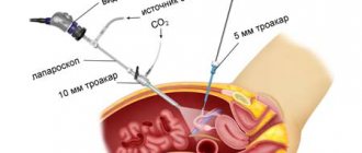
The ultrasound procedure itself, which fits into the following scheme, does not raise any special safety questions. An ultrasonic emitter (it is also a scanner for receiving signals reflected from body tissues), in which sound with a frequency of 20 kHz to 1 GHz is concentrated into a narrow beam and directed to the desired location. The doctor moves the emitter over the skin at the observation site, which, in the role of a scanner, receives signals and transmits them to the control panel. And it converts ultrasonic waves into an image on the monitor.
That's it, the session is over, be it a regular two-dimensional ultrasound or a 3D ultrasound. But the “cinema” will be different. And the future of “actors” raises many more questions among specialists when using 3D. Two dimensions, three or four?
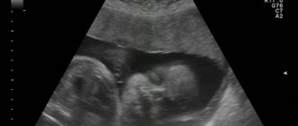
First, a regular two-dimensional ultrasound. This is a flat picture on the screen, essentially a black and white photograph that does not provide any information to a non-professional. Only a specialist can understand the intricacies of jagged dots and dashes.
3D and 4D ultrasound
3D and 4D ultrasound. The latter is the same as 3D, but differs in that time is added as a fourth dimension to the length, height and depth of the picture. If the three-dimensional image is static, then the four-dimensional image shows the object in motion in real time, allowing recording on various media. By the way, most ultrasound devices operating in 3D mode can also use 4D.
With 3D ultrasound, the picture is completely different: firstly, the image is three-dimensional and color; secondly, the baby’s appearance is visible in every detail.
When and at what time is it better to do it?
According to the plan for screening examinations of pregnant women, ultrasound examination is carried out three times: in the first, second and third trimester of gestation. Why shouldn’t they all be replaced with modern three- and four-dimensional examinations? Simply because in the early stages, 3D ultrasound and 4D ultrasound are impractical.
Reference! The optimal period, from the point of view of the information content of the method, is 20 weeks, and after this, such a study can be carried out at the request of the mother.
When is ultrasound used?
During pregnancy, a large number of situations may arise in which ultrasound examination will be able to help understand and decide on further actions. In addition, it is extremely important for pregnancy management to know whether the weight, height and other parameters of the fetus correspond to the period of its development. In general, ultrasound during pregnancy is most often performed for:
- pregnancy diagnosis;
- determining the age of the fetus;
- diagnosis of congenital anomalies;
- determining the position of the fetus;
- diagnosis of multiple pregnancy.
The ultrasound mode during ultrasound is safe for the expectant mother and her baby. This is confirmed by clinical studies and many years of experience in using ultrasound in pregnancy management. But the fact remains obvious that this diagnostic method should be used only as prescribed by a doctor, and not guided by one’s own desire.
You should try to perform ultrasound during pregnancy within strictly regulated periods (when its diagnostic value is especially high). An individual ultrasound schedule for each pregnant woman is drawn up by a gynecologist.
Two, three or four dimensions?
At first, only a two-dimensional conventional ultrasound is performed, during which you will see a flat picture on the screen, similar to a black and white photograph, and you are unlikely to be able to “read” any information from it. A detailed description and interpretation of everything seen on the monitor will be given only by the specialist who performed the ultrasound.
What is the difference between ultrasound and 4D ultrasound?
Three-dimensional and four-dimensional ultrasound examinations are special procedures. To carry them out, the most modern equipment is used, and on the screen the pregnant woman sees a clear three-dimensional image. 4D ultrasound allows the mother herself, without difficulty, to see not only the gender of the baby, but also his facial features, as well as count the fingers or toes.
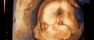
The image of the child, usually in golden tones, is displayed on the monitor and requires small medical clarifications, while it is difficult to understand the image of a two-dimensional ultrasound without a doctor. 4D ultrasound is no longer a photo, but a video image of the baby. After such a procedure, a woman will receive as a souvenir not just a picture from the monitor, but an entire disk with a mini-film about the intrauterine life of a child.
A significant difference between two-dimensional, three-dimensional and four-dimensional methods and in the timing of the examination:
- traditional ultrasound can be performed from 3-5 weeks of pregnancy;
- 3D and 4D examinations are best carried out from 20 to 33 weeks.
In the early stages, it is not very advisable to replace the usual four-dimensional ultrasound, but from the 20th week of pregnancy this procedure becomes more interesting for the mother and more informative for the doctor leading the pregnancy.
How is the examination carried out?
The 4D ultrasound procedure is no different from a regular examination. The patient lies on her back and a transabdominal ultrasound is performed. Conductive gel is applied to the abdominal area to ensure tight contact of the sensor with the skin.
A characteristic difference and disadvantage from a conventional ultrasound is that during a 4D examination it is important to lie still. The slightest movement can blur the picture. Also, a lot depends on how skillfully the doctor handles the ultrasound sensor.
Photo gallery
The photo shows what the baby looks like on a 4D ultrasound.
Difference between conventional 2D ultrasound and 4D ultrasound
Baby in 4D ultrasound picture
What does this examination show?
The latest ultrasound can help your doctor determine:
- genetic pathologies and their manifestations;
- pathologies of heart development or congenital defects;
- find out or clarify the gender of the child;
- brain structures, as well as assess their development;
- some anomalies and developmental features, for example, cleft lip;
- fetal presentation and location of the placenta, which plays a significant role in the possibility of spontaneous childbirth;
- transparency of the amniotic fluid to exclude the presence of infectious diseases.
Usually, a doctor performs a four-dimensional ultrasound at the request of the parents, and not on a referral from the clinic.
Reference! This procedure is paid and available only in large perinatal centers and medical clinics.
But there are also indications that give the attending doctor reason to prescribe 3 or 4D ultrasound as planned:
- An infection suffered by a pregnant woman that could seriously affect the condition of the child.
- Suspicion of genetic pathology, hypoplasia, severe defects of the nervous system or skeletal dysplasia.
- Established developmental anomalies or hereditary pathologies.
- In vitro fertilization or surrogacy.
- Pregnancy with twins or triplets.
Possible results
First of all, such an examination is done in order to assess the condition of the fetus and the fullness of its development. It is possible to more accurately determine the sex of the child. If a regular ultrasound may be uninformative due to the fact that the child has turned or covered the genital area with his foot or hand, then with 4D diagnostics this problem can be easily solved.
With 4D ultrasound, you can see not only the baby from all sides, but also its movements in the uterine cavity
With a comprehensive examination, the accuracy of diagnosing developmental anomalies is much higher. The sooner they are detected, the sooner a decision will be made whether to continue or terminate the pregnancy. At what time to do such an ultrasound is not important, but if there is a high risk of developing fetal anomalies, it is better to diagnose earlier.
A woman can now get more than just a photograph, as was the case with a classic ultrasound. 4D ultrasound makes it possible to make a video recording, which will show a three-dimensional image of the fetus in the uterine cavity and all its movements there.
What can be seen in the photo of the fetus?
The difference between 4D ultrasound is that this procedure reflects the dynamics of the baby’s movements in real time. On the monitor screen you can see how the baby sucks its thumb, waves its arms, turns, and swallows amniotic fluid.
Ultrasound examination plays a big role in the formation of the psycho-emotional attachment of mother to baby, even at the stage of pregnancy. And four-dimensional ultrasound strengthens this connection many times over.
On the disc received after the procedure, the expectant mother will be able to see her baby in all details, feel that very soon she will be able to hold him in her arms and feel how the fear of childbirth goes away, giving way to the positive expectation of a new, loved one.
What can interfere with the procedure?
- Scars on the uterus in a pregnant woman;
- Overweight mother of the child;
- A small amount of amniotic fluid, since in this case it is a conducting medium;
- Position the child with his back to the sensor.
It is important to remember that the advantages of 4D ultrasound methods are enormous: performing such an ultrasound takes much longer than a regular ultrasound. It requires forty-five to fifty minutes, and for a traditional ultrasound, 15 minutes is enough. It can be difficult for a pregnant woman to stand for almost an hour without moving.
How much does it cost and where is it made?
4D ultrasound is rarely prescribed in clinics; the initiative to undergo such a procedure most often belongs to parents and is currently available only in large cities of our country. Perinatal and diagnostic regional centers, as well as large private clinics, provide the service of four-dimensional computer ultrasound for 3500-5000 rubles.
Most commercial medical institutions additionally offer:
- video disc;
- printout of the baby's photo;
- a plaster cast of the baby’s face according to the parameters from the monitor.