How to do an ultrasound of the kidneys
Kidney ultrasound requires preparation. A few days before the test, doctors will advise you to take anti-flatulence medications and follow a diet. Immediately before the test, an hour before the procedure, the patient drinks a liter of water.
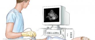
When performing ultrasound diagnostics, the patient is placed on his side or stomach. The surface of the body in the projection to the kidneys is thickly lubricated with a special gel.
Ultrasonic waves are transmitted through the surface of the body and reflected from obstacles of varying densities.
Decoding is done based on the picture, the organ is visible on the monitor screen. Echogenicity is distinguished by shades of gray, by which the doctor judges the normality or pathology of the organ structure.
It is possible to make a diagnosis immediately during the examination; the patient receives a conclusion about the condition of the organ on the same day.
Such a study receives mobile data on the patient’s health status and begins therapeutic measures as soon as possible.
Kidney ultrasound
What are the symptoms that a woman has chronic pyelonephritis?
The apparent reasons are varied. If you have chronic pyelonephritis, you may not know about it for some time (before diagnosis). Pain is felt in the lumbar region. Aching or dull and weak. When it's cold or damp outside, they get worse. Women experience frequent urination and even urinary incontinence. Blood pressure increases in patients. Women feel pain when urinating.
How intense will the disease manifest itself? It depends on whether it is 1 kidney or both and how long ago? If a woman has chronic pyelonephritis, then during the period of remission she will not feel much pain and will decide that she is healthy. Painful sensations will become noticeable during the acute stage of the disease.
What causes the exacerbation? Visible reasons: people have weak immunity. It happens after eating spicy foods, if you often drink alcohol in any form, or if you get hypothermic somewhere. Symptoms of the disease:
Your temperature is above +38 °C; You feel a nagging pain in your lower back. There are also pains in the peritoneal area, but less often. If you stand somewhere for a long time or play sports, they will remind you of themselves. You get tired faster than usual and often feel weak; Headache; Muscle pain is felt; You feel sick; The face and limbs swell; Urination becomes more frequent, persistent frequent urge; You feel pain when urinating; Urine is cloudy; There was blood in the urine.
Diagnosis of acute pyelonephritis
Acute inflammation of the renal pelvis is focal or diffuse. In the focal type, inflammation has clear boundaries within which the pathological process occurs.
The diffuse type of disease is diffuse in nature, and pathology will not show clear boundaries.
The acute form of the disease is characterized by the fact that the kidney affected by the pathology becomes less mobile when inhaling.
Doctors ask the patient to take a deep breath, as a result of which the organ moves slightly, whereas normally the mobility is pronounced.
Also, a focal lesion provokes an increase in echo density in a certain area, this is the focus of infiltration. At the same time, the kidney remains of normal size.
With the diffuse type, the picture is different. The kidney is larger than it should be. The echo density of the organ with pyelonephritis is reduced, and diffuse signs of damage can be clearly identified on the device’s monitor. With an extensive pathological process, the organ loses the distinctness of its layers.
Who is more likely to be prescribed an ultrasound by a doctor?
For what symptoms will the doctor give you a referral for an ultrasound examination:
If you feel pain in the abdomen and lower back. For no apparent reason, you have a high body temperature for quite a long time. A general blood test shows leukocytosis, increased ESR, leukemia is shifted to the left, anemia is observed; Biochemical analysis shows that creatinine has increased, as has urea, potassium, and blood serum. These indicators are especially important for making a diagnosis if you do not yet know exactly which organ is affected? The kidneys' ability to remove urine is impaired. At night you have a frequent urge to urinate. At the same time, you feel pain. Over the course of a day, there was less or more urine, and swelling appeared. There is more or less urine, but its specific gravity is less than it was before. Blood with a high protein content, a lot of bacteria, urate salts, with phosphates, and a lot of leukocytes appeared in the urine.
How is an ultrasound examination of the kidneys performed? The patient is asked to take off his clothes. expose your back. They put sensors on the place where the kidneys are located, move them and look on the screen in what condition is the organ?
"Advice. Take a deep breath and continue to breathe deeply. Then the picture of the kidney examination will be most complete and clear.”
Now you know how the kidneys are examined and that pyelonephritis is visible on ultrasound. It can be in acute or chronic form. All that remains is to be examined using ultrasound equipment and treated. How long will the course take? It's different for everyone.
Click the Sign up button and we will find you an ultrasound specialist or another doctor in 10 minutes.
You can read about laboratory diagnostic methods and the most important analysis for diagnosing pyelonephritis in the corresponding article on tests. In this article we will talk about instrumental methods for diagnosing pyelonephritis.
Echo signs of pustular pyelonephritis
If acute pyelonephritis can still be seen using ultrasound, then pustular lesions of the organ, or apostematous form, are difficult to diagnose.
Signs of pustular pyelonephritis are not visible on ultrasound. An echography of such a condition will more closely resemble acute diffuse pyelonephritis, but the patient’s condition with pustular inflammation is worse.
This visualization is maintained until the pustules merge into a single focus and form a carbuncle.
The echo density of the organ will be reduced in the area, although the focus itself will have a homogeneous structure, but without clearly defined contours.
Signs of a carbuncle will vary depending on the stage of development of the pathology at which the ultrasound examination was performed.
At the recovery stage, ultrasound shows the normal size of the organ, a decrease in the thickness of the renal parenchyma, and the layers of the organ will begin to be visualized.
In the place where there was previously a purulent cavity, scarring forms, which will also be visible during the study in the form of a hyperechoic formation, with some inward depression in this place.
With timely treatment of the disease after acute pustular pyelonephritis, there may be no signs of the pathological process suffered earlier.
Ultrasonic Characterization
Morphologically, pyelonephritis is an inflammatory disease of the renal pyelocaliceal system. This process can be of two types: primary - without previous pathology on the part of the urinary organs (the infectious agent enters through the blood from other foci) and secondary - against the background of diseases that manifest themselves locally in the pyelocaliceal system. The line between these types is quite thin. This disorder is often unilateral.
It is important to pay attention to the fact that ultrasound results cannot always clearly distinguish between chronic and acute inflammatory processes, primary and secondary infectious lesions. The conclusion on interpretation of ultrasound therapy is not a diagnosis - only a description of the echostructure. Therefore, ultrasound results always become an addition to the overall picture of the patient’s condition, medical history, symptoms, the presence of other diseases (heart), and laboratory data.
Chronic pyelonephritis: ultrasound diagnosis
Chronic pyelonephritis is the result of a sluggish, long-term inflammatory process lasting more than six months.
Ultrasound signs of chronic pyelonephritis are nonspecific, and therefore the disease often cannot be diagnosed based on ultrasound alone.
The final decision is made after the doctor receives the results of the urine and blood tests.
Using ultrasound diagnostics, signs of advanced chronic pyelonephritis, in which nephrosclerosis has already developed, are determined.
On ultrasound it will look like thinned parenchyma, the echo density is increased. The contours acquire lumpy outlines, and the kidneys themselves decrease in size compared to the norm.
There is an expansion of the renal pelvis. In some cases, nodular tumor formations are visible, the boundaries of which extend beyond the kidney area - doctors suspect kidney tuberculosis or the presence of helminthic infestations.
Indications for the study
For pyelonephritis, the study can be performed several times. The doctor will prescribe an ultrasound in the following cases:
- the presence of clinical signs of pyelonephritis: fever, pain in the lower back, changes in the nature of urine;
- results of laboratory tests of blood and urine characteristic of pyelonephritis;
- suspected formation of stones in the kidneys or urinary tract;
Suspicion of kidney stones - indication for diagnostic ultrasound
- the need to monitor the effectiveness of treatment measures for kidney inflammation;
- routine preventive examination for chronic forms of the disease;
- checking the restoration of urine outflow after surgery to remove stones.
Ultrasound of the kidneys - video
Identifying complications
A complication of the disease is glomerulonephritis. This process takes place in two kidneys. In this case, an ultrasound examination shows signs of swelling of the organs, thickening of the walls of the parenchyma, and an increase in size.
At the same time, the edges are clearly visualized, smooth and even. If glomerulonephritis becomes chronic, patients develop renal failure.
On ultrasound, symptoms are manifested by a decrease in organ size, tuberous contours, wrinkling of organs, and thinning of the parenchyma.
In this case, the ultrasound diagnostic report contains a diagnosis of diffuse changes.
Ultrasound of the kidneys for pyelonephritis is not always informative; this diagnostic measure helps to see other pathologies of the organ, which is important for assessing the pathological process and the patient’s health status.
For example, an ultrasound scan shows stones in the kidneys. This can be both a consequence of a pathological process and a cause.
Formidable pathologies such as an abscess and carbuncle are also visualized. It is worth recognizing that ultrasound examination of the kidneys is extremely valuable for a patient with pyelonephritis.
Types of disease
Young women are more often affected. Pyelonephritis can be acute or chronic. Sometimes it is mistaken for a worsening condition in other infectious processes.
Acute pyelonephritis becomes chronic due to improper or untimely treatment. This form causes virtually no complaints from the patient, but the kidney tissue gradually degenerates and ceases to perform its functions. Over time, the signs of pathology become more and more apparent, and exacerbations occur more often.
To identify pathology, X-ray and ultrasound examination methods are used. Using ultrasound, any form of the disease is diagnosed for pyelonephritis. CT and NMR (nuclear magnetic resonance) provide better opportunities to see subtle changes, but these methods are expensive.
Ultrasound is a minimally invasive procedure. It has virtually no contraindications. The method does not carry radiation exposure, so it is prescribed to children and pregnant women throughout pregnancy. The study is widely used for people at risk, for example, hypertensive patients, patients with diabetes mellitus. The interpretation and conclusion after the examination is made only by a specialist in this field.
Encyclopedia of Ultrasound and MRI
Ultrasound diagnostics is one of the most popular instrumental methods due to its indicativeness, safety, the ability to save photos and video documents, as well as the relative ease of implementation. Ultrasound of the kidneys for pyelonephritis provides a lot of information to determine the diagnosis and further tactics for managing the patient. This procedure can be performed on pregnant women during their gestational age; it must be done on a child in the first year of life.