Any woman has to examine her breasts for preventive, diagnostic purposes during pregnancy and lactation.
In some cases, a mammogram is done, in others they are sent for an ultrasound. Many women wonder: what is better – mammography or ultrasound of the mammary glands, which study to prefer? To get an answer to this question, you need to know how ultrasound of the mammary glands fundamentally differs from X-ray examination - mammography, when they are prescribed and on what basis. Each method has its own advantages and disadvantages and specific indications.
The principle of operation of mammography and ultrasound examination
Ultrasound examination is based on the ability of tissues to reflect ultrasound. It depends on the density of the structures. As a result of signal processing, an image is obtained that reflects all pathological foci.
The mammography method works on the same principle as ultrasound examinations. Only instead of scanning tissues with ultrasonic waves, X-ray irradiation is used. The rays also pierce the tissue and, as a result, an image is displayed on the screen. The examination is most often used to detect early stage cancer.
Comfort
Ultrasound of the mammary glands does not require special preparation. The procedure itself is absolutely painless, but it takes some time so that the specialist can conduct a more thorough examination.
In order to have a mammogram, no additional preparation is required. This process also takes a short amount of time. However, during the process, painful sensations may occur when the breast gland is compressed between the plates at the moment when the picture is taken. In addition, the doctor may ask you to hold your breath a little to get a clearer picture.
Types of ultrasound and mammography
To compare diagnostic methods, you need to know their types and features. To decipher the results of each examination, a specialist of a certain qualification is required.
Mammography
Mammography is an x-ray examination method. Diagnostics can be analog, when the results are recorded on film, and digital, when saved on electronic third-party media. The latter option has a number of advantages:
- high resolution images;
- short research time;
- creating the required number of copies;
- image analysis using special programs;
- minimum radiation dose;
- the ability to work with the image (for example, enlargement, adjusting contrast and brightness).
Another type of mammography is tomosynthesis. It allows you to obtain 3D images. The X-ray tubes themselves move around the chest. The result is photographs taken from different angles.
Galactography (otherwise ductography) is the second subtype of mammography. The examination is carried out using a contrast agent if various formations are suspected in the gland canals. Ductography helps to examine pathological areas, determine the structure, nature and thickness of the canals, and evaluate even the smallest neoplasms (for example, papillomas).
Ultrasound
Ultrasound examination, like the X-ray method, is simple and accessible, but is used more often due to the absence of harmful radiation. During diagnosis, tissues and their structure, pathological areas are also visualized, but at the same time their echogenicity is assessed. Ultrasound scanning is most informative for lipomas, cysts, fibroadenomas and formations containing fluid. However, it is not suitable for assessing cancer as it may give a false result.
The second type of ultrasound is with contrast, when the Doppler method is used. Such a scan allows you to evaluate the vascular system of the mammary glands, the speed of blood flow, and compare these data with healthy breasts. Using an ultrasound examination, a more accurate puncture is made.
General characteristics of the studies
In order to understand the difference between ultrasound and mammography, and which method is preferable in different situations, you need to understand the physical basis of each.
Mammography
This is an x-ray examination of the mammary glands. The chest area is exposed to ionizing radiation in small doses and an image of the internal structure of the organ is obtained. Modern radiology offers several options for mammography:
- traditional with obtaining an image on X-ray film in several projections;
- digital mammography, in which X-rays are converted into an electrical signal and recorded on photographic film;
- digital tomosynthesis, when a three-dimensional image of sectors of the mammary gland is obtained, which makes this study the most accurate and informative.
Mammography technique
According to WHO recommendations, all women over 40 years old should have mammography every year. This is a mandatory study during clinical examination of this population group.
Ultrasound of the mammary glands
Traditional sonography involves studying the mammary glands using ultrasound. It is performed on all women under 40 years of age for preventive purposes and if alarming symptoms are detected during palpation. The method is absolutely harmless and has virtually no contraindications. Another difference between ultrasound is its cost (several times cheaper than mammography).
Types of ultrasound examination:
- conventional ultrasound examination - obtaining a two-dimensional image of an organ;
- 3D study, when using special computer programs a three-dimensional image of the structure of the mammary gland is constructed;
- Doppler ultrasound, which is used to determine blood flow parameters.
Breast ultrasound technique
The safety of the method expands its use for preventive purposes. It is sonography that is used as mass screening during dispensary observation of young patients.
Advantages and disadvantages of methods
Ultrasound scanning and x-ray methods have advantages and disadvantages. What is common in both cases is the accuracy of the results (90-95 percent).
| Diagnostic methods | Advantages | Flaws |
| Ultrasound | · you can assess the condition of the glands from any angle; · even small tumors are detected; · the condition of regional lymph nodes, the absence (or presence) of metastases are determined; · the examination is informative, regardless of breast size; · when taking a puncture, ultrasound is preferable to the x-ray method; · Ultrasound diagnostics can be done regardless of age, disease, or stage of pregnancy; · the examination is absolutely safe and can be performed as many times as necessary (even daily); · blood flow in glands and tumors is checked; · The method is ideal for inflammatory pathologies and chest trauma. | Sometimes additional diagnostic methods are required to clarify or deny a preliminary diagnosis (for example, the malignancy of tumors is determined only indirectly). The accuracy of decoding depends on the quality of the equipment and the experience of the diagnostician. For example, only an experienced doctor will be able to notice signs of cancer. |
| Mammography | · any neoplasms (even minor ones) are identified, their nature, shape, size can be assessed; · accuracy of microcalcification detection; · the condition of the gland ducts is well determined; ideal for identifying cysts; · in the digital type of diagnostics, the impact of radiation is reduced to a very minimum and does not have a negative impact on health. | The disadvantages include, although small, radiation. Therefore, diagnostics cannot be done often, preferably only once a year. The data obtained may not always be accurate, especially before the age of 30, when the glandular tissues are still very dense. |
Both diagnostic methods are carried out after the end of the cycle on days 5-10. At this time, tissue density decreases.
Safety
When comparing the safety of methods, ultrasound is preferable. Ultrasonic waves cause little harm to the body, even to the fetus in the womb. Mammography uses X-rays. Despite the minimal radiation dose, the method is not recommended for frequent use, especially in the presence of cancer.
Comfort and side effects
Both methods are painless and do not require special preparation. However, unlike ultrasound, during mammography the patient may experience some discomfort, sometimes mild pain due to compression of the plates between which the breast is fixed. Also, as directed by the doctor, you will need to hold your breath for a few seconds.
The methods have almost no side effects, with the exception of allergic skin redness during ultrasound (reaction to the gel). If the techniques are performed with contrast, short-term nausea, slight discomfort, and skin rashes may occur. However, to avoid the appearance of such symptoms, allergy tests are performed before the examination.
Price
Ultrasound and mammography are performed free of charge in all clinics during medical examination, before treatment and during therapy. If diagnostics are carried out in private medical institutions, the cost of ultrasound will be significantly lower.
Comparison by accuracy, information content, safety, comfort, number of shortcomings, price
Comparative analysis of mammography and ultrasound according to the main parameters:
| Comparison points | Mammography | |||
| Analog | Digital | Tomosynthesis | Ductography | |
| Comfort | During the procedure, the patient is in a standing or sitting position. The breasts are located between the X-ray plates, causing discomfort and sometimes tenderness. For accurate results, you need to hold your breath. | It is carried out with painkillers, since the process of introducing a contrast agent is painful (the composition is administered through the milk ducts). | ||
| Safety | The methods are safe as there is no tissue damage. But in the presence of injuries, this examination method is not used. Procedures are not used during pregnancy. When breastfeeding, clarification is required from the treating specialist. | If a specialist is negligent, an infection may enter the milk ducts. Does not apply during pregnancy and breastfeeding. | ||
| Accuracy percentage range depends on the modernity of the equipment | 90% | 90-95% | 95-99% | 90-95% |
| Informativeness | Allows you to identify cancerous and benign formations, as well as salt deposits. With ductography, you can monitor the functioning of the milk ducts. | |||
| Disadvantages and advantages | Pictures cannot be reproduced or image quality increased. Minimal radiation exposure. | The information on the media can be reproduced, the image can be enlarged or reduced, and the contrast can be changed for a clearer image. | The radiation is higher than with other mammography methods. Allows you to obtain a 3D image with high accuracy of localization of the pathological formation. | The procedure is painful. The only method to determine the condition of the milk ducts. |
| Cost (minimum in Russia in rubles) | 1500 | 2200 | 3200 | 1500 |
| Efficiency up to 40 years | They are ineffective because the mammary glands consist of denser tissue, distorting the results of the research. | |||
| Efficiency after 40 years | The effectiveness of the procedure is high (when compared with ultrasound). | |||
| Efficiency after 50 years | The effectiveness is also high, but the risk of damage to glandular tissue by radiation is reduced. | |||
| Efficiency after 60 years | It is more convenient to use, since there is no need to wait for the required period in the menstrual cycle for examinations. Also, tissue damage from rays is minimal at this age. | |||
| Comparison points | Ultrasound |
| Comfort | The procedure is performed in a lying position and does not cause physical discomfort. |
| Safety | The integrity of the skin is not compromised. There is no chance of infection entering the glands. Examination during pregnancy and lactation is allowed. |
| Accuracy percentage range depends on the modernity of the equipment | 85-90%. |
| Informativeness | For cancer, the procedure is ineffective, but it allows you to determine the condition of the glands in the presence of silicone implants. It is also possible to determine the condition of blood vessels and the quality of blood supply in the glands. Ultrasound allows you to determine abnormalities in the lymph nodes. |
| Disadvantages and advantages | Does not detect cancer. But the procedure is effective for identifying benign formations at any age. The frequency of procedures is not specified. Repeated examination is allowed without delay. Examination is allowed in the presence of an inflammatory process in the chest. |
| Cost (minimum in Russia in rubles) | 300 (using Doppler 3500) |
| Efficiency up to 40 years | Allows you to better identify changes in the breast. |
| Efficiency after 40 years | Allows to identify neoplasms, but mostly benign ones |
| Efficiency after 50 years | Used to identify non-oncological formations. |
| Efficiency after 60 years |
When undergoing an ultrasound or mammography, preference must be given to examination using modern equipment and to specialists with certificates that confirm their qualifications. When examined in government institutions, this choice is not possible, but then the procedure is carried out free of charge.
Indications for methods
If you choose which is better - mammography or ultrasound, then it is preferable to do the second method before the age of 40, and after that - the first. The examination must be carried out every 24 months. After 50 years - at intervals of a year. Main indications for:
- gynecological diseases in chronic form;
- changes in the shape of the glands (for example, irregularities, depressions, various protrusions);
- various discharges (mucus, pus, etc.) from the nipples;
- changes in the skin;
- the appearance of pain, swelling, compaction in the glands;
- change in the shape or color of the nipples;
- monitoring the effectiveness of therapy or the development of the disease.
If a woman is at risk of cancer, then an X-ray method is done every 12 months from the age of 35. Indications may include hereditary predisposition, mastopathy, hormonal imbalance, and surgical operations on the glands.
Ultrasound is performed for the following indications:
- inflammation of the glands;
- redness or peeling of the skin on the chest;
- checking the integrity of implants and tissues that come into contact with them;
- identifying the causes of enlarged regional lymph nodes;
- nipple discharge;
- control over cyst growth;
- examination of the breasts after injuries.
Ultrasound is also necessary to clarify the structures of dense neoplasms that were discovered during other examinations.
Ultrasound examination of the breast
A modern and highly informative research method using ultrasound is most widely used in the diagnosis of various breast diseases. It has the widest indications and is not contraindicated for anyone.
When is an ultrasound performed?
Ultrasound examination is done in the following cases:
- when a lump in the mammary gland is detected by touch;
- if there are external changes - deformation, increase in size, ulcerations on the skin, discharge from the nipple;
- with an inflammatory process in the gland (mastitis);
- if milk stagnation is pronounced, lactation is difficult;
- for pain in the glands;
- after breast surgery for control;
- for chest injuries;
- when a mammography reveals an area of tissue compaction - to clarify the diagnosis;
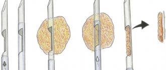
- for diagnostic and therapeutic puncture (taking a biopsy, administering medications);
- in children - with too early development of the mammary glands in girls, gynecomastia in boys and men.
Research principle
The method is based on the property of living tissues to transmit (absorb) or reflect ultra-high frequency sound waves - ultrasound. The scanner device consists of an ultrasound generator, a sensor through which the generated waves are sent to the organ under study, a wave catcher and transmitter, a digital analyzer and a display.
The diagnostic principle is as follows: the denser the tissue, the less it transmits waves; they are reflected and enter the analyzer, which projects the image on the screen in the form of a light spot. Such areas are called echo-positive. Rarefied tissue, cavities and liquid, on the contrary, absorb these waves and reflect them to a lesser extent, less of them enters the analyzer, and on the screen such objects appear as dark, black areas - echo-negative.
The study is computerized and allows you to accurately determine the location, shape and size of various pathological formations. New scanning technologies make it possible to obtain more information - to examine an object in a 3-dimensional or 4-dimensional image.
Pros and cons of ultrasound examination
The advantage of the ultrasound method is that it is highly informative and absolutely harmless to the body. After all, sound waves are essentially mechanical vibrations of a medium of a certain frequency, and do not pose any danger to the patient. Ultrasound can accurately determine the nature and parameters of the object being diagnosed, down to dimensions of 1-2 mm.
The disadvantage of the method is that it cannot accurately diagnose cancer, but only suggest its presence. It is also necessary to observe the timing of the study, depending on the duration of the menstrual cycle.
When ultrasound is indispensable
The ultrasound method is indispensable when there are contraindications to radiation examination: in pregnant women, breastfeeding women, and children. It is also very valuable for periodically monitoring treatment - how effective surgery, radiation or chemotherapy is.
Preparation for mammography and ultrasound and their implementation
Both methods do not require special preparation. The main condition is that before the examination, gels, ointments and other cosmetics should not be applied to the chest or armpits. This can greatly distort the results - false spots and shadows may appear in the pictures. The examination is carried out on days 7-14 of the cycle. If you need to get results urgently, then at any time. Also, diagnostic time is not calculated for women with menopause.
Conducting surveys
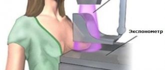
Mammography is done using special equipment. The device is equipped with special plates in which the female breast is clamped. The examination is carried out while standing. The breast is compressed, which evens out its thickness and allows you to get rid of some defects during decoding and more clearly visualize the tissue. Then the mammary gland is fixed. The abdomen and reproductive system are protected with a lead apron. The beam of rays is directed in direct and oblique projections. To examine the second breast, the body position changes and the scanning process is repeated.
During an ultrasound, the patient lies down on a couch. The chest is lubricated with a special gel, through which the diagnostician runs a sensor along specific lines. Diagnostics is carried out in real time - an image immediately appears on the monitor. If necessary, the doctor can stop the sensor in a certain area for a more detailed study. In both cases, the results are issued in 1-2 days, if necessary, immediately in hand.
What is breast mammography?
The examination does not require special preparation. The only condition is clean skin without cosmetics in the chest and armpits, since applied creams or lotions create unnecessary shadows and can reduce the information content of the study. In this case, the resulting image will be blurry, and it will be very difficult to determine whether the patient has the disease. The risk of an error increases. Such a shadow may well be mistaken for a tumor.
For preventive purposes, this examination is carried out at least once a year, and more often if there is a need to monitor the development of compaction. In this case, the examination is carried out in a small area limited by the contour of the proposed compaction. The study is made possible thanks to the presence of special tubes.
Comparison of information content of methods
Since up to 30 years of age the glands are distinguished by good density, mammography may not give results at all, therefore, up to this age, ultrasound will be preferable and more informative. Ultrasound allows you to examine even very large breasts. Mammography does not always provide a complete diagnosis for large sizes.
Ultrasound is indispensable for monitoring the growth of tumors or the effectiveness of treatment. A big plus is that diagnostics can be carried out repeatedly, even for pregnant women. Ultrasound is more effective for dense tissue in the glands, assessing blood flow, lymphatic system, and metastases. However, the information content (especially for detecting cancer at an early stage) is higher for mammography. It allows you to evaluate neoplasms of any size (even inside the ducts), and is indispensable for microcalcifications.
Ultrasound and mammography are similar diagnostics, but are used for different indications, although they also have common ones. Each examination is universal, and more accurate results are achieved if both breast scanning methods are used together.
- PET CT
- The test is positive, but the ultrasound does not show pregnancy
- Ultrasound of the pancreas
- What will a heart ultrasound show? What diseases will it detect?
- Ultrasound of neck vessels
Safety
From a safety point of view, many consider the ultrasound method to be the best, of course. This is a study using ultrasonic waves that does not cause any harm to the body. Experts prefer to prescribe this method to young women under the age of 40. In addition, it can be used during pregnancy without fear of causing any harm to the baby.
Mammography involves irradiating the breast with X-rays. This is a harmless study, the second name of which is “screening method”. 4 photographs of the breast are taken. Older women are most often sent for this procedure. In addition, after 40 years of age, experts recommend having a mammogram at least once a year in order to prevent the occurrence of malignant neoplasms in time. However, despite the fact that the radiation dose is small, you should not abuse this method. During pregnancy, this method is used only in very extreme cases, if malignant neoplasms are suspected.
What is ultrasound?
Several centuries ago, no one imagined that it was possible to look inside a living organism thanks to a certain device; at the moment this does not pose any difficulties.
Ultrasound is a method through which specialists examine the internal organs of a person.
It is completely painless and safe, requiring a minimum of your time, allowing you to reveal a complete picture of the condition of your internal organs. Ultrasound, abbreviated as its full name (ultrasound examination) is the examination of the inside of a person using ultrasound. Ultrasound begins to penetrate the tissues of the body and allows one to determine the shape, size, and structure of any organ. This procedure helps to timely identify various types of pathologies and diagnose the stages of their progression.
Comparison of methods by main characteristics
Often, ultrasound and mammography are used in combination, which increases the likelihood of establishing an accurate diagnosis with further selection of the maximum treatment regimen.
The patient should refrain from independently choosing an instrumental examination - she must consult a doctor.
Because mammography and breast ultrasound have some differences, as well as individual pros and cons, it is important to obtain a professional opinion from a specialist. Indeed, in some cases, one or another method may be uninformative, so it is recommended to pay attention to the following:
- For women under 40 years of age, ultrasound is more suitable than mammography.
- For patients over 50 years of age, it is better to have a mammogram, since X-rays are less dangerous for this age than at a young age.
- The presence of substances such as calcifications is determined only by mammography.
- The presence of cysts is well revealed by ultrasound.
- If a cancerous tumor in the breast is suspected, the doctor must send the patient for a mammogram.
The table provides a brief comparative description of the differences between the two diagnostic procedures:
| Criteria for evaluation | Differences | |
| Ultrasound | Mammography | |
| Security level | It is absolutely harmless for pregnant and lactating women. | Not recommended during pregnancy, especially in the first trimester, as there is a risk of harm to the baby. Relatively contraindicated during lactation. |
| How often can you do it during the year? | Of necessity. | X-rays are used, so the procedure can only be done once a year. For women who are predisposed to breast cancer, examination 2 times a year is acceptable. |
| Information content percentage | Up to 80% | Up to 95% |
| Dependence on the monthly cycle | From 5 to 10 days of the monthly cycle. If an urgent examination is necessary, the procedure can be performed at any time. With the onset of menopause - any day. | |
| Degree of difficulty | It is simple and accessible. | Doesn't have any difficulties. Duration – up to 20 minutes. |
| Comfort | In most cases it does not cause any discomfort. If there are nodes in the chest or mastopathy, unpleasant discomfort may occur. An individual allergy to the gel used during the procedure is possible. During ultrasound with a contrast agent, there is a short-term manifestation of nausea, rash, and minor discomfort. | To take a photo, you need to perform some preliminary manipulations. Such actions can bring discomfort and mild pain to the patient. There is a need to hold your breath for a couple of seconds. |
| Features of preparation for the procedure | Before diagnosis, you should refrain from applying deodorants, gels and other cosmetics to the armpits and chest area. Such substances can degrade image quality (false spots and shadows appear in the picture). | |
| At what age is it relevant? | Up to 40 years old. | After 40 years. |
| Price in private clinics | Including lymph nodes – 2000 rubles. With puncture – up to 3500 rubles. | Classic - from 1500 to 2500 rubles. Digital – from 3500 to 4000 rubles. |
conclusions
It is practically difficult to say which is better – mammography or ultrasound. Each method has its own individual diagnostic advantages. They perfectly complement each other in terms of research, allowing doctors to obtain more extensive information about the nature of the pathological process in the mammary gland.
It follows from this that both types of diagnostics are complementary techniques that help specialists establish an accurate diagnosis and prescribe the most effective treatment.
(
1 ratings, average: 5.00 out of 5)
Who is suitable for mammography?
Who is suitable for mammography?
As already mentioned, the need for mammography before the age of 40 is determined only by a doctor (usually based on ultrasound results). After 60 years, the opposite is true: if you suddenly need an ultrasound, you will be informed about this additionally. And it’s not a matter of radiation exposure at all. Although you are probably thinking about her. How big is it?
Indeed, mammography is an X-ray examination, but the radiation exposure is quite low, 7.5 times less than during conventional fluorography.
Perhaps you should take into account that the radiation exposure during fluorography, as well as during mammography, falls on the breast area - it would be reasonable to separate these two examinations in time.
For what conditions is research relevant?
The doctor can prescribe tests at the following clinic:
| Mammography | Ultrasound |
| Chronic gynecological diseases. Discharge of pus and unusual fluid from the nipples. Deformation of the mammary gland with the manifestation of depressions, protrusions and other irregularities. The patient has a predisposition to cancer. Against the background of obvious modification of the skin of the mammary glands. Pathological change in the shape of the nipples or the color of the nipple skin. The effectiveness of medical or surgical treatment must be determined. | There is a suspicion of an inflammatory process in the chest, or its obvious signs are present. It is necessary to find out how actively the enlargement and growth of cystic formations in the breast occurs. There is a disruption in the menstrual cycle. The patient has reached menopause. Monitor the successful survival of breast implants. Pathological nipple discharge is observed. The postoperative period after removal of a tumor in the breast, when it is necessary to assess how successful the operation was and whether the risk of relapse is possible. |
