- What is computed tomography
- Advantages and disadvantages
- Contraindications to CT
- PET CT scan of the breast
- Conclusion
Computed tomography is one of the methods of radiation diagnostics that allows one to obtain layer-by-layer X-ray images of the examined organs and tissues.
Most patients consider CT to be one of the most accurate diagnostic methods. In fact, its information content, as well as the information content of any other examination methods, varies greatly depending on the organ being examined and the nature of the pathological changes. Thus, computed tomography of the mammary glands is far from the first place among the diagnostic methods used in examining this area. And it’s not just the relative high cost of this method.
Breast CT
Just a few years ago, CT or multislice computed tomography (MSCT) was not used to diagnose breast pathology. It was used only to search for metastases in the lungs, bones and lymph nodes in breast cancer.
Modern CT devices have shown their informative value in breast cancer. It is necessary to take into account that for a high-quality examination (in addition to the device itself), qualified trained personnel and the motivation of this personnel to realize all the capabilities of the equipment, their knowledge and experience are still needed.
Other diagnostic methods
List of alternative diagnostic methods:
- physical examination, palpation;
- thermometry of breast formations;
- analysis of secretions for tumor cells;
- laboratory tests for tumor markers;
- Ultrasound (gray scale and Doppler ultrasound);
- Ultrasound elastography;
- X-ray mammography, incl. with ductography;
- MRI with gadolinium-based contrast;
- PET, SPECT, scintigraphy;
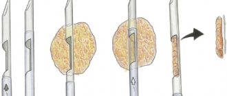
- aspiration fine- or thick-needle biopsy;
- excision with pathomorphological examination;
- tomosynthesis.
Please note: the final diagnosis is established only through pathological examination.
Advantages and disadvantages of computed tomography
Advantages:
- speed of research;
- possibility of comprehensive assessment;
- possibility of using contrast.
Flaws:
- low information content for dense glands (without contrast);
- low specificity;
- ionizing radiation;
- risks of an invasive procedure (with contrast).
Computed tomography of the breast
MSCT of the mammary glands is part of a CT scan of the chest. In this case, not only the mammary glands are assessed, but also all groups of lymph nodes where the tumor can metastasize (supraclavicular, subclavian, axillary, parasternal, mediastinal and contralateral lymph nodes). The skin of the mammary glands, subcutaneous tissue, nipple, pectoral muscles, bones, lungs, vessels of the chest organs and intrathoracic lymph nodes are also necessarily evaluated. All possible changes are identified.
CT scan of the mammary glands for cancer
The use of CT with a contrast agent for breast cancer allows one to obtain the same information as with MRI of the mammary glands.
In the absence of breast cancer, doing a computed tomography scan of the mammary glands for the purpose of “checking it out” is not logical. Mammography , MRI and are traditionally primarily used for these purposes .
A chest CT scan for breast cancer can be performed if there are various metal structures in the body, which is a contraindication to MRI.
Indications for use
Computed tomography is usually performed as an addition or clarification to the diagnosis. If the nature of the tumor in the breast is unclear, computed tomography is the best diagnostic method. It can help more effectively evaluate situations where ultrasound and mammography do not provide complete information.
If the presence of any neoplasm is detected, the procedure is carried out as a determinant of the malignancy of the tumor. So, with actual confirmation of a tumor formation in the breast, a CT scan can either confirm or refute the diagnosis of cancer.
When cancer progresses and metastases appear, CT scans are performed not only of the chest, but also of the abdominal cavity, as well as the brain. When metastases spread to other organs, CT results reveal the operability of the tumor.
The examination helps determine the possibility of surgical intervention for tumor formation. At the same time, thanks to a clear picture of the tumor, you can immediately determine the course and extent of the operation.
What does a breast CT scan show?
In the hands of an experienced specialist, a breast CT scan will help:
- identify the tumor, determine its size and clearly localize its location relative to the skin, nipple and quadrant of the gland. CT scan can detect a tumor with a diameter of 5 mm.
- assess the number of tumor nodes. Often the patient is examined with suspicion of a single tumor node in the gland, but CT reveals several nodes or damage to both mammary glands
- assess the condition of the skin and subcutaneous tissue above the tumor (edema, degree of tumor invasion)
- assess signs of involvement of the fascia of the pectoralis major muscle in the tumor process
- identify and describe the degree of swelling of the skin around the areola
- identify lesions of the nipple of the mammary gland
- identify and describe signs of metastatic lesions of all groups of lymph nodes in the examination area (comparing them with the lymph nodes of the opposite side) and not only in the axillary region, but also interpectoral and parasternal, supra- and subclavian nodes
- identify and describe signs of bone metastases in the scanning area (vertebrae, ribs, sternum) - areas of destruction or sclerosis, depending on the type of metastases (which is sometimes more informative than MRI)
- identify and describe metastases of breast cancer to the lungs and pleura (their number, size and location) - which is generally impossible to see on MRI at an early stage of the disease
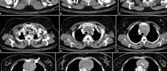
| CT scan of the mammary glands with contrast. In the left mammary gland at the border of the outer quadrants, only a CT scan with a contrast agent reveals a tumor 9 mm in diameter (indicated by a red arrow) | |
| CT scan of the mammary glands with contrast. A tumor differentiates in the left mammary gland at the border of the outer quadrants. The tumor is connected by a “path” to the skin and nipple of the gland (red arrow). In the left axillary region, one metastasis to the lymph node is detected (blue arrow). | |
For breast cancer, we recommend following the following examination schedule:
Use of contrast agent
For most patients, an iodine-based contrast agent is recommended. Before use, it is mixed with saline and administered via a dropper or a one-time injection: the choice depends on the degree of spread of metastases to the lymph nodes and the need to identify the bloodstream of the tumor in the mammary gland. The examination is carried out according to 2 schemes:
- For a small affected area, 1 single dose injection is sufficient.
- The patient receives the contrast agent gradually through an IV. The radiologist monitors the scan result, increasing or adding the dose if necessary. This scan lasts from 40 to 60 minutes.
After the study, the patient is recommended to drink more water without gas during the day in order to remove the drug from the body. If you are prone to allergies to iodine or medications, you can take any antihistamine for 1–2 days.
CT chest
We recommend performing a chest CT scan for breast cancer before a tumor biopsy : after it, edema and hematoma may appear in the breast tissue. This will make diagnosis difficult and tumor assessment impossible.
The lymph nodes of the axillary groups may respond to trauma (biopsy) - this may worsen the picture on computed tomography as a suspicion of their metastatic lesion.
If OSG (osteoscintigraphy) reveals accumulation of radiopharmaceuticals in the bones (which fall within the scanning area of a chest CT scan), show this conclusion to a CT specialist. He will be able to examine these bones more closely to look for metastatic disease in these areas.
How the research is carried out
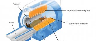
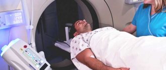
The procedure is performed by a radiologist, who is located in the next room to avoid exposure to radiation.
The patient lies down on a special table, which slides into the CT machine. There is a special sensor and emitter inside the device. These devices scan the patient's chest and transmit information to the PC. There it is processed, after which pictures appear on the screen showing different sections of breast tissue.
The specialist monitors the receipt of data and gradually gets a complete picture of the situation. The duration of the study is from half an hour to an hour.
Breast CT price
Computed tomography for breast cancer with contrast is on average 3 times cheaper than MRI of the mammary glands with contrast. In this case, CT will be able to evaluate not only the mammary glands, but also the lungs and bone tissue in the scanning area for metastases, but MRI will not.
Breast CT is not included in the compulsory medical insurance program, therefore (especially when examined by a competent specialist) be prepared to do it for a fee.
It is better to find out the price of research from the performing doctors:
Specialists at the University Breast Center recommend being examined by trusted specialists.
Preparing for CT
This examination does not require special preparation methods. In this case, you need to adhere to some rules and recommendations:
- If a medical facility asks you to change out of your clothes into a sterile gown, then this rule must be followed. This precaution helps to avoid malfunctions of the tomograph.
- Jewelry, metal objects, and a telephone must either not be taken for examination at all, or left in a specialized place in the hospital. This information should be clarified at the reception desk or with a doctor before the examination.
- Sometimes diet adjustments are required. The doctor will warn you about this during your consultation before the CT scan. In some cases, a fasting diet is required for 12 hours before the examination.
- If you suffer from claustrophobia, you need to warn your doctor about this. You will be given a sedative to calm you down.
- If there are foreign objects in the body, you need to notify the radiologist. Foreign bodies that affect the course of computed tomography include implants, metal plates, neurostimulators, shunts, and pacemakers.
After completing all points of the recommendations, you can proceed directly to conducting a breast examination using CT methods.
Whole body PET CT
Of course, PET CT of the whole body can immediately replace all other regulated examinations for breast cancer, except for biopsy: mammography, ultrasound of the breast and abdomen, ultrasound of the abdominal cavity and pelvis, X-ray or CT of the chest, osteoscintigraphy.
But there is a “minus” of such diagnostics: it “ties” the patient to PET CT. That is, to assess the dynamics of the process in the future, you will have to do a PET CT scan again (and not everyone can afford this).
Of course, patients who can financially afford this examination more than once can perform it both before the start of treatment (for primary operable breast cancer) and for monitoring after treatment.
Main tasks of CT
CT does not belong to the category of screening research methods.
The main purpose of performing a computed tomography scan after confirming breast cancer is to clarify the preliminary diagnosis. The study is also prescribed to study the nature and extent of the damage, the characteristics of the disease. The use of CT scans during annual preventive examinations is unacceptable. This X-ray examination can aggravate the health condition and provoke the transformation of a benign formation into a malignant one.
Considering the main tasks of CT, we can list the indications for this type of diagnosis:
- surgical intervention planning;
- clarification of tumor location;
- performing a biopsy;
- examination of lymph nodes near the location of the tumor;
- tracking the dynamics of therapy.
Based on the information received, the doctor selects or changes the treatment.