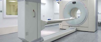
This device is currently the most effective means of monitoring the condition of various tumors in the body. It is most often used to detect cancer. Diagnosing cancer using MRI allows you to take clear pictures, shows the nature of the tumor, and it is practically harmless. The tomograph must have a certain field strength so that the test results can be successfully interpreted.
MRI is an effective method for diagnosing cancer
What is the role of MRI in diagnosing cancer?
MRI is the most effective procedure for identifying cancer in the body. It allows you to detect cancer in the human body at various stages of its formation. Analysis of the results in conjunction with other studies allows you to see the full picture of the disease and successfully perform the operation.
This device can be used to conduct studies of almost any organ with the exception of lungs and bone tissue. Oncology on MRI is well identified in 90-95% of cases. With its help, you can see even very small tumors. Unlike computed tomography (CT), it is more harmless and does not have a negative effect on the tumor.
Cancer and viruses
Let's look at the geography of tumor distribution and their racial “preferences.” For example, liver cancer. It is most widespread in China, African countries (Mozambique, Zimbabwe, South Africa), and Japan. And it immediately becomes clear that the most important cause of this cancer is the hepatitis virus, since it is very common in these countries.
Viruses that can cause cancer include, in addition to the human papilloma virus and hepatitis viruses, the Epstein-Bar virus (in ordinary life, the causative agent of infectious mononucleosis), the herpes virus, the immunodeficiency virus and many others. And here the mechanism is quite clear. Viruses (and this is actually a chain of DNA or RNA with a minimum of other structures) tend to invade a cell and penetrate its nucleus - (they don’t have one of their own!) - and cause mutations of previously normal genes.
What are the advantages of the procedure?
The main advantages of diagnostics using this device are:
- high resolution and clarity of images;
- monitoring the dynamics of tumor development;
- the ability to determine the nature of the neoplasm;
- no harmful effects.
MRI is suitable for people of all ages as it is completely safe
Unlike research methods such as ultrasound, x-rays, and radiation diagnostics, MRI provides clearer and more informative images. It is indispensable for studying the spinal cord, head, heart muscle and joints.
Do you know how much radiation you are exposed to during a CT scan?
To understand how dangerous CT exposure is, consider that over the course of one year, you are naturally exposed to about 3 millisieverts (mSv) of radiation due to background radiation from space. Compare this to undergoing a single CT scan of the head, which could expose you to about 2 mSv of radiation, or a full abdominal scan, which would expose you to more than 30 mSv of radiation. This is 10 times the natural radiation you are exposed to in one year!
Radiation exposure between 5 and 125 mSv is already considered “statistically significant” due to the fact that such radiation doses greatly increase the risk of developing cancer. However, people who have already been diagnosed with cancer tend to be exposed to even higher levels of radiation than healthy people due to the frequency of scans for diagnosis and follow-up purposes.
Are tumors visible on MRI?
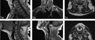
On a tomograph you can see formations measuring only 0.1 mm. Much depends on the quality of the device itself and the qualifications of the specialist analyzing the images. Thin sections—images of the patient’s diseased area—allow you to create a three-dimensional model of the tumor on a computer. The doctor can clearly examine the features of its location, structure, and development dynamics.
However, an accurate result can be obtained only after examining part of the material from the diseased area (biopsy) for histology.
A tomograph allows you not only to examine a tumor at an early stage, but also to determine whether it is a fibroma, lipoma or oncology based on the state of the fluid in it.
Magnetic resonance imaging in oncology is used in the following cases:
- Cancer prevention. If there are formations that can turn into cancer in the future, the doctor often prescribes a preventive MRI examination for the patient once every six months. This allows you to monitor the internal state of problem areas and detect the initial stages of cancer in time.
MRI accurately shows the size and location of the tumor
- Detection of a malignant tumor in the initial stage. With this device you can even see small malignant tumors (schwannomas). This allows you to stop the disease at the very beginning, so as not to have to use radiation and chemotherapy later.
- Determination of tumor status. An MRI will show cancer with high accuracy. From the image you can understand whether the tumor is benign or not. There are a number of signs that allow you to do this.
- Before surgery. Typically, studies of the cancer process in the body using a tomograph are carried out before and after surgery. This allows not only to successfully perform the operation, but also to make the recovery period easier.
- To monitor and control the condition of the tumor. If there is a benign tumor in the patient’s body, doctors often prescribe regular testing. This allows you to keep the development and condition of the tumor under control. In case of negative dynamics, regular monitoring will allow timely initiation of treatment.
Based on the results obtained, the doctor will decide on the need for surgery.
Is it possible to detect metastases?
When metastases form, the patient can remain functional and not notice changes. Therefore, doctors recommend undergoing a routine examination several months after surgery or cancer treatment. Diagnostics is carried out using different methods:
- If an ultrasound or CT scan shows problem areas, shadows with unclear contours, only the organs or the area where the development of secondary cancer is suspected (chest, abdominal cavity) are scanned.
- In the remission stage, a full body examination with MRI is often recommended. This allows for timely detection of metastases or dense calcifications.
When diagnosing cancer and searching for metastases, oncologists pay more attention to several areas where secondary tumors are most often detected:
- thoracic region (lungs, bronchi, thyroid gland);
- peritoneum (liver, spleen, intestines, stomach);
- pelvis (genitourinary organs);
- head (nasopharynx, eye orbits);
- brain or spinal cord.
Magnetic resonance imaging is practically not used if the cancer has metastasized to the bones. In such a situation, it is better to use computed tomography with x-rays.
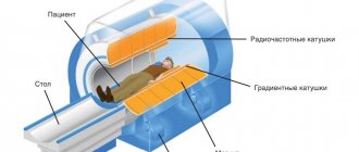
Is it possible to see cancer on an MRI if the machine emits low magnetic doses? If oncology is suspected or when searching for metastases, it is recommended to use a high-field tomograph with a power of at least 1.5–2 Tesla. It makes thinner sections and has increased sensitivity. Otherwise, the diagnosis may be inaccurate.
What signs indicate cancer
The following signs of cancer on MRI can be identified:
- Uneven, lumpy structure. The heterogeneity of a schwannoma tumor on MRI indicates its aggressive nature. She “plans” to metastasize, which grows from them. Pathogenic cells actively grow and kill the body.
- Presence of dark spots, knots, seals. A benign tumor has a smooth and even structure. It lacks various calcifications, darkening and irregularities.
- Blood vessels form a network. In a cancerous tumor, blood vessels intertwine with each other, forming an uneven, lumpy lump that is well supplied with blood.
To make the image of the tumor clearer, doctors use special contrast. The common belief that MRI with contrast causes cancer is unfounded. It spreads through the vessels and illuminates the tumor, making the images more informative.
Causes of Spinal Cancer
The etiology of spinal cancer and the causes of its occurrence still remain a mystery to scientists. It has been proven that a third of episodes are the result of improper behavior - smoking, insufficient consumption of fresh vegetables and fruits, and low levels of physical activity. In addition, the main risk factors that provoke spinal cancer include :
- • age;
- • burdened heredity (it is not the gene responsible for the appearance of cancer that is inherited, but a predisposition to it);
- • contact with carcinogens at home and at work;
- • exposure to ionizing radiation, which leads to mutations;
- • psychosomatics.
No pathogens causing spinal cancer have been detected. It is not formed from a hernia either. The pathology is not contagious, is not transmitted by airborne droplets or other means, so it is impossible to infect others.
Tumors from the prostate and female genital organs metastasize to the spine.
Features of MRI for blood cancer
Diagnosis of blood cancer using MRI is one of the leading methods for analyzing the condition of a patient with such a disease. Using a contrast agent, the doctor can see the state of the patient's circulatory system. Understand the degree of development of the pathology and possible treatment options. The main symptoms of leukemia are:
- weakness and loss of strength;
- the appearance of hematomas on the skin;
- poor sleep;
- dizziness;
- gum problems.
With this procedure, the use of Gadolinium is not always possible, since patients with leukemia do not tolerate it well.
Startling statistics show long-term dangers of CT scans
In a study published in 2013 in the British Medical Journal, researchers looked at about 11 million subjects, ranging from people born in the 1980s to young adults. Within this group, the researchers were able to identify 680,000 people who had been diagnosed with a CT scan at least once.
Those who had a CT scan at some point in their childhood had a 24% increased risk of developing cancer compared to those who did not have the procedure. The larger the body area subjected to CT scanning, the greater the risk of developing cancer.
The increased level of risk remained for a long period of time after the CT scan. The researchers also found that the more CT scans a person had, the higher their risk of developing cancer. The increased risk of cancer did not decrease even after several days. The researchers found that the increase in cancer risk remained at:
- 35% during the first four years after exposure to radiation during a CT scan
- 25% for the period from 5 to 9 years
- 14% in the period from 10 to 14 years
What errors can occur during MRI?
The use of this device may not always be effective and has its negative aspects. These include:
- Low quality photos. Oncology research devices with field strengths less than 1.5 Tesla should not be used. The pictures are not entirely clear, and it is difficult to understand the location and nature of the tumor. Similar devices can be used for other studies that require less clarity.
There may be problems when examining bones
- Low qualification of personnel. It is important not only to take a good photo, but also to interpret it correctly. A medical error can cost a patient not only his health, but also his life.
- Errors in the study of oncology in bones and lungs. MRI is based on scanning the conditions of various fluids and soft tissues in the body. The lungs are filled with air, and the bones have a dense structure and contain little fluid, so their examination is difficult. If necessary, CT scans are used to study these organs.
Read also: what is the difference between CT and MRI.
The table shows the frequency of detection of cases of errors during examination on a tomograph per 1000 patients.
| Errors on MRI | |||
| Test error rate per 1000 patients | Poor quality pictures | Unqualified personnel | Bone and lung studies |
| 6 | 3 | 1 | |
Tumors and carcinogens
The list of carcinogens—substances that can cause cancer—is quite extensive. Even 250 years ago in England, they noticed that chimney sweeps often suffered from scrotal cancer and associated this with constant exposure to soot from chimneys. (It’s not that they rubbed their scrotum on the soot - but they inhaled and swallowed soot particles!)
Carcinogens include oil overheated in a frying pan and smoke from the grill (lung cancer is an occupational disease for cooks of oriental cuisine!), in general, smoked food and meat (fish) from the grill should be treated with caution.
Also all kinds of dyes (classic bladder cancer among dye factory workers), food additives (many of them are simply banned as proven carcinogens), the notorious nitrates and much, much more.
Asbestos, a type of silicate, is one of the most potent carcinogens known. Until recently, it was widely used in construction. After identifying its harmful abilities, it was first abandoned in the USA, in 1995 in France, and since 2005 it has been banned throughout Europe. In other countries, including Russia, this material is still used...