What is screening
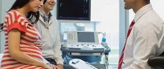
It's actually quite simple. This term refers to a control comprehensive examination of a pregnant woman at a certain period. The concept includes ultrasound diagnostics, in which the doctor draws conclusions about the condition of the expectant mother, her reproductive organs, and also evaluates how correctly the baby is growing and developing.
Also included in the mentioned complex is a biochemical blood test for very specific hormones, the level of which is of great importance in the diagnosis of possible genetic disorders in the fetus.
Screening is prescribed by a doctor during a normal, physiological pregnancy three times during the entire period - every three months.
Who needs it
This diagnosis is carried out not only for pregnant women who are suspected of having problematic fetal development. Therefore, I say again: if you are appointed, do not immediately faint.
It is also recommended to conduct a similar study for women in position who:
- over 35 years old;
- in the past or in the first 3 months they suffered from hepatitis, rubella measles, or had herpetic rashes;
- during pregnancy they contracted a bacterial disease (inflammation of the maxillary sinuses, pneumonia);
- have relatives with genetic diseases;
- previously gave birth to a sick baby;
- are related to the father of their child;
- underwent therapy during pregnancy that could affect the development of the fetus;
- had premature births and miscarriages.
Sometimes couples want to make sure the baby is healthy and go for this procedure without a doctor’s recommendations. And well done!
What is ultrasound
Ultrasound during pregnancy is an indispensable method of visual observation of what is happening inside the uterus. It is carried out using sensors, one of which is elongated and of small diameter, which the doctor inserts into the woman’s vagina in the first trimester. Thanks to the ultrasonic waves emitted by the device, the doctor sees on the monitor an image of the uterus and the embryo in it. The proximity of the sensor to the organs being studied allows for the most accurate analysis of how the very beginning of pregnancy proceeds.
Subsequently, at any period after 12 weeks, another – wide – sensor is installed on the woman’s stomach. However, the method is the same: the doctor evaluates the picture, measures and compares the parameters, tracking the progress of the pregnancy.
Conducting screening
The first planned screening study contains two procedures: a biochemical blood test and an ultrasound examination.
During the procedure, pathologies may be identified, including a defect in the development of the neural tube, Edwards syndrome, Down syndrome, omphalocele, and others.
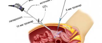
The first screening is performed in one of the following ways: transabdominal, transvaginal. The first method is carried out through the woman’s abdomen using a special sensor.
Video:
To undergo the procedure, a woman must prepare. Before starting the test, you need to drink 1.5 liters of water to ensure your bladder is full.
READ We observe the development in the photo of the size of the fetus, ultrasound at 14 weeks of pregnancy
A transvaginal scan is performed using a probe that is inserted into the vagina. For this procedure, the bladder must be empty.
After the ultrasound is completed, the doctor examines the results obtained, then a transcript is drawn up.
This should include the following information about the child:
- CTE - this indicator is extremely important, since it tells not only about the development of the fetus. By measuring the distance between the crown and the coccyx, one can judge pregnancy, the level of a woman’s hormones, and chromosomal pathologies in newborns;
- the size of the collar zone indicates the absence/presence of chromosomal pathologies of the fetus;
- the size of the nasal bone must be normal, otherwise this is a signal of the presence of Down syndrome;
- fetal head size;
- heart rate.
The second screening study is similar to the first. It also includes two procedures: a blood test and an ultrasound scan.
The only difference is that the study is done only transabdominally, through the abdomen. The woman no longer needs to fill her bladder, so preparation for the study is minimal.
When scanning with ultrasound during pregnancy 18 - 21 weeks, the results of the first procedure are confirmed or refuted.
Photo:
In addition, a study is carried out for the presence of other abnormalities of the fetus. The face, limbs, kidneys, heart, and other areas are studied for compliance with standards.
The transcript of the second screening contains:
- fruit size;
- description of facial structure;
- description of the baby’s internal organs, their correspondence to anatomy;
- lung maturity level;
- compliance with the standards for the number of fingers and toes.
The transcript also contains not only fetal parameters, but also descriptions of the woman’s pelvic organs. The doctor examines the cervix and uterus, measures the thickness of the placenta, describes its structure, and checks the volume of amniotic fluid.
For those whose pregnancy is between 30 and 34 weeks, the doctor prescribes a third screening. Its purpose is to determine the position of the fetus, study the placenta and amniotic fluid.
Such a study allows the doctor to understand how to carry out childbirth: cesarean section or advancement of the fetus along the birth canal.
During this period, the woman undergoes a set of procedures:
- ultrasound scanning;
- cardiotocography - studies the rhythm of the fetal heart;
- Dopplerography - assesses blood flow in the vessels of the female organs and the baby.
READ Features of fetal development on ultrasound at 16 weeks of pregnancy
Photo:
Bipariental size (between the temporal bones) – BPR or BRGP; Thigh length – DlB; Chest diameter - DGrK
The transcript contains:
- baby parameters, which includes measuring height, weight, head circumference, and other measurements;
- results of the functioning of the circulatory system organs;
- description of the fetal face;
- data on the size of bones and spine;
- description of the baby’s internal organs;
- characteristics of the placenta, amniotic fluid.
Cardiotocography is a cardiogram of a child's heart. The procedure is performed using an ultrasound machine.
On the monitor, the doctor sees the heartbeat curve. Such a study makes it possible to determine whether the baby has hypoxia or not.
Dopplerography allows you to assess the condition of the uterus and placenta of a woman, calculate the blood velocity of mother and baby, and assess the patency of all blood vessels.
Sometimes a doctor may prescribe Doppler ultrasound for a woman when undergoing a second screening ultrasound.
What do screening procedures and ultrasound have in common?
What to do - ultrasound or screening - there is no such question. During pregnancy, an ultrasound scan prescribed for a general examination of the fetus and the expectant mother will also be called screening.
In the first trimester, a biochemical blood test as part of screening will be mandatory. You cannot skip it, so as not to miss the development of possible congenital diseases. In the remaining two trimesters, during repeated comprehensive examinations, a blood test for hormones will be prescribed if the first time any indicators alerted the doctor.
Ultrasound examination of the initial period of pregnancy is no less mandatory. The doctor will determine whether the embryo is positioned correctly in the uterus and whether it has begun to grow. In addition, in this phase of gestation, the fetus’s heart begins to beat. This is an important moment both for the doctor and, undoubtedly, for the expectant mother.
What kind of ultrasound is done during screening and what is the difference?
Ultrasound can be performed using several different methods, hence the question: “How is ultrasound screening done?”
In gynecology, it is mainly used transabdominal and transvaginal. Transabdominal ultrasound is performed through the anterior abdominal wall, and transvaginal ultrasound is performed by inserting a sensor into the vagina. Both types of diagnostics are normally painless and do not cause any harm.
- During pregnancy, the type of ultrasound is determined by the stage of pregnancy. In the earlier ones, transvaginal ultrasound is used for ultrasound screening, and in later ones - transabdominal, however, some doctors prefer not to use transvaginal ultrasound at all, due to irritation of the woman’s reproductive organs, which, under unfavorable circumstances, can even lead to early termination of pregnancy. But if there are indications for such a diagnosis, it should be performed immediately.
- From the point of view of information content, transvaginal ultrasound in the early stages is superior to transabdominal, but later, when there is enough amniotic fluid, ultrasound waves calmly pass through them and transabdominal ultrasound is superior to transvaginal.
Ultimately, the decision on which method of diagnosis should be made should be made by the gynecologist, and not by the pregnant woman. During ultrasound screening, the choice of the day of examination remains with the doctor.
The difference between ultrasound and screening
Since ultrasound is included in the concept of screening during pregnancy, it is impossible to say which is better. How to determine the differences between these procedures.
The only thing that needs to be noted is the different content of the comprehensive examination in different trimesters. As already mentioned, it is not necessary to take blood tests in the second and third trimesters if absolutely no abnormalities were revealed in the very first trimester.
However, an ultrasound examination must be done every trimester - three times. If blood counts determine the presence of genetic disorders, then ultrasound reveals anatomical abnormalities and disorders of the formation of various organs and systems of the fetus. They look to see if the baby has enough oxygen supplied from the mother through the arteries of the umbilical cord. And during the final examination, the doctor also checks the child’s readiness to leave the mother’s womb: location (legs to the birth canal or head), absence of umbilical cord entanglement, proportions of the heart and lungs.
As a result, the question of what is the difference can be answered this way: ultrasound diagnostics can be performed on a pregnant woman outside of screening, if necessary. For example, to clarify the gender of the unborn baby. But screening always includes a mandatory ultrasound for general monitoring of the progress of pregnancy.
Screening also differs from a simple ultrasound in that any ultrasound examination of the internal organs of the expectant mother is not part of the screening. It only includes ultrasound of the uterus and fetus, the vessels supplying them, in specific weeks of the observed pregnancy.
And, finally, if any irregularities were discovered during the complex, or the doctor suspected that something was not going as it should, I can send the woman for a repeat ultrasound, and also take blood for analysis again. In this situation, an additional examination will not be considered screening.
What is screening?
During pregnancy, a woman undergoes an ultrasound examination.
This procedure is planned and allows the doctor to clarify the timing of pregnancy, examine the woman and newborns, and monitor their health.
Using a scan, you can see the condition of the uterus, ovaries, and other organs. In addition, amniotic fluid and the condition of the placenta in the expectant mother are monitored.
Photo:
An ultrasound scan checks the baby’s physiological data, monitors development, and prevents the appearance of pathologies in newborns.
Screening ultrasound differs from regular ultrasound, as it has more precise goals and objectives.
Screening is a complex procedure with which you can track the occurrence of pathologies in newborns, identify chromosomal abnormalities and other disorders.
Doctors recommend that expectant mothers undergo screening ultrasounds several times.
The first procedure should take place at 11–14 weeks, the second should be done at 18–21 weeks, and the final one should be performed at 30–34 weeks of pregnancy.
Routine ultrasound and screening are not done at the same time. The last test will be scheduled several weeks earlier than usual.
This is no coincidence - the doctor will be able to refer the woman for a second scan if the results of the ultrasound scan are questionable or inaccurate. In exceptional cases, the doctor may recommend that the woman terminate the pregnancy.
Parameters 1 of ultrasound screening of pregnant women:
The first screening of a pregnant woman is a very informative procedure that allows you to measure and identify:
- the size of all the baby’s bones;
- symmetry of the cerebral hemispheres;
- size and position of the heart and stomach;
- risks of developing Down disease, Patau syndrome, neural tube pathology, and other chromosomal disorders.
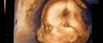
Also during the procedure, you can receive the first photo of the baby and record the ultrasound on an electronic medium. Any mother wants to see her baby as soon as possible - the ABIA clinic provides such an opportunity. After the screening, you will be left with photos and videos of this wonderful period as a keepsake.
What it is?
Ultrasound examination is a method that allows you to obtain a visual image of the unborn baby while he is still in the womb. This examination is prescribed by an obstetrician-gynecologist. During pregnancy, at least 3 such studies are carried out. In pathological cases, this examination may be required more frequently.
Doctors call screening a whole complex of diagnostics, which makes it possible to identify various abnormalities in the intrauterine development of the fetus or anomalies in the mother.
Screening includes more than just an ultrasound. Some biochemical tests are also used to carry it out. In each period of pregnancy they are different.
At what time is the 2nd screening performed?
The examination is carried out after the 14th week. But depending on the expectant mother’s doctor’s visit schedule or other factors, it may be postponed for some time. An examination carried out before the 27th week will be informative, but it is better not to delay its implementation.
According to statistics, from the 24th week in women who are overweight and have a predisposition to diabetes, the risk of developing gestational diabetes increases. Its early detection reduces the risk of disease progression and helps protect mother and child from dangerous consequences: premature birth, the development of preeclampsia and complications when the baby is born.
During this period, problems in the development of the fetus can be identified, including congenital heart defects and tumors of internal organs. Early detection of severe pathologies allows parents to be informed about them in a timely manner, who can make an informed decision about the advisability of continuing the pregnancy.
Indications for diagnosis
Prenatal screening is recommended for all women during pregnancy, in accordance with global and Russian standards and recommendations.
An ultrasound is performed on all women; it not only looks at the correct anatomical structure of the child, but also examines for Down syndrome.
In-depth fetal screening, which includes laboratory tests, is not performed on all women during pregnancy. Indications for it:
- mother’s age is from 35 years, father’s age is from 40 years;
- the woman has children with anomalies and developmental defects;
- miscarriage, undeveloped fetus, history of spontaneous miscarriage;
- the woman suffered an infection in the 1st trimester;
- taking medications from the 1st to the 13th obstetric week that have a teratogenic (disturbing normal development) effect on the fetus (antibiotics, hormones, psychostimulants);
- consanguinity with the future father of the child;
- the effect of ionizing radiation on the mother or father shortly before pregnancy;
- the presence of genetic abnormalities in relatives of the mother or father;
- pregnant women working in hazardous conditions;
- IVF using sperm from an unknown donor;
- the woman smokes and drinks alcohol.
How is 1st screening carried out, preparation
Ultrasound in the first trimester can be performed in two ways:
- transvaginally;
- abdominally.
In the first case, no preparation is needed. When performing an abdominal ultrasound, it is necessary that the woman’s bladder is full - for this purpose, it is recommended to drink 2 glasses of water half an hour before the procedure.
You need to donate blood for biochemical analysis on an empty stomach - you cannot eat at least 4 hours before the procedure. This is especially true for sweets, fatty foods, meat and seafood. Failure to comply with this diet may lead to errors in the analysis.
What are the differences between screening and ultrasound diagnostics?
During pregnancy, women are recommended to undergo various types of examinations to regularly monitor the development of the fetus. Screening differs from a regular ultrasound in that it is a set of diagnostic methods during the period of pregnancy, carried out with the aim of determining negative changes in the development of the unborn baby. Ultrasound examination also makes it possible to identify existing pathological abnormalities using special technologies.
What research does it include?
Before undergoing screening, it is useful for a woman to arm herself with knowledge about how to prepare for tests as part of screening during pregnancy, what information about family and relatives should be provided, and what additional studies may be required if abnormalities are suspected.
First
The very first screening is carried out in the week of pregnancy in which a planned ultrasound is performed, that is, at 10-14 weeks of obstetrics. The first screening complex includes 2 procedures.
- Ultrasound, which allows you to see the fertilized egg attached to the uterus and the baby inside, calculate its gestational age (accuracy: +/- 3 days), evaluate the anatomical features of the fetus, including the thickness of the nuchal translucency, which is a marker of one of the trisomies.
- Biochemical analysis of venous blood for the determination of free beta-hCG and plasma protein A (pregnancy associated plasma protein A, or PAPP-A), which does not require special preparation.
The blood test during the first screening test is called a double biochemical test of the 1st trimester of gestation. Its results make it possible to calculate the risk of developing Down or Edwards syndromes in the fetus. If the answers are positive (that is, the risk of chromosomal abnormalities is confirmed), the woman will be asked to undergo an invasive procedure (with penetration into the uterine cavity) - a chorionic villus biopsy - to clarify the diagnosis.
Second
The timing of the second screening during pregnancy coincides with the time of the second planned ultrasound. The study takes place at 16-18 weeks of gestation and is called triple biochemical screening. As in the first case, this study is preceded by an ultrasound, and blood from a vein is taken on the same day to monitor growth dynamics:
- total hCG or free beta subunit of hCG, as part of hormonal screening during pregnancy;
- free (unconjugated) estriol (E3);
- alpha fetoprotein (AFP).
The results of the triple test allow us to calculate the risk of developing Down and Edwards syndromes, as well as spinal canal cleft and other fetal neural tube defects.
Screenings are carried out non-invasively, that is, without entering the uterine cavity, so they do not pose any danger to either the baby or his mother.
Third
By third screening, many obstetricians mean prenatal examination, which is mandatory for all future mothers. There are several procedures in terms of examination:
- Ultrasound, the timing of which coincides with the timing of prenatal screening in the 3rd trimester of pregnancy - at 32-34 weeks;
- fetal Doppler (ultrasound with Doppler), which allows you to evaluate the blood circulation of the uterus, umbilical cord, and fetus;
- biochemical blood and/or urine tests;
- cardiotocography – assessment of heart rate (pulse), rhythm of heartbeats and cardiac reflexes of the fetus.
In this list of procedures, ultrasound is the basic examination; the rest are prescribed as additional procedures for medical reasons.
How much does the research cost?
Three screenings during pregnancy are performed free of charge with a birth certificate. If desired, a woman can undergo them in a private clinic.
The cost of an ultrasound scan for the first screening is about 2,000 rubles, if you add hormones to it, then 4,000 rubles. Screening at 20–24 weeks of pregnancy costs from 1,000 to 9,000 rubles. For the 3rd, the price will be from 2000 rubles.
Carrying out 3 screenings during pregnancy is recommended for everyone. This allows you to identify developmental defects, examine the baby and mother, and choose the type of delivery.
When were you screened during pregnancy? Share this article with your friends.
How to prepare for research
Preparation for ultrasound screening of the first trimester is as follows:
- The day before the test, you should not eat food that causes severe gas formation in the intestines. Due to gases, the ultrasound result is distorted.
- If an ultrasound scan of the fetus is performed transabdominally, through the abdominal wall, the bladder should be full. Otherwise, when an ultrasound is performed with a transvaginal probe, this organ is emptied.
- You need to buy a disposable diaper to put on the couch. If desired, buy napkins if you need to wipe the gel off your stomach.