Screening translated from English means “sorting” - in fact, this examination plays the role of a “sorting”, dividing pregnant women into those whose indicators fit into the normal framework, and those who should undergo additional diagnostics. What kind of analysis is this, and what is blood taken for? Biochemical screening of the 1st trimester (specifically 11-13 weeks) is a set of diagnostic measures consisting of ultrasound and biochemical blood tests.
It is used to identify the risk of developing gross pathologies of the fetus, as well as genetic abnormalities and severe congenital diseases.
The unknown is scary, so let’s try to understand the concepts so that on the threshold of the biochemical part of screening of the first third of gestation you feel confident and competent.
Diagnostics
Every woman expecting a child has the right to decide independently whether she needs to undergo screening. From the point of view of the law, this type of examination is recommended by doctors, but the expectant mother has the right to refuse tests.
However, this is an unwise approach; from the point of view of the health of the mother and baby, it is better to find out about any possible deviations or disorders as early as possible.
The results of 1 screening provide doctors with a lot of useful information about the health and development of the unborn baby. As a rule, a standard set of studied parameters is used, which makes it possible to detect the following deviations:
- Risk of developing pathological changes in the fetus
- Down syndrome
- Patau syndrome
- Disturbances in the development and pathology of the neural tube of the fetus
- Abnormal set of chromosomes
- Physiological defects of the internal organs of the developing fetus
A screening test does not make it possible to establish a diagnosis or determine with certainty whether the child will be sick. Based on the results of the analysis, one can judge the degree of risk for the development of a particular deviation. Women who are at risk may be offered additional examination. Using an invasive test, amniotic fluid is taken for analysis.
1st trimester screening on an empty stomach or not – Magazine about everything
Screening is a comprehensive examination of the expectant mother. There are certain deadlines, as well as a list of studies carried out. In the first trimester, data collection is aimed at identifying possible genetic pathologies, as well as monitoring the condition of the female body and the development of the embryo.
Includes the first screening during pregnancy, a biochemical blood test (sampling from a vein), as well as an ultrasound examination.
Features of the first trimester
The planned first examination is carried out at 11-13 weeks; this is the optimal time frame for obtaining objective data.
On an individual basis, they can be prescribed earlier or later than this period, most often when there are threats of miscarriage, as well as suspected ectopic pregnancy. This is not a mandatory study and a woman can write a refusal to undergo it.
It is worth remembering that uncontrolled pregnancy is associated with high risks that are dangerous to both the health of the mother and her baby.
Screening during pregnancy in the 1st trimester includes:
- ultrasound diagnostics;
- biochemical blood test.
The order in which it is carried out is also important; the ultrasound should be done first, and only after that the blood should be donated. It is this sequence of test results that matters. Ultrasound helps determine the exact period of gestation; after it is clarified, it will be possible to compare blood test data according to the norms.
To prepare for a superficial ultrasound, you need to drink at least 1.5 liters of water within an hour before visiting the office, or do not visit the toilet for 3 hours, so that your bladder is full. With the transvaginal method, on the contrary, it is necessary to completely empty the bladder. The sensor will be inserted into the vagina; this method will only be performed during the first screening.
The expectant mother needs to know how a biochemical study is done. To obtain accurate tests, blood is drawn from a vein on an empty stomach.
Also, a few days before delivery, you need to exclude fried, spicy, fatty foods from your menu. Also, do not consume known allergens - nuts, citrus fruits, seafood, chocolate. These are the basic conditions that must be met to obtain reliable screening data.
Many pregnant women do not know when the first ultrasound is performed. The most accurate period for conducting the study is the period from 11 to 13 weeks, counting from the day of the last menstruation. It is especially important for women under the age of 18 and after 35 years of age if miscarriages, fetal death, abortions have already occurred, or hereditary diseases and genetic abnormalities have been observed in the family.
Ultrasound standards in the first trimester:
- measurement from the parietal point to the coccyx - CTE, at 11 weeks - 40-58 mm, 12 weeks - 47-73 mm;
- BPR - measurement of the distance between the protruding lobes of the crown - biparietal size, by the end of the 11th week 14-17 mm, 22 mm by 12, by the end of the 13th week - 25-26 mm;
- the length of the nasal bone, up to 11 weeks it can be determined, but it is difficult to estimate its size, at 13 weeks it corresponds to approximately 3 mm;
- Head circumference;
- the cranium should already be closed, the symmetry of the brain hemispheres is determined;
- TVP at 11 weeks is 1.4-2.3 mm, at 12 weeks – 1.7-2.4 mm, at 13 – 1.8-2.9 mm;
- the size of the heart muscle, the largest vessels, as well as the frequency of contractions are determined, normally at 11 weeks – 153-178, at 12 weeks – 150-172, at 13 weeks – 140-170 beats per minute;
- limb length;
- examination of the placenta;
- volume of amniotic fluid;
- umbilical cord and number of vessels;
- The condition of the uterus, tone, and cervix are assessed.
When performing a biochemical blood test, human chorionic gonadotropin indicators deserve special attention. If immediately after conception its quantity increases several times every day, then by the time of the first screening it has more stable indicators.
An increase is a signal of the development of genetic abnormalities, and can also be observed in diabetes mellitus and severe toxicosis. If the indicators are below normal, the cause may be fetal fading, the threat of miscarriage, the development of an ectopic pregnancy, or pathologies in the formation of the child.
Norm β-hCG in the first screening:
- from 10 to 11 weeks – 18-130 ng/ml;
- from 11 to 12 weeks – 13-126 ng/ml;
- at week 13 – 14 -115 ng/ml.
Each laboratory has its own hCG standards; the numbers may differ significantly; the consistency of the results depends on the established indicators in a given clinic. You cannot evaluate them yourself; only a doctor can do this.
The protein produced by the placenta, which determines its condition, is called protein-A (PAPP-A). If its indicators are significantly lower than normal, this may indicate a threat of miscarriage, genetic abnormalities - Edwards syndrome, Down syndrome. A slight increase is considered normal and does not have much significance for the diagnostic results.
What does the norm of protein-A (PAPP-A) show:
- from 10 to 11 weeks 0.8-4.7;
- from 11 to 12 weeks 1.2-3;
- at week 13 1.3-8.7.
Not only the amount of protein matters, but also the dynamics of growth. As pregnancy progresses, its amount should also increase.
What may affect the results
Some individual characteristics of pregnancy may influence the decoding:
- in the first trimester, the weight of the expectant mother affects the indicators; if you are overweight, the amount of hormones will be higher than normal; if you have an asthenic physique, it will be lower than normal; however, pregnancy can proceed without complications;
- with in vitro fertilization, PAPP will be lower than normal, the concentration of human chorionic gonadotropin, on the contrary, will be higher, and on ultrasound the fronto-occipital size will also be increased;
- for multiple gestations, standards for biochemical screening have not yet been established;
- collection of amniotic fluid (amniocentesis) carried out within a week affects the reliability of the blood test results;
- with diabetes, the amount of hormones will be below normal;
- a woman’s emotional state also affects the reliability of research.
First screening results
Carrying out the first comprehensive examination helps not only to assess the overall development of the fetus. Thanks to the collection of data, it is possible to confirm or refute the following developmental deviations:
- disturbances in the formation of the neural tube;
- trisomy 21 chromosome - Down syndrome;
- formation of internal organs in the hernial sac;
- slow heart rate, the nasal bone is not visible;
- Patau syndrome - developmental deviations leading to the death of a child in the first months after birth;
- triploidy – tripling the set of chromosomes;
- pathological metabolic processes due to genetic abnormalities lead to mental retardation and other complications;
- absence of the brain and skull bones, the fetus dies in the third trimester or after birth.
After data collection, the MoM coefficient is determined. When calculating, the following factors must be taken into account: the age of the expectant mother, weight, the presence of chronic diseases, general medical history, lifestyle, bad habits. The degree of deviation during a normally developing pregnancy should not exceed 0.5-2.5, during pregnancy with twins (or after IVF) up to 3.5.
Source: https://zzyrnal.ru/skrining-1-trimestra-natoshhak-ili-net.html
Preparing for analysis
Proper blood sampling and preparation for it ensure the reliability of the results obtained. Blood for research is taken from a vein, but usually the collection of material is completely painless. The procedure is performed in a doctor's office or laboratory. Some private medical institutions can collect material at home for an additional fee:
- The main question that causes a lot of controversy: are tests performed on an empty stomach or not? You should donate blood strictly on an empty stomach, even if the feeling of hunger is very strong, you cannot eat, you can only drink clean water. You can have breakfast immediately after blood sampling, so many pregnant women take sandwiches or other snacks with them so as not to be hungry.
- On the eve of the test, it is not recommended to eat chocolate, fatty meat and seafood.
- When women donate blood, a calm atmosphere is needed. Anxiety, fear, and fast walking to the doctor's office can trigger the activity of certain chemicals and enzymes inside the body.
- 2-3 days before the test, doctors recommend excluding intimacy.
Test parameters
The blood test is performed in a laboratory setting. When a blood sample is given, a test is performed and the patient or her doctor receives a form with the results. Any questions should be consulted with a physician. One measure is called beta human chorionic gonadotropin, or beta-hCG. This is a hormonal substance that is responsible for the normal development of the fetus in the earliest stages. At first the indicator increases significantly, then gradually decreases.
Standards have been established for β-hCG at various stages of pregnancy for screening:
- Week 10: 25.8–181.6 ng/ml
- Week 11: 17.4–130.3 ng/ml
- Week 12: 13.4–128.5 ng/ml
- Week 13: 14.2–114.8 ng/ml
It is important that the β-hCG level is within the established norms. High levels of the hormone may indicate the development of Down syndrome in the fetus. This indicator also increases with multiple pregnancies, early manifestations of toxicosis and established diabetes mellitus in the expectant mother. Low β-hCG levels may indicate the development of Edwards syndrome, placental insufficiency, or be a sign of an ectopic pregnancy.
When hormone levels are low, the risk of miscarriage is very high.
The second main indicator is protein A or PAPP-A. This substance is protein in nature and is responsible for the normal development and functioning of the placenta. Exceeding the norm of this indicator is not considered a violation of the body’s functioning. A decrease in the concentration of PAPP-A leads to the threat of miscarriage and may be a sign of the development of Down syndrome, Cornelia de Lange and other pathologies:
- Week 10-11: 0.45–3.73 mIU/ml
- Week 11-12: 0.78–4.77 mIU/ml
- Week 12-13: 1.03–6.02 mIU/ml
- Week 13-14: 1.47–8.55 mIU/ml
Based on the data obtained, the laboratory calculates another parameter: MoM - coefficient. This value shows the deviation of the values obtained as a result of the patient's blood test in comparison with the average values established as the norm. For a healthy woman and a normally developing fetus, MoM should be in the range of 0.5-2.5. During multiple pregnancy, the rate increases to 3.5.
What can you eat before your first trimester screening?
In any case, a girl needs to adjust her diet during pregnancy so as not to harm the health of the unborn baby. A special diet has also been developed before screening in the 1st trimester.
A woman needs to limit some types of foods in her diet for 2-3 days and increase the consumption of others. Preference should be given to:
- dishes made from lean meat;
- vegetable oils;
- butter;
- porridges made from buckwheat, rice, millet, cooked in water;
- fermented milk products (kefir, fermented baked milk);
- vegetables;
- honey or homemade jam;
- dried white bread;
- weak tea;
- green apples, plums;
- compotes and still waters.
There are no special requirements for cooking. General recommendations regarding diet before the first and second screenings are the same.
Allowed food table
| Products | Protein content*, g | Fat content*, g | Carbohydrate content*, g | Calorie content*, kcal |
| Boiled vegetables | ||||
| potato | 2 | 0,4 | 17-19 | 79-81 |
| carrot | 0,8 | 0,3 | 4-6 | 24-26 |
| Fruit desserts | ||||
| apricot, peach | 0,9 | 0,1 | 10-11,5 | 40-47 |
| watermelons and melons | 0,6 | 0,1 – 0,3 | 5 – 7,5 | 25 – 33 |
| Cereals and porridges | ||||
| buckwheat | 4-5 | 2,3 | 25,0 | 132 |
| semolina | 3 | 3-3,5 | 15-15,5 | 98 |
| oatmeal | 3 | 1,5-2 | 15 | 88 |
| flakes | 11-12 | 7-7,5 | 69-70 | 366 |
| pearl barley | 3 | 0,4 | 22-22,5 | 108-110 |
| rice | 2-2,5 | 0,5 | 24,9 | 116 |
| Pasta | ||||
| spaghetti | 12 | 3,5-4 | 60 | 321-323 |
| Flour products | ||||
| made from wheat flour | 7,5-8,5 | 1-3 | 48-51 | 242-264 |
| Confectionery | ||||
| jams | 0,3 | 0,2 | 63 | 262-264 |
| marshmallows | 0,5-1 | – | 78-79 | 303-305 |
| marmalade | 0,5 | – | 80-81 | 309-311 |
| products with honey | 0,5-1 | – | 81-82 | 328-330 |
| Sauces | ||||
| dairy | 2 | 7-7,5 | 5-5,5 | 83-85 |
| Meat (fillet) | ||||
| chicken | 29,5-30 | 1,5-2 | 0,5 | 136-138 |
| turkey | 25 | 1 | — | 129-131 |
| Beverages | ||||
| still mineral waters | – | – | – | – |
| compotes | 0,5 | – | 19-20 | 80-82 |
| juices | 0-1 | 0,1 | 9-9,5 | 37-41 |
| jelly | 0-0,5 | – | 16,5-17 | 67-69 |
* the data indicated in this range is calculated for one hundred grams of products
Reliability of analyzes and risks
Interpretation of blood test results for screening should only be carried out by a qualified physician who is familiar with the patient’s medical history. It should also be taken into account that screening for twins will show a completely different result. The second child increases the levels of certain chemicals in the mother's body. Every doctor has information with recommended standards for the parameters under study.
However, the individual characteristics of the body and the composition of the patient’s blood before pregnancy should be taken into account.
With the screening analysis and the correctness of the research at all stages, the reliability of the result is estimated at 95%. However, the analysis cannot accurately determine whether the fetus will have a pathology or not. The degree of risk is assessed.
The results contain numerical designations and verbal expression:
- “Low”/1:10000 (and below this mark), this means there is a low degree of risk for the development of any pathological conditions of the fetus.
- “Medium”/1:1000 means that there is an average degree of risk, additional examination may be required.
- “High”/1:380 means that there is a high probability that the fetus will have developmental disorders. Additional tests are recommended.
- “Extremely high”/1:100 means that the woman needs additional examinations and consultation with a genetic specialist. In some cases, the doctor may recommend termination of the pregnancy.
After receiving the results, a good or bad prognosis is possible. Based on the results of the first prenatal examination, some types of pathologies can be excluded. What to do if the screening is bad? First of all, don't worry, stress can harm you much more than the possible risks. You will have to decide how prepared future parents are for the birth of a child with special needs.
Interpretation of screening results. Norms and deviations
Having received test results in their hands, many parents are worried and cannot wait to see a doctor who would explain the meaning of the obtained numbers. Standards will help dispel fears, thanks to which you can independently decipher the results of the first screening during pregnancy at 11-13 weeks. For convenience, they are presented in a table.
Despite the opportunity to compare your data with the presented standards, you must contact a specialist. And do not be alarmed if the indicators do not coincide with the norms. This test is not an accurate diagnosis of genetic disorders, but only a prerequisite for a more thorough examination in case of doubt. If the test results are concerning, additional tests are prescribed.
As for deviations, sometimes directly from an ultrasound the doctor can determine any physical defects of the fetus: the absence of organs or body parts, their incorrect location. So, with Down syndrome, the doctor will see an increased thickness of the nuchal space, a too small maxillary bone and too large bladder, deformed facial features, will not be able to visualize the nasal bone and will record tachycardia - increased heart rate - and disturbances in the blood flow. These may be obvious conclusions or suspicions that will require further examination.
Blood biochemistry will indicate violations in the following cases:
- decreased hCG levels (Edwards syndrome, placental pathology, ectopic pregnancy);
- high level of hCG (diabetes mellitus in a pregnant woman, multiple pregnancy, Down syndrome, severe toxicosis);
- reduced level of PAPP-A protein (high probability of genetic pathologies);
- its elevated level may be a variant of the norm.
The indicators themselves do not provide an accurate answer to the question of the presence of pathologies. To interpret them, a special program called PRISCA, based on a special algorithm, calculates the level of risk. If it is 1 in 380, it means there is nothing to worry about. A smaller value, for example, 1 in 250, should cause concern. But, again, this is a reason for further thorough examination.
So, if the results are poor, the pregnant woman will need to undergo other examinations: amniocentesis (study of amniotic fluid by puncturing the amniotic sac), chorionic villus biopsy, umbilical cord blood test, consultation with a geneticist. A second screening will be required in the second trimester. Only on the basis of these procedures can the final diagnosis be confirmed or refuted.
Triple test
Blood is donated at the second screening at 20-25 weeks of pregnancy. You should take the test, just like the first one, on an empty stomach. After the woman has done the analysis, a consultation with a doctor is held. The second test includes an analysis of those key indicators: hCG, alpha-fetoprotein and free estyrol.
The reliability of the second screening is lower compared to the first.
In medicine, it is generally accepted that this type of research is 80% effective. However, the reliability of the “2nd screening” test is only possible if the exact date of conception is known. Otherwise, the reliability of the results obtained drops to 20%, since the standards for hormones and chemicals change with each week of pregnancy.
For blood, screening is carried out several times, at the same time it is necessary to do an ultrasound examination. Modern diagnostic and research methods of medicine make it possible to identify pathological and genetic changes in the heart in the early stages of pregnancy. If necessary, consultation with specialists and treatment are carried out.
1st trimester screening, what is it and when should it be carried out? Is it mandatory and how accurately are the results interpreted? This examination is a combination of two – an ultrasound and a blood test, which must take place on the same day.
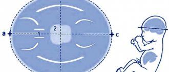
Prenatal screening of the 1st trimester is considered reliable if 2 conditions are met. 1. It was performed by an experienced specialist in a period of 11 to 13.6 weeks. In this case, the CTE of the fetus should not be less than 45 mm, otherwise the measurement of the TVP (nuchal translucency space) cannot be considered reliable for calculating the risk of having a child with chromosomal pathologies.
2. Risks need to be considered, not relying only on ultrasound data, and not looking only at biochemical screening of the 1st trimester. This work must be performed by a special program. And it will give results on risks for various genetic and chromosomal diseases. The risks will be described as average (where only age is taken into account) and individual. So, 1st trimester screening is bad if individual risks are higher than the basic (age-related) ones. In this case, the doctor may refer you to a geneticist for consultation. And he will probably prescribe a repeat ultrasound as soon as possible, only an expert class and (or) invasive diagnostics (cordocentesis, chorionic villus biopsy or amniocentesis). Recommendations will largely be based on ultrasound data. Sometimes at this stage the ultrasound diagnostic doctor sees very severe developmental defects in the fetus that are incompatible with life. In this case, a repeat ultrasound is prescribed and interruption is recommended. Invasive diagnosis is usually not necessary.
If the 1st trimester ultrasound screening is generally normal, but the individual risk of having a sick child is high, then the doctor may recommend waiting for the second screening or conducting invasive diagnostics, the result of which can be used to say for sure whether the child is genetically healthy.
In the early stages, a chorionic villus biopsy is performed - this is a rather risky procedure, in terms of a high risk of miscarriage after it. The doctor takes cells from the placenta for genetic analysis; this procedure is also called a placenta biopsy.
After 16 weeks, amniocentesis is performed. Amniotic fluid is taken for analysis. This analysis is considered very informative and safer than chorionic villus sampling and cordocentesis. Women usually agree to the first one in order to avoid prolonged gestation of a non-viable fetus. After all, after 12-13 weeks you need to wait until doctors can induce artificial labor. And this is approximately 18 weeks.
But if you do not meet the 1st trimester ultrasound screening standards, and do not want to wait for the second screening, much less invasive diagnostics, there is the opportunity to do a non-invasive test. It is not yet widespread in Russia. And very expensive. The analysis costs about 30,000 rubles. But its reliability is approximately the same as amniocentesis. There is no risk of spontaneous abortion.
Screening in the 1st trimester involves examining the levels of two hormones in the blood of the expectant mother - hCG and PAPP-A. A high hCG may indicate a high risk of Down syndrome in a child, and a low hCG may indicate a high risk of Edwards syndrome. At the same time, PAPP-A also turns out to be low. But the final interpretation of the 1st trimester screening results is performed only together with an ultrasound scan.
The results of blood tests can be affected by a situation where a woman has a clear threat of miscarriage if she is taking progesterone drugs. But many pregnancies are at risk. In addition, it plays a role whether a woman is carrying one fetus or several, whether she is overweight or underweight, and whether the conception was natural or in vitro fertilization. Therefore, it is completely wrong to try to interpret and worry about blood tests separately.
When is it better to do screening in the first trimester? The gynecologist can and should calculate it directly. This is why it is important to register for pregnancy before 12 weeks. It is advisable to visit a doctor for the first time no later than 8-9 weeks, since there is a shortage of coupons for free screening in many Russian regions. You may need to wait a little longer to receive your voucher. You will have extra time just for this waiting.
But the timing of 1st trimester screening has been determined. All that remains is to donate blood and do an ultrasound on the specified day. Many women believe that to make the result more reliable, some measures need to be taken. Should preparation be made for the first screening during pregnancy and what kind? There is no need for any preparation as such. Some experts recommend following a diet a few days before the examination, but there is no great need for this. The expectant mother does not need to limit herself in nutrition. And one more important question - how is screening of the first trimester done on an empty stomach or not, what is better? In general, it is advisable to take all blood tests on an empty stomach. Including this one. But sometimes it happens that the analysis is scheduled for the second half of the day. Then, of course, you should not go hungry since the evening of the previous day.
An ultrasound is performed. Moreover, sometimes the doctor requires not only abdominal access, but also vaginal access. The indicators that the doctor is especially interested in are the thickness of the collar space, visualization and size of the nasal bone, and the speed of venous blood flow. In addition, the doctor examines all organs and systems of the fetus, the size of the limbs and head. Measures a woman's cervix in order to early diagnose isthmic-cervical insufficiency, if present.
The results of the 1st trimester biochemical screening are usually ready within a few days. And all this time the expectant mother is in suspense. Especially if you have read or are personally familiar with bad stories related to this examination. Typically, a woman visits a doctor once every 2-3 weeks, and if a high-risk screening result comes back before she should attend an appointment at the antenatal clinic, the doctor or midwife calls her and invites her to come. If the screening is good in the first trimester, you don’t have to worry and don’t have to take anything additional or visit a geneticist. It should be noted that young women have a much lower risk of chromosomal abnormalities.
Screening for the 2nd trimester consists of taking three blood parameters. But it is usually prescribed only to those women who either did not pass the first screening or whose results were unsatisfactory.
And in conclusion, it is necessary to say how necessary the first screening is in general. Is it possible to do without it? Of course, yes, it all depends only on the desire of the woman. An ultrasound scan at 12 weeks can reveal severe malformations. Well, high or low risk does not guarantee the birth of a sick or healthy child.
However, classical screening is recommended for women over 35 years of age and those who already have children with genetic abnormalities, or cases of bearing children with chromosomal abnormalities, if there are abnormalities in the genetic code.
Screening is a diagnostic procedure that allows the doctor to obtain information about the woman’s condition, and also allows him to see chromosomal mutations in the fetus and its developmental defects in the early stages. When going for the first screening during pregnancy, women, as a rule, do not know what not to do and what to do. Proper preparation for the examination guarantees reliable results. The main question that worries patients is whether they can eat before screening. The answer to this question will depend on the duration of pregnancy and the specific goal that the doctor sets for the study.
Screening is a completely painless procedure that is performed to monitor the condition of a woman who is at risk of having a baby with disabilities. Screening combines two procedures at once - ultrasound diagnostics and blood tests.
. A biochemical blood test allows the doctor to determine the presence of a special placental protein, pregnancy-related plasma protein A, as well as human chorionic gonadotropin.
A change in the level of these substances in the patient’s blood indicates the risk of developing various chromosomal and non-chromosomal mutations. Even if a woman has differences in protein subunits from the norm, this is not a guarantee of pathology; such indicators are only a reason for a doctor to carefully monitor the course of a particular patient’s pregnancy.
The research results are compared, and the doctor draws conclusions about the development of the fetal physique, as well as deviations from the norm. The first screening is carried out from 10 to 13 weeks. If during the study there is a high risk of chromosomal abnormalities, the woman is referred for a chorionic villus biopsy.
Indications for perinatal screening are:
- the age of the pregnant woman is over 35 years;
- women who have already given birth to children with genetic disorders and Down syndrome;
- women who are carrying a child from a relative, that is, there is mixing of blood;
- women who were exposed to radiation and X-ray examinations early in pregnancy;
- women who have had spontaneous miscarriages and stillborn children;
- women who used toxic drugs or underwent chemotherapy before pregnancy, as well as those who suffered viral diseases in the first trimester of pregnancy.
A woman can undergo an examination at her own request to ensure the full health of her baby and its normal development. The second screening is carried out at 18-21 weeks of pregnancy. The gynecologist must mentally prepare the woman for possible results, because they will more clearly reflect possible malformations of the baby in the womb. During the examination, de Lange syndrome, Smith-Opitz syndrome, Down syndrome, Patau syndrome, Edwards syndrome, pathologies of neural tube formation, omphalocele, and triploidy may be detected.
All these diseases are incurable, and future children require special education and care. It should be noted that the examination results do not provide a 100% guarantee of the presence of malformations and mutations; they only suggest such a probability, which is indicated as a percentage.
What is determined?
A woman donates blood biochemistry twice during the entire period of bearing a baby - at 11-13 weeks and 16-20 weeks. To do this, venous blood is taken from the patient on an empty stomach.
What does this show?
The first screening is also called a “double test”, since during the analysis two main indicators are determined in the blood: hCG and PAPP-A. Let's try to find out what each of them is.
Reference! Human chorionic gonadotropin (more precisely, its free unit) is a hormone secreted by the cells of the chorion (embryonic membrane).
It is determined almost immediately after conception. This indicator increases, reaching its highest values by the end of the first three months of pregnancy. Then the number that determines hCG decreases, stopping at one value after a certain gestational age.
Reference! PAPP-A is a protein secreted by the outer layer of germ cells.
During all nine months, its amount should increase in proportion to the period. Functionally, PAPP-A is responsible for the immune response of the expectant mother during pregnancy, and also determines the development of the placenta and its normal functioning.
Video
The video below will help you get an idea of what biochemical screening is and why it is done.
How to prepare for the examination?
In order for the study to give the most accurate and reliable results, preparation for screening is necessary. Few people know how to prepare, so they make a number of mistakes, due to which there is a need to undergo diagnostics again. The very first thing a woman should consider is to refuse medications that were prescribed for other reasons. Some medications may interfere with the results. If a woman has any chronic or systemic autoimmune diseases that require constant correction, additional consultation with a doctor is necessary. He will tell you individually which medications you will need to stay off for a day, and which you can continue to use, since they will not show up in your blood quality.
Doctors do not recommend having sex with a partner the day before you go for screening. Before the examination, as well as the day before the procedure, be sure to reduce physical and emotional stress. The woman should come to the examination rested and in a calm state.
It is necessary to take a bottle of water with you; it is likely that the doctor will want to additionally examine the woman’s genitourinary system and kidneys. You should drink water only after the doctor’s instructions; the procedure should be carried out strictly on an empty stomach; you should not eat at all. Is it possible to eat before screening? Absolutely all women who are preparing for tests are interested. It should be noted that the pre-screening diet is only required the first time so that the blood test is as accurate as possible.
When the second screening is done the evening before the study, a light dinner is allowed, possibly even with meat and fatty dishes, but in small quantities. It is important not to eat 4 hours before the test before screening. Returning to how to prepare for the first examination, and whether it is possible to eat heavy foods, it should be noted that there is a clear list of foods that doctors have allowed and which have been prohibited.
How are the results assessed?
When assessing the results, the doctor relies on the norms of fetal ultrasound and biochemical analysis, corresponding to certain stages of pregnancy. If a deviation from the standards is detected at one of the screening stages, additional examination is carried out. The final diagnosis is made only on the basis of several studies. The table shows the norms of ultrasound and blood tests for different periods of screening.
| Gestation period, weeks | Ultrasound indicators | Blood test parameters, mU/ml | |||||
| KTE, mm | TVZ, mm | BPR, mm | Nasal bone | Heart rate, beats/min | RARR-A | hCG | |
| 11 | 42-50 | 1,6-2,4 | 17 | not measured | 153-177 | 0,78-4,77 | 20000-95000 |
| 12 | 51-59 | 1,6-2,5 | 20 | more than 0.3 cm | 150-174 | 1,03-6,02 | 20000-90000 |
| 13 | 62-73 | 1,7-2,7 | 26 | 147-171 | 1,47-8,55 | 15000-60000 | |
When assessing the PAPP-A and hCG indicators, a coefficient is determined that indicates the level of deviations from average norms - MoM. At the first screening it should be in the range of 0.5-3. The risk of genetic pathologies is calculated using a special electronic program. The possibility of complications should not be less than 1:1380.
Mandatory diet
The diet should exclude highly allergenic foods, such as:
- seafood and all types of fish;
- chicken eggs,
- cow and goat milk;
- fruits and berries that are red or orange in color;
- coffee and cocoa;
- alcohol-containing and carbonated drinks;
- smoked and canned products;
- chocolate and foods high in sugar;
- mushrooms;
- nuts;
- legumes;
- wheat;
- photo products made from herbs;
- processed beans and watermelon.
A woman should give up all of the above products three days before the test. Please note that if a pregnant patient belongs to the group of hypersensitive individuals, then throughout pregnancy it is better for her to avoid these foods or eat them in small quantities. Low-allergenic products that are allowed for consumption three days before testing include kefir, fermented baked milk and similar fermented milk products, lean pork and beef, greens, chicken meat, vegetables, olive oil, dried fruits, semolina, pearl barley and rice porridge, liver .
Eating a portion of the above products will be very useful for a woman who is carrying a baby, because all of them are not only low allergenic, but also very rich in nutrients and macroelements.
Separately, I would like to note the moment of heat treatment of food; a day before taking a blood test for biochemical screening, the products chosen for consumption should be boiled or stewed. Baking or frying dishes in sunflower oil is prohibited. The best options would be mashed potatoes, cereals and light salads with fresh vegetables. After the woman has had the analysis, she can return to her usual diet without limiting herself to her favorite foods.
Whether fasting screening is carried out or not, now you know, and you will be aware of how to properly prepare if the need suddenly arises to undergo this type of perinatal diagnosis. Perinatal screenings cause a lot of controversy and controversy, some consider them inappropriate, because they once managed without it, others have a positive attitude towards such diagnostics. In any case, modern methods of examining women cannot be underestimated, because it is thanks to correct diagnosis that the correct treatment can be started in a timely manner.
A woman has the right to decide for herself whether she is ready to raise a child with genetic abnormalities and developmental defects, or whether it is better to terminate the pregnancy in the early stages. You can undergo perinatal screening at any medical institution without a doctor’s referral.
Biochemical screening during pregnancy - this word refers to a venous blood test, which determines special hormones that are markers of chromosomal pathologies of the fetus. It is the results of this examination that determine, without being a diagnosis, how high the risk of developmental defects is in a growing fetus. This is what a biochemical examination is in the “interesting period”, and it should only be carried out in conjunction with an ultrasound examination at the same time.
What is a survey and what is its purpose?
1st trimester screening includes ultrasound and biochemical blood test. Its main goal is the early detection of congenital pathologies of fetal development. Based on the examination results, doctors can assume that the child has chromosomal abnormalities such as Down, Edwards, Patau, and De Lange syndromes. You need to undergo screening from 11 to 14 weeks of pregnancy.
Ultrasonography
During the first screening, ultrasound is performed in 1 of 2 ways:
- Transvaginally. The sensor is inserted shallowly into the vagina.
- Transabdominal. The examination is done through the abdominal wall.
The first method is preferable because it allows you to get a clearer picture. The second method is used in the following cases:
- the patient is allergic to latex (since a latex condom is placed on the vaginal sensor);
- refusal of the expectant mother to undergo the procedure due to moral and religious beliefs.
When performing an ultrasound at 12-13 weeks, the specialist evaluates the site of attachment of the fertilized egg, as well as the following fetal parameters:
- size from coccyx to crown (KTR);
- characteristics of the nasal bone;
- thickness of the collar zone (TVZ);
- development of the brain hemispheres;
- head parameters (circumference, interparietal size (IPD), length from forehead to occiput);
- measurements of formed tubular bones;
- location and characteristics of internal organs;
- abdominal circumference.
Based on data on the physiological parameters of the fetus, the doctor makes a conclusion about the expected date of birth. If necessary, a specialist assesses the condition of the mother’s internal organs. The results of the study enable doctors to determine further tactics for pregnancy management.
Blood chemistry
The first screening blood test involves determining the level of the hormone human chorionic gonadotropin (hCG) and pregnancy-associated plasma protein A (PAPP-A). Each gestation period corresponds to a certain level of these substances.
Activation of hCG production occurs at the moment of conception. By the presence of this hormone in the blood, the approximate duration of pregnancy can be determined. The hCG level also indicates possible genetic abnormalities of the fetus. However, assumptions about violations cannot be made only based on the indicators of this hormone. A cumulative comparison of hCG and PAPP-A has diagnostic value.
PAPP-A is a plasma protein that is produced by the outer layer of the embryo when it implants into the uterine cavity. This indicator during pregnancy is advisable only until the end of the first trimester, that is, up to 12 weeks. In the second trimester, the level of PAPP-A in all pregnant women is approximately the same. The purpose of studying this indicator in the early stages of pregnancy is to identify chromosomal abnormalities of the fetus.
In addition to child development disorders, analysis for hCG and PAPP-A reveals:
- threat of miscarriage;
- ectopic embryo attachment;
- death of the embryo.
Since during perinatal screening, ultrasound is performed first, if a missed abortion is detected, analysis for PAPP-A and hCG is not performed. However, in the case where a blood test indicates that fetal development has stopped, the diagnosis is confirmed by ultrasound examination.
For analysis, 10 ml of venous blood is taken from the pregnant woman. The laboratory uses the resulting serum. The most reliable result is obtained by studying fresh material, so the blood is examined within 24 hours after donation.
Do pregnant women need biochemical screening?
The study can be carried out for all women who want to be sure that their child will be born healthy (meaning, without chromosomal pathology that cannot be treated). But there are also strict indications, taking into account which the gynecologist at the antenatal clinic gives a referral for biochemical screening. These are the following situations:
- future parents are close relatives
- This woman has already had a stillbirth or was fading from pregnancy
- mother over 35 years old
- there is already 1 child with some kind of chromosomal pathology
- there is a long-term risk of spontaneous miscarriage
- there was a single or repeated case of miscarriage or premature spontaneous birth
- the pregnant woman suffered a viral or bacterial pathology during or shortly before pregnancy
- it was necessary to take medications that were not approved for pregnant women
- before conception, one of the couple was exposed to ionizing radiation (X-rays, radiation therapy)
- ultrasound diagnostic results that are questionable regarding developmental defects.
Perinatal diagnostics for hormone levels is carried out only in conjunction with screening ultrasound during pregnancy.
What pathologies are determined by blood tests in pregnant women?
These are the following diseases:
- Edwards syndrome
- neural tube defect
- Down syndrome
- Patau syndrome
- de Lange syndrome
Edwards, Patau and Down syndromes are collectively called “trisomy”. Moreover, each cell contains not 23 pairs of chromosomes, but 22 normal pairs and 1 “triplets”. In which particular pair the “trinity” was formed, that’s how the disease is called.
For example, if screening for Down is performed, then there are three (not two) chromosome 13 (the syndrome is written “Trisomy 13”). These conditions should be identified prior to 3rd trimester screening.
Preparation for analysis for chromosomal pathologies
Preparation for the diagnosis of the 1st trimester for markers of chromosomal diseases is that the day before the test you exclude from the diet:
- spicy food
- fatty and fried foods
- smoked meats
- chocolate
- citrus.
Why is it important
Not so long ago, only those women who were at risk were invited to the first screening. And these are not only patients who have problems with bearing a baby - miscarriages, early births, frozen embryos, but also those who are in a close family relationship with their spouse.
Also at risk are:
- children of relatives who have genetic abnormalities or whose first baby was born with such abnormalities;
- sick during pregnancy with pathologies of a viral or bacteriological nature;
- taking medications that are contraindicated during pregnancy.
But today the first screening is shown to every woman who wants to become a mother.
If you have questions about the first study, be sure to read the book by A.A. Strelnikova, A.G. Obrezan, E.V. Shaydakova “Screening and prevention of current diseases.”
Here you will learn what the screening standards are, which may vary according to the age of the patients. This book will be useful not only to ordinary people, but also to novice obstetricians-gynecologists and other specializations, whose activities are, in one way or another, related to screening.