Screening - translated from English, this word means sorting or selection.
In short, perinatal screening is a special set of tests, tests, and studies that can give a clear idea of possible deviations in the development of the unborn child. All screening is divided into the number of trimesters, since during each period of gestation the expectant mother is required to undergo the planned tests.
Screenings are divided into double, triple and quarter tests, demonstrating certain hormonal abnormalities during all periods of pregnancy.
The main goal of screening is to separate risk categories for the development of congenital defects in the fetus: Down syndrome, Edwards syndrome, neural tube defects. Based on the indicators of the ultrasound examination and the results of a blood test taken from a vein, the result is calculated.
Naturally, when processing information, the woman’s personal information is taken into account (from age, weight, bad habits to the use of hormonal drugs during pregnancy).
What screening tests should be taken during pregnancy?
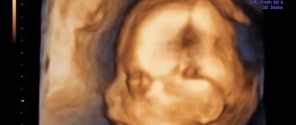
An ultrasound should examine the thickness of the nuchal translucency (nuchal translucency).
Its coefficient, if it exceeds 2-2.5 cm, indicates the possible presence of Down syndrome in the child. TVP is measured at strictly limited periods of pregnancy - from 11 to 14 weeks, more precisely - up to 12 weeks. Later, the fetus will grow up and the TVP indicators will lose their information content.
In the first trimester, blood is donated for the hormones b-hCG and PAPP-A.
The second screening (16-18 weeks) does not include an ultrasound scan - the indications for it are taken from the first. And blood must be donated for the hormone b-hCG, the alpha protein AFP and estriol - that is, the so-called “triple test”.
Screening test results
You must wait about three weeks for results.
Analysis indicators are expressed not in numbers, but in MoM, which means multiplicity in medicine. The median is the statistical average for the given marker. According to the norm, MoM should be within the range of 0.5-2.0. If, based on the tests, a deviation from the norm is revealed, it means that there is some pathology in the development of the fetus. Elevated hCG may indicate the following abnormalities: chromosomal developmental defects, multiple births, Rh conflict. Reduced hCG indicates an ectopic pregnancy, threatened miscarriage, or undeveloped pregnancy. An increase or decrease in AFP indicates probable chromosomal abnormalities. The sum and combinations of deviations in the proportions of hormones can also indicate the presence of pathologies. Let's say that in Down syndrome, the AFP indicator is underestimated, and hCG, on the contrary, is overestimated. The hallmark of an unclosed neural tube is increased levels of alpha protein (AFP) and decreased levels of the hormone human chorionic gonadotropin (hCG). In Edwards syndrome, the test hormones are reduced.
If there is a high risk
If the risk is high, the woman is referred for consultation to a genetic specialist.
Here you need to make a very important decision in life. The malformations indicated by your measurements cannot be treated. Here you will be given information that you will most likely have a “different” child. The geneticist will study your indicators, information about your pedigree, clarify whether hormonal treatment was used to maintain the pregnancy (Utrozhestan, Duphaston) and will certainly warn that there is no way to find out with one hundred percent accuracy whether the baby has pathologies, except by invasive methods. These methods are not very harmless: chorionic villus biopsy, amniocentesis (taking amniotic fluid through a puncture in the abdomen), cordocentesis (puncture from the fetal umbilical cord). There is a certain risk in conducting invasive research.
Unfortunately, today screenings provide little information. The unreliability and fallibility of non-invasive studies is quite high. Some doctors even argue about the advisability of such procedures.
First screening during pregnancy
In the first three months of pregnancy, absolutely all women undergo this painless procedure.
First screening during pregnancy
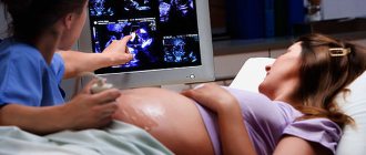
provides an opportunity to recognize pathologies in fetal development. It consists of an ultrasound examination and blood tests. To carry out diagnostics, all personal data of a woman are taken into account (from age, weight, presence of chronic diseases to bad habits). Blood is taken from her vein and an ultrasound is performed.
Cost of the study
In the early stages, the price of an ultrasound examination in non-state clinics is 700–4000 rubles. For multiple pregnancies, the procedure will cost 600–8000 rubles. The cost of the planned first screening starts from 300 rubles. In budget antenatal clinics, ultrasounds are performed for pregnant women at the place of registration free of charge.
The first ultrasound in the early stages evaluates the condition of the uterus, the presence and location of the embryo. From the twelfth week, the structure of the embryo is studied.
The reason for the identified deviations is the low technical ability of the device or improper preparation for ultrasound, inexperience of the diagnostician, or another human factor. To exclude pathologies, it is better for a woman to undergo screening with a top-class sonograph and consult doctors at an independent medical institution.
Comment on the article, share your experience of examination at the beginning of pregnancy, spread information on social networks when you do your first ultrasound diagnosis. All the best.
Timing of the first screening during pregnancy
All these actions are carried out at 10-13 weeks of gestation.
Despite such a short period, they help identify genetic and chromosomal abnormalities in the fetus. All conclusions about the development of the unborn child are made based on the results of a sum of research and analyses. If the diagnosis determines a high probability of abnormalities in the formation of the baby, the woman is sent for amniocentosis and IVS.
Risk group:
- Women who are whiter than 35 years old.
- Those expectant mothers who had children with Down syndrome or other genetic abnormalities.
- Pregnant women who have already given birth to children with disabilities or who have had miscarriages in the past.
In addition, screening is mandatory for women who suffered from viral diseases in the first trimester and took contraindicated medications for therapy.
Enlist the support of loved ones
As you know, friends are known in trouble. Now more than ever you need their support and participation.
“Sometimes the presence of another person, even silently, helps to cope with despair” (Marc Levy).
I absolutely agree with the author of the quote. Tell us about your problem and you will feel better. Most likely, your loved ones will offer to provide you with all possible help. Don't refuse, but don't take too much either. Don't forget to thank them warmly and, if possible, do something good for them in return.
How does the first screening process work?
The first stage
is biochemical. This is the process of blood testing. Its task is to identify such abnormalities as Down syndrome, Edwards syndrome, and defects in the formation of the brain and spinal cord in the fetus.
The results of a blood test during the first screening do not provide reliable data for making a diagnosis, but give rise to additional research.
Second phase
- This is an ultrasound scan of the first three months of pregnancy. It determines the development of internal organs and the location of the limbs. In addition, measurements of the child’s body are taken and compared with age-appropriate standards. This screening examines the location and structure of the placenta and nasal bone of the fetus. Usually at this stage it is visible in 98% of children.
Norms for first screening during pregnancy
Also, the first screening determines a multiple pregnancy if all indicators exceed the norm.
- If the test results are higher than normal, then the risk of Down syndrome in the unborn child is high. If they are below normal, then Edwards syndrome is possible.
- The PAPP-A norm is another coefficient for the first screening during pregnancy. This is plasma protein A, the level of which increases throughout pregnancy, and if this does not happen, then the unborn child has a predisposition to diseases.
- If PAPP-A is below normal, the child has a high risk of developing abnormalities and pathologies. If it is higher than normal, but other research results do not deviate from the norm, then do not worry.
The norm at the first screening allows the mother to independently decipher the results of the tests obtained.
Knowing their norms, a pregnant woman will be able to determine the risks for the development of pathologies and diseases of the unborn child. To calculate the indicators, it is necessary to use the MoM coefficient, which indicates deviations from the average. During the calculation process, adjusted values are taken that take into account the characteristics of the female body.
If you have any doubts about the results of the screening, repeat it by taking the same blood tests and ultrasound again in another laboratory. This can be done up to 13 weeks of pregnancy.
What to do when you've had a Caesarean
After the “caesarean” procedure, the state of blood circulation and restoration of sensitivity are monitored, recommendations are given on nutrition and physical activity, and treatment of the suture. Upon discharge, treatment is prescribed according to indications; after resumption of sexual intercourse, contraception is required.
Surveys
During the first hours after surgery, the woman in labor is examined:
- blood pressure is measured, since it often drops or changes;
- monitor the condition of the suture, the presence of vaginal discharge, the height of the uterine fundus and its tone;
- determine pulse, respiratory rate, volume of urine excreted.
An example of a suture on the uterus after an ultrasound
If the woman’s condition is satisfactory, after 8 hours she can sit on the bed, and by the end of 12 hours she should try to get to her feet.
The doctor observes how sensitivity of the lower extremities is gradually restored after spinal anesthesia. If pain occurs in the lower back or lower abdomen, painkillers are used. If necessary, drugs to prevent thrombosis and antibiotics that stimulate intestinal contractions are recommended.
On days 4-6, an examination by a gynecologist, ultrasound, and blood and urine tests are scheduled. If there are no complications, the woman is discharged for observation at her place of residence. After 7-10 days, you must undergo a doctor’s examination and the necessary examination (ultrasound, smears, blood tests).
Suture after caesarean section
Restrictions in the first days
During the first day, you are initially allowed to drink only water, and then non-acidic kefir and diluted juice.
The next day, the list of dishes expands to include:
- low-fat chicken broth with white croutons;
- curdled milk, yogurt without sugar and fruit;
- steam omelette.
From 3-4 days, a gentle diet is prescribed with pureed porridge, fish, meat, and puree soup. By the end of the week, you can switch to a regular table, but with the exception of fried, fatty and spicy foods. It is important for a woman to prevent constipation.
For this we recommend:
- sufficient intake of drinking water;
- vegetable juices;
- boiled pumpkin;
- plum puree.
Seam processing
Until healing, I lubricate the postoperative suture with solutions of iodine, brilliant green, and colorless Fukortsin. At home, antiseptics are recommended for care: Chlorhexidine, Miramistin, Decasan. To speed up healing, ointments (Solcoseryl, Traumeel) and silicone patches (Mepiform, Dermatix) are prescribed.
Behavior at home
In order not to disrupt the healing process of the scar on the uterus and skin, you must refrain from:
- lifting weights over 3 kg (3 months);
- intense physical activity, sports (8 weeks), abdominal exercises (4 months);
- sexual intercourse (2 months);
- hot shower (1 month, warm allowed), bath (2 months);
- visits to the bathhouse, swimming pool (3 months).
When restoring at home, you need to know which symptoms you need to see a doctor urgently (ambulance), routinely (gynecologist in consultation), and which are acceptable (see table).
| Call an ambulance immediately | Contact the clinic | Not dangerous signs |
| Sudden chest pain, difficulty breathing, bluish skin | Pain, distension in the mammary gland, redness, palpable lump | Aching pain in the lower abdomen |
| Sharp pain in the leg, swelling, redness | Pain when feeding a baby, cracked nipple | Pain in the lumbar region when moving after spinal anesthesia |
| Temperature above 38 degrees, purulent vaginal discharge with an unpleasant odor | Lack of milk or its release in drops | Pain throughout the abdomen, bloating, tendency to constipation |
| Increased headache, nausea and vomiting | High blood pressure, swelling of the legs | Unpleasant sensations in the suture area in the absence of inflammation |
| Bleeding from the genital tract (2 pads get wet in an hour) | Constant weakness, irritability, tearfulness | Vaginal discharge is bloody and spotting (in the first week) |
| Rash, headache, cramps | Exacerbation of varicose or hemorrhoidal disease | |
| Redness of the postoperative suture |
Contraception
After the gynecologist allows you to resume sexual intercourse, you should take care of the method of contraception. When a woman fully breastfeeds her baby after a natural birth, but does not have periods, then in 98% of cases this gives a fairly reliable contraceptive effect.
During cesarean section, due to the lack of a mechanism for physiological stimulation of lactation, 75% of patients have little milk. By the end of 3 months, many women already have their cycle restored, so it is important to use contraceptives from the first days of sexual relations, regardless of breastfeeding or the presence of menstruation.
It is preferable to use condoms, as they help further protect against infection and there is no risk of side effects, as with hormonal pills. A woman may also be prescribed medications in cases where it is necessary to treat endometriosis, functional ovarian cysts, or prevent active growth of the endometrium. Exluton, Charozetta, Lactinet are used.
2-4 months after cesarean section, an intrauterine device can be inserted. Repeated births are allowed no earlier than after 3 years, since the scar on the uterus must be fully formed.
Timing of the second screening during pregnancy
A secondary comprehensive study is carried out to determine the likelihood of the formation of chromosomal abnormalities in the fetus: at this time their probability is quite high.
There are three types of second screening:
- ultrasound (detection of abnormalities using ultrasound),
- biochemical (blood parameters),
- combined, where the first two are used.
An ultrasound is usually performed as a screening test at the end of the second trimester and consists of a blood test for various signs.
The results of the ultrasound examination, which was carried out earlier, are also taken into account. The system of sequential operations during this set of studies is as follows: after blood is donated and an ultrasound examination is performed, the woman fills out a questionnaire indicating personal data, which will be used to determine the duration of pregnancy and the likelihood of developing defects. When taking into account the duration of pregnancy, tests are performed. After this, the information received is processed by a computer program to calculate risks. However, even the results obtained cannot be considered a final diagnosis, an absolute guarantee of the presence of risk, if it is established. To obtain more accurate and detailed information, the pregnant woman is sent to undergo additional tests and consults with a geneticist. Second screening during pregnancy
is a biochemical study of the blood of the expectant mother using certain tests.
More precisely, according to the so-called “triple test”, which studies the level of proteins and hormones, such as: human chorionic gonadotropin (hCG) in the blood, alpha-fetoprotein (AFP), free estyrol. The test becomes “quadruple” when this secondary set of studies also involves taking blood to determine the level of inhibin A.
Studying the concentrations of these hormones and proteins in the blood makes it possible to judge with a high degree of probability the possibility of a child developing Down syndrome, Edwards syndrome, and neural tube defects.
The conclusions of a repeated set of studies may be an indirect indicator of the defective state of the child’s formation and exacerbations of the course of pregnancy. For example, an abnormal level of hCG indicates abnormalities in chromosomes, the danger of the formation of preeclampsia, or the presence of diabetes mellitus in the expectant mother.
Reduced hCG levels may indicate disturbances in the development of the placenta.
Increased or decreased AFP and inhibin A in the blood serum of a pregnant woman is a sign of a disorder in the natural formation of the baby and possible congenital anomalies - open neural tube defects, possibly Down syndrome or Edwards syndrome. If alpha-fetoprotein increases sharply, the fetus may die. If the level of the female steroid hormone, free estriol, changes, the activity of the fetoplacental system can be disrupted: its deficiency suggests probable abnormal functioning of the child.
If the results of a repeated set of studies turned out to be unfavorable, you should not worry ahead of time. They only talk about the estimated risks of deviations, they do not constitute a final diagnosis. In the case where at least a single component of the secondary screening does not fit into the norm, it is necessary to carry out additional research. The indicators of a screening study may be influenced by several reasons: in vitro fertilization, a woman’s weight, the presence of diabetes mellitus, bad habits, such as smoking.
Progress of the operation
First, drugs for general or regional anesthesia are administered, the skin and uterus are cut, the baby and placenta are removed, the tissue is sutured, and the patient is transferred to the recovery room.
What kind of anesthesia will it be?
The vast majority of women (about 90%) use spinal or epidural anesthesia. It is carried out in this way: a solution of the drug is injected between the lumbar vertebrae with a thin spinal needle or a set (needle + catheter) for epidural anesthesia.
There is a complete loss of sensitivity from the lower back to the feet, which allows you to perform all the necessary manipulations without pain and discomfort. The woman in labor is conscious all the time, she is separated from the surgical field by a screen, but can maintain contact with the surgeon. After the baby is born, you can immediately put it to the breast.
If such regional anesthesia is contraindicated, or there is a need for a long operation, then general anesthesia is prescribed. The drugs are first injected into a vein, then, after the onset of drug-induced sleep, a tube is installed into the trachea through which the gas mixture is supplied. With the help of these medications, complete relaxation of the muscular system and loss of consciousness occurs.
How is a second planned caesarean section performed?
The second cesarean section differs from the first in that the incision is made close to an existing scar.
All other stages of the operation are similar:
- The incision in the skin of the subcutaneous layer (for most women with a planned cesarean section, it is horizontal above the pubis, no more than 10 cm, this protects against hernias and heals faster).
- Dissection of the uterus in the lower part.
- Opening the membranes, removing amniotic fluid.
- Removing the child.
- Crossing the umbilical cord.
- Removal of the placenta.
- Layer-by-layer suturing of tissues.
- Fixing a protective bandage on the skin and placing an ice pack on top.
After the second, and especially the third, caesarean section, the woman is offered tubal ligation surgery. This is a fairly reliable way to prevent unwanted pregnancy in the future, since carrying a fetus with scars on the uterus is very dangerous.
Possible complications and their prevention
With a caesarean section, especially a repeat one, there are risks of complications:
- blood loss exceeding the norm (up to 500-750 ml is allowed) - the uterine cavity, due to strong muscle relaxation during anesthesia, can be difficult to contract; to prevent severe anemia, blood or its components are injected, and if the bleeding cannot be stopped, then removal of the uterus is necessary ;
- adhesive disease - any operation in the abdominal cavity during healing causes the formation of adhesions, that is, fibers that connect the organs of the adhesive disease (for example, intestinal loops, bladder with the body of the uterus), which later lead to the appearance of chronic pain in the abdominal area, and if they affect the patency of the fallopian tubes, they become the cause of ectopic pregnancy and infertility; treatment – physiotherapy, therapeutic exercises;
- inflammation of the inner layer of the uterus (endometritis) - occurs due to the penetration of bacteria into the uterine cavity; for prevention, antibiotics are mandatory before surgery;
- infection of the suture - accompanied by redness, pain, discharge, fever, to prevent this, the woman is prescribed treatment with antiseptic solutions, and at home must be lubricated with wound-healing ointments (Levomekol, Bepanten plus);
- the formation of a rough scar at the site of skin stitching - Contractubex and Dermatix are used to soften and form a thin seam;
- divergence of the edges of the wound - occurs when there is an understanding of the severity, strong straining during bowel movements, hacking cough in the first days, as well as when an infection is attached; medications are prescribed for treatment; surgical debridement of the wound and re-application of suture material may be necessary.