The second screening is not a mandatory procedure; doctors prescribe it to pregnant women to confirm or refute the results of the first examination.
The data obtained during screening is studied carefully and checked for compliance with normal indicators, because from it parents will reliably know how correctly and fully the child is developing in the womb. If the results of the second study are positive, then the embryo is absolutely healthy, has no genetic pathologies, and feels good; if negative, then most likely the child suffers from a serious illness. Having identified the disease, the doctor either begins intrauterine treatment of the small patient or artificially induces premature birth. That is, screening of the 2nd trimester during pregnancy can be called a responsible and important procedure, which cannot be taken lightly or without regard.
At what stage is 2nd screening performed during pregnancy?
The possibility of screening to detect possible developmental problems in an unborn child appeared not so long ago - it began to be performed on expectant mothers only in 2000. Typically, the first screening is done at 10-13 weeks of pregnancy: this study helps to identify the risks associated with the presence of chromosomal abnormalities in the fetus. The second examination is usually carried out at 16-20 weeks of pregnancy, but doctors tend to schedule such screening before the 19th week, since diagnosis at this period will show more reliable results than at a later time. Screening may be complete or incomplete, including, for example, only ultrasound examination (ultrasound): in this case, it may be prescribed later, at 19-22 weeks of pregnancy.
Why is 2nd screening performed during pregnancy? The main task of the study is to either refute the results of the first screening (usually negative) or confirm them. The obtained indicators are subjected to a thorough analysis, after which they are compared with the values accepted as the norm. When compared, it becomes obvious how the fetus develops and grows.
Indications and contraindications
An ultrasound scan at twenty weeks of gestation is performed for all women. There is only one indication - pregnancy. If for some reason the examination was carried out at -19 weeks, then if there is no need to monitor the condition of the mother or baby, an ultrasound scan is not performed at 20 weeks.
There are no absolute contraindications to ultrasound.
A relative contraindication is an allergy to the latex from which the condom is made, which is placed on the ultrasound sensor. In this case, you need to warn your doctor in advance.
A woman’s reluctance to be examined for personal reasons is the only condition for which an ultrasound is not performed at 20 weeks of pregnancy. In this case, the refusal is made in writing and attached to the medical documentation.
To watch a medical video review about this stage of pregnancy:
When is screening necessary in the second trimester of pregnancy?
- if both parents have reached the age of 35+;
- when there are hereditary genetic diseases in the family;
- if a pregnant woman has had a viral infection in the early stages: some seemingly “harmless” diseases can negatively affect the health of the unborn child;
- if the woman’s history has already included cases related to frozen pregnancy, miscarriage, or stillbirth;
- when the pregnant woman used drugs that have a teratogenic effect;
- if the expectant mother or father has an alcohol or drug addiction;
- if chromosomal abnormalities were detected in children born from previous pregnancies;
- if the expectant mother has chronic or autoimmune diseases that can affect the development of the unborn child and its growth;
- in cases of detection of malignant neoplasms in a woman who is already in the third month or later of pregnancy;
- when one of the parents of the unborn child, shortly before the time of conception or during this period, worked in an industry where there is radiation exposure or has a “harmful” status.
The second screening during pregnancy allows you to see those problems and pathologies that could not be detected in the early stages. In addition, screening helps to determine the expected risks of dangerous conditions that may develop in the near future, as well as to see deviations in the functioning of the main systems of the fetal body. And although doctors consider the studies conducted during the first screening to be more informative, the importance of the second screening cannot be underestimated. After all, many pregnant women are looking forward to confirmation or refutation of information received earlier.
What will an ultrasound scan show in the second trimester?
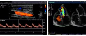
The baby in the womb develops so quickly that by the time of the second trimester screening, you will notice how the baby’s appearance and physical parameters have obviously changed. An ultrasound examination will allow you to see all this: thanks to it, the doctor will assess whether the fetus’s physical data meets the standards and will look to see if there are any signs of pathological conditions. Also, during the ultrasound, the doctor will assess the condition of the placenta and uterus, and if there is a need to clarify data on the state of the circulatory system, an ultrasound examination with Doppler will be performed.
During an ultrasound scan during the second screening during pregnancy:
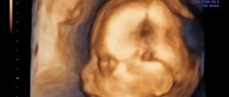
- the doctor will evaluate the structure of the head and facial contours of the fetus: special attention will be paid to the size and structure of the nasal bones, the absence (or presence) of a cleft between the nasal and oral cavities, and the process of formation of the eyeballs;
- the doctor will thoroughly determine the size of the fetus, consider whether the number of fingers and toes is normal;
- the specialist will also determine how mature the fetal lungs are and whether their development corresponds to the obstetric stage of pregnancy;
- problems with the condition and formation of the heart and the development of the spinal cord and brain will be noticeable;
- many internal organs should already be formed by the time of the second screening, so their location, structure and condition are clearly visible on an ultrasound examination;
- the doctor will also note in what volume of water the fetus has to develop, what tone the uterus has at the moment, in what condition the woman’s appendages are;
- the thickness of the walls and the level of maturity of the placenta are important;
- the doctor will pay attention to the organization of blood flow in the vessels of the uterus, as well as the functioning of the umbilical arteries and the middle vessel of the child’s brain;
To determine how functional the uterine vessels are, the doctor will conduct separate examinations of the left and right arteries, since when examining only one side, the results are usually false. How can we explain this? The fact is that when a state of gestosis (late toxicosis) occurs, blood flow is disrupted only in one artery of the uterus. This condition is considered one of the dangerous pathologies, quite often leading to premature birth or even the death of the newborn.
If the fetus is successfully located in the uterus, then at the second screening during pregnancy you will already find out the sex of the unborn child, because during this period the primary sexual characteristics will already be obvious.
The second ultrasound does not require special preparations or procedures. However, the day before the procedure, try not to eat foods containing potential allergens - chocolate, cocoa, seafood, citrus fruits, fried and fatty foods.
It should also be remembered that the condition for performing an ultrasound is an unfilled bladder. To obtain high-quality results of this examination, an abundant amount of amniotic fluid in the uterus is quite sufficient.
How to prepare for the second perinatal diagnosis
To conduct screening in the second trimester, no special preparation is required, but still, to obtain reliable results, a woman must follow some rules.
- Blood must be donated on an empty stomach. Consumption of any food, as well as liquid, can distort the analysis results. It is best to take a blood test in the morning.
- 1 day before donating blood, you should avoid sweet foods, as well as foods that can cause an allergic reaction.
- Ultrasound does not require any preparation. It is not necessary to fill your bladder. Diagnostics can be carried out at any time of the day.
Many doctors in the field of obstetrics and gynecology assure that a woman needs moral preparation and the support of relatives. It is important for a woman to tune in to positive results, to hope and believe in good results.
Indicators of normal ultrasound
Ultrasound at the second screening during pregnancy is performed transabdominally, that is, in a superficial way. This technique allows you to see not only the condition of the fetus, but also to evaluate the internal organs of the pregnant woman - the possible presence of tumors (provided that they are not small in size), and to assess the general picture of the condition of the pelvic organs. The technique does not require special preparation, is absolutely harmless to the body of the mother and the unborn child, and is also painless. The doctor will apply a special gel to the abdomen, which will allow the ultrasound signal to produce a clearer image. To obtain it and examine the necessary organ of the fetus or mother, the doctor moves a sensor over the skin.
All necessary assessments and measurements are made based on the data in the monitor picture. Also, looking at the general “video”, the doctor visually assesses the condition and appearance of the child. To assess the uterine vessels, the ultrasound doctor will use a special resistance index: at the time of the second screening, 0.38-0.7 units will be considered normal. However, to conduct such a study, a condition is necessary: the child must be at rest and not make rapid movements. At the same time, the baby’s pulse is also measured, which should be in the range of 119-161 beats per minute.
To exclude any pathological aspects on the ultrasound, the doctor first of all pays attention to the condition of the middle artery of the brain - there should be no hypoxia in this area. This problem can be detected by a decreased pulsation index. If a sharp increase in this indicator is diagnosed, it may indicate a cerebral hemorrhage. Normal pulsation index values during the second screening are in the range of 1.35-2.32 units.
Helpful information
The starting point for such tests is considered to be 16 weeks, but you can familiarize yourself not only with the features of screening, but also with how your baby develops at 16 weeks of pregnancy.
Many may also be interested in information about how often an ultrasound can be done during pregnancy. In the future, the doctor may suggest you undergo a third screening, so now we invite you to learn more about these tests in order to be aware of everything.
Also, if you have already passed 30 weeks, you may need information about exercises in the third trimester of pregnancy, thanks to which you can not only support your body, but also prepare for the upcoming birth.
Have you ever undergone such an examination? Share your story and useful tips that may be useful to those who are just planning to undergo screening, and also leave your feedback.
What indicators are taken into account in a blood test?
The first of these is the level of human chorionic gonadotropin (hCG, it is estimated in mg/ml). HCG is one of the main tests for pregnant women; it is no coincidence that it is called a “pregnancy hormone level study.” Human chorionic gonadotropin is an active hormone, the production of which begins during the formation of the embryo and then continues throughout pregnancy.
The second important indicator is the level of estriol (estimated in mmol/l). Estriol (E3) is otherwise called estrogen. This is a low-active minor female sex hormone that is produced by the follicular apparatus of the ovaries in women.
ACE (angiotensin-converting enzyme, estimated in IU/ml) is a special enzyme that is found in small doses in the epithelial tissue of the kidneys, and in large quantities in the human lungs and blood serum. The ACE level is important when the doctor determines how normal electrolyte and water metabolism are in a woman’s body.
Sometimes these three general indicators are supplemented with a fourth, establishing the level of another hormone - inhibin A. It is produced in female follicles and shows how sufficient a woman’s follicular reserve is.
What data in a blood test give rise to suspicion of pathology?
- if Down syndrome is suspected in the fetus, the level of hCG turns out to be elevated, while the levels of other hormones are decreased;
- Edwards syndrome should be suspected if all hormone levels decrease;
- liver necrosis, a defect in the formation of the neural tube or Meckel's syndrome will be indicated by an increased concentration of E3 and AFP, and hCG will maintain a normal concentration;
- if an infection occurs in the fetal area or placental insufficiency is detected, the level of free estriol will decrease; the same indicator can be seen as a result of a pregnant woman taking antibiotics.
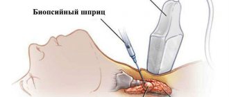
Serious deviations from standard values in a biochemical blood test give the doctor a reason to perform an amniocentesis procedure. This is a puncture in the abdominal wall to collect a small amount of amniotic fluid, which is then sent for testing in a laboratory. Such a diagnosis is highly reliable (about 99%) and makes it possible to make a final conclusion about the presence or absence of pathology in the fetus.
It is necessary to take into account that a biochemical blood test during period 2 of screening during pregnancy is carried out on an empty stomach. It is important not to experience stress before the study and to be in a calm state of mind, as well as not to be physically overloaded and to refrain from sexual contact.
Alpha-fetoprotein (AFP) level assessment
AFP is a blood plasma protein. First, its production occurs in the yolk sac, and then in the fetal liver. If the level of this compound is low, one can assume Down syndrome or Edwards disease in the baby. In addition, low alpha-fetoprotein indicates:
- diabetes mellitus in women;
- low location of the placenta;
- hypothyroidism.
Hypothyroidism is one of the reasons for a decrease in AFP levels
An elevated AFP level indicates:
- damage to the fetal nervous system;
- neural tube/abdominal cavity defect;
- abnormal functioning of the child’s kidneys;
- complete or partial absence of the brain in the fetus;
- oligohydramnios;
- Rhesus conflict;
- intrauterine fetal death;
- high risk of miscarriage.
At the same time, a high AFP is considered normal when it comes to multiple pregnancies.
Negative results of the second screening during pregnancy
Despite the certain reliability of the second screening, these studies are still not 100% accurate. Therefore, even if the results of an ultrasound and biochemical blood test showed the risks of chromosomal abnormalities in the fetus, there is hope that the baby will be born completely healthy. However, this would be a lucky coincidence rather than an exception to the rule. Therefore, if a geneticist and gynecologist inform you about the high risks of pathologies in the fetus, be prepared for any outcome.
In some cases, a study may show false results:
- if the fruit is large enough;
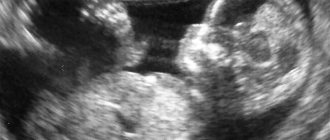
- when a pregnant woman is carrying twins or triplets;
- if the conception date was set incorrectly;
- when the family used IVF methods;
- if the expectant mother has too little body weight or, on the contrary, is obese;
- when a woman suffers from diabetes.
Negative results: what to do?
The period of the second screening occurs in the middle of pregnancy, so doctors no longer talk about terminating it for medical reasons. Sometimes there are cases when fetal pathology can be corrected by medical methods - in this case there is hope, and doctors will offer the pregnant woman a choice of possible manipulations and interventions. If the negative results of the second screening show a high risk of developing anomalies (in a ratio of 1:360) without hope of reversing the process, then the woman is asked to decide on an artificial birth.
In any case, both parents will have to make the difficult decision to continue or terminate the pregnancy. When there is a chance to correct the pathology, the expectant mother is explained in detail: what modern methods exist to correct the situation, how risky they are, whether complications are possible during the intervention process, and what are the overall chances of having a healthy baby.