What is ultrasonic scanning: the essence of the method, modes
Translated from English, the word “duplex” means double. In this case, the use of two ultrasound modes is implied:
- B – mode – the essence of the method is that the sensor emits ultrasonic waves of a certain frequency passing through the patient’s tissues. Then, at different densities of the tissues being examined, ultrasound is reflected and the signal returns back to the sensor. The device emits ultrasonic waves and receives a return, reflected signal in a pulse mode, that is, at different time intervals. For example, if it is necessary to examine the most distant areas (of an echogenic structure), much more time passes between the ultrasound radiation and the reflection of the signal than when diagnosing structures located closer to the device. The unique properties of the sensor allow it to emit waves at different angles, with different time intervals. Modern technology allows you to scan and visualize a two-dimensional image of the vessels of the brain and neck vessels in a matter of minutes.
- Doppler - this technique is based on the Doppler effect: ultrasound directed at the area under study, when colliding with a moving object, not only sends a response signal, but also changes the frequency and, accordingly, the wavelength of the radiation. When diagnosing blood vessels, the main object from which ultrasound is reflected are red blood cells. This allows you to identify the exact spectrum of blood flow speed.
Also, using the innovative method of color Doppler scanning (CDC), it is possible to build a color cartogram of blood flow in the vessel under study.
Color Doppler scanning method (Color Doppler scanning) Advertising partners
What does ultrasound scan show?
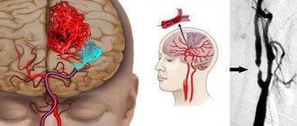
Duplex scanning of the vessels of the brain and neck allows you to study in detail their structure, condition and direction of blood flow. Each of the above modes determines the following data about the vessels of the head:
- Ultrasound examination (B-mode) determines the location of blood vessels in the human body, their diameter, and the condition of the vascular lumen is assessed; the technique allows you to identify various pathologies that impede normal blood flow (for example, blood clots formed on the walls, cholesterol plaques); The most innovative devices are capable of visualizing a layer-by-layer image of the vessel.
- The Doppler mode allows you to study in detail the hemodynamics of blood movement through the vessels, the direction of blood flow, and its speed.
Duplex scanning of the vessels of the head is a procedure that allows you to fully study almost all parameters of the blood vessels and make the most accurate diagnosis.
Duplex scanning of head vessels
The technique is universal when it is necessary to identify pathological processes in the vessels of the brain and neck. In addition, the study refers to non-invasive diagnostic methods, which implies its complete safety and the ability to perform ultrasound scanning the required number of times.
Duplex scanning of neck vessels
During the diagnosis, not only blood vessels are visualized, but also nearby tissue structures. Intracranial duplex scanning is used to study the cerebral vascular beds. If it is necessary to obtain a color image, transcranial duplex scanning is used.
How it goes
The study of fetal blood flow is carried out in the same way as a conventional gray scale ultrasound. Most often, these two types of research are performed simultaneously.
Ultrasound procedure
The woman should free her stomach from clothing and lie down on the couch. It is advisable to lie straight on your back, but if it is difficult for a pregnant woman to remain in this position for a long time or the fetus is in some atypical position, it is permissible to lie on your side. The doctor applies the gel to the area of the uterus and places an ultrasound probe on the pregnant woman's abdomen at various points, looking for the main vessels needed for study.
With each Doppler examination, the condition of both uterine arteries, the umbilical artery, and the middle cerebral artery of the fetus must be examined. According to indications, a study of the ductus venosus, thoracic aorta, renal arteries, umbilical vein, inferior vena cava, and intracardiac blood flow of the fetus can be performed.
The doctor displays the required vessel on the screen, first in gray, then turns on one of the Doppler modes, registers and studies the necessary blood flow parameters, and then enters them into the study protocol.
The time for an ultrasound with Doppler ultrasound is slightly longer than without it and depends on the position and motor activity of the fetus. The calmer the child behaves, the faster and easier it is for the doctor to register his blood flow.
In what cases is a study prescribed?
Duplex ultrasound scanning is used for various diseases and pathological conditions of cerebral vessels:
- congenital vascular malformations;
- deformations of head vessels;
- atherosclerosis;
- injuries;
- when blood clots form in the vessels of the brain (thrombosis);
- if you suspect the presence of an aneurysm (expansion of part of the vascular wall);
- inflammation of blood vessels (vasculitis);
- damage to blood vessels (angiopathy);
- with discirculatory encephalopathy (brain damage that occurs due to circulatory failure);
- in the presence of diseases in nearby adjacent tissues and structures.
Diagnostic measures are also prescribed for patients who have the following symptoms:
- migraine;
- constant feeling of heaviness in the head, accompanied by tinnitus and
- frequent headaches;
- dizziness;
- motor coordination disorders, unsteadiness when walking;
- fainting;
- feeling of weakness and numbness in the legs and arms;
- speaking disorders.
Duplex scanning is prescribed to patients who have previously suffered a stroke or surgery on the vessels of the brain, as well as people suffering from chronic cerebrovascular accidents. In this way, the condition of blood vessels and blood flow is monitored. Duplex scanning most informatively reveals secondary pathological changes in the vessels of the head in the presence of various diseases (for example, with a persistent increase in blood pressure or in the presence of diabetes mellitus).
The study is prescribed in the presence of large tumors inside the brain (cystic, cancerous formations and hematomas). In this case, the goal of diagnosis is to evaluate the tumor and its blood supply. Ultrasound scanning is also required before surgical intervention on cerebral vessels or to determine methods of conservative treatment.
Duplex scanning is of high importance in neurological practice and makes it possible to diagnose cerebral circulatory disorders in the early stages of development. This allows you to take timely measures to prevent an acute process. In this case, the possibility of a detailed study of cholesterol plaques and their density is of great importance.
When is it prescribed for pregnant women?
In the third trimester of pregnancy, Doppler sonography is performed on all pregnant women during the 3rd screening in addition to the results of CTG. For some women, Doppler sonography is indicated already in the second trimester of pregnancy, since during this period it is still impossible to assess the well-being of the fetus by any other methods.
Indications for ultrasound examination of the fetus may include concomitant diseases or certain conditions of the mother: arterial hypertension, threat of miscarriage or premature birth, high degree anemia, large uterine fibroids, genital infection, respiratory and heart failure.
Fetal circulation, color Doppler mapping
,Indications for Dopplerography may also include changes detected in the placenta: changes in the structure of the placenta identified by ultrasound, discrepancy between the thickness or degree of maturity of the placenta and the gestational age, placental presentation.
In addition, Dopplerography is indicated if there are signs of any abnormalities in the development of the fetus itself: IUGR or vice versa a large fetus, Rh conflict, ultrasound signs of infection in the fetus, signs of hypoxia or heart failure, congenital defects of the heart, great vessels, kidneys, 2 and more loops of umbilical cord around the neck.
There are no contraindications to Doppler ultrasound in pregnant women.
At-risk groups
Duplex scanning is prescribed to people who are predisposed to developing diseases of the vascular system. The following categories of patients are at risk:
- long-term smokers;
- people who are overweight;
- people leading a sedentary lifestyle, which as a result leads to physical inactivity;
- exposed to frequent stressful situations;
- age category after 45 years;
- patients with a history of diabetes mellitus of any stage;
- in the presence of elevated cholesterol levels in the body.
Preparing for Duplex Scanning
No special preparation is required for the ultrasound duplex scanning procedure. Diagnosis is carried out at any time of the day, but more often in the morning.
On the day of ultrasound scanning, patients are advised to avoid cigarettes, alcohol and some drinks that affect the tone of the vascular walls (coffee, strong black tea, various energy drinks).
If the patient takes medications such as Phezam, Betaserc and some others, it is necessary to consult with your doctor about taking them on the day of the procedure.
Immediately before the examination, all possible jewelry should be removed from the head and neck area. At the end of the procedure, you should thoroughly wash your head and neck to completely remove any remaining gel.
May be of interest:
|
Is Doppler harmful to the fetus?
There are many opinions about Doppler ultrasound.
The harm of this study has not been proven. However, most expectant mothers are suspicious of this procedure. Doctors call for this method of diagnosis, because it makes it possible to identify most fetal diseases at an early stage. Moreover, neither mother nor child feel any discomfort during ultrasound scanning.
Therefore, stories on the Internet from mothers that the baby was twisting around a lot during this procedure, or was wrapped in the umbilical cord, do not indicate in any way that this happened due to the Doppler. Yes, the baby may move during the Doppler ultrasound, but this will also happen during the ultrasound, as the baby feels the cool gel being applied to the abdomen.
Features of the event
Since ultrasound is a non-invasive diagnostic method, it does not cause pain or discomfort, and therefore does not require the use of anesthesia. The procedure is performed in a sitting or lying position, depending on which area of the head is being examined.
Then the specialist applies a transparent gel to the skin of the examined area, which acts as a conductor between the human body and the ultrasound sensor. The sensor is installed on the required area of the head; more often, certain areas with the thinnest bones of the skull are used for this purpose. Thus, ultrasonic waves freely penetrate inside the skull.
The duration of the diagnosis takes between 20-30 minutes, during which the specialist may ask the patient to hold his breath or change his body position. The procedure is well tolerated and does not cause discomfort.
Will the procedure harm the baby?
There is an opinion among expectant mothers that ultrasound harms the baby.
Many argue that every procedure is stressful for the unborn child. Therefore, pregnant women try to do as little research as possible. But this opinion is erroneous, since ultrasound and Doppler sonography do not pose a risk to the health of the expectant mother and her child.
All devices have recommendations for use. The specialist who conducts Doppler sonography knows how the equipment works. During the examination, he makes sure that the tissues are not overheated and unpleasant consequences do not occur.
The procedure takes a short amount of time, ultrasound does not cause significant harm to health. And the results obtained can save the baby’s life.
If a problem in the fetus’s blood circulation is detected in time, this will help eliminate the problem. Take the necessary measures to maintain pregnancy and the safe birth of a full-fledged child.
Author: Lina Valerievna, gynecologist Specially for the site kakrodit.ru


