The pancreas is a very important organ in the human body, which is responsible for the digestion of carbohydrates, fats and proteins, metabolic processes, and the supply of insulin into the blood. It is located behind the stomach; the pancreas is directly connected to the duodenum by the pancreatic ducts. Through these ducts, amylase, lipase and protease enter the duodenum - secretions that take an active part in the digestive process.
Diagnosis of pancreatic diseases
The pancreas in the human body has two different functions: digestive and endocrine (due to the secretion of enzymes that help break down food into compounds that are easily absorbed by the body, and insulin).
The work of this gland is closely interconnected with the activity of the liver, therefore liver pathologies are often the cause of disruption of the pancreas. The presence of feedback is also known - pancreatitis and pancreatic tumors can disrupt the functioning of the liver.
The most commonly diagnosed diseases of the pancreas are:
- benign and malignant neoplasms;
- stones in the pancreatic ducts;
- acute form of pancreatitis;
- chronic form of pancreatitis.
Diagnosis of pancreatic diseases at the Yusupov Hospital is carried out using various methods, the most informative of which are MRI, CT without contrast, CT of the pancreas with contrast. The price of each study depends on the complexity of the procedure and the medical equipment needed to perform it.
Contraindications
Such research is prohibited for pregnant women and children, as X-rays are used. Contraindicated for people with multiple myeloma. Contraindications to the procedure with contrast would be the patient's renal failure, heart disease, endocrine system disorders, and an allergy to iodine. Claustrophobia, the patient’s serious condition, and mental illness will prevent the patient from remaining completely still, so sedatives can be used.
CT or MRI of the pancreas: which is better?
Today, at the Yusupov Hospital, magnetic resonance and computed tomography of the pancreas are most often used to diagnose pathologies. These studies provide multiple magnification of the resulting image, thanks to which the doctor assesses the area of interest in the layer, the size and characteristics of the pathological formation.
A more informative and detailed image display is achieved through the use of artificial color contrast.
Both magnetic resonance imaging and computed tomography of the thyroid gland are the most informative and popular examination methods that have the same indications. However, CT can be used in situations where certain reasons make it difficult to perform an MRI. Most often, computed tomography of the thyroid gland is prescribed to patients with pancreatitis, suspected neoplasms and frequent digestive disorders.
X-ray radiation used in computed tomography is the reason for limiting the use of the procedure - the intervals between CT scans should be quite long, at least six months, despite the insignificant radiation dose and the absence of negative effects on the patient’s body.
The information content is approximately the same for CT or MRI of the pancreas. Which of these methods is better cannot be answered unequivocally. The advantages of magnetic resonance imaging include the ability to more accurately determine the location of the tumor. However, MRI, due to its higher cost, is slightly less common.
Both magnetic resonance and computed tomography of the pancreas have the same ability to detect diffuse changes in the organ and clarify their localization.
Unlike ultrasound diagnostics, both MRI and CT make it possible to obtain the highest quality three-dimensional picture, determining not only the presence of changes or neoplasms, but also measuring their size and clarifying their nature.
Decoding
A radiologist interprets the computed tomography data. He evaluates the obtained tomograms to ensure that the organ’s indicators correspond to the norm. If the diagnostician finds anatomical abnormalities, he describes them in detail in terms of structure, size, extent and degree of invasion. If a contrast examination was performed, the doctor additionally notes the behavior of the tissues in terms of accumulation and release of the contrast agent. He draws up all his findings in the form of a written report and accompanies them with CT images recorded on electronic media or printed on film. This entire package of documents is transferred to the patient with recommendations on further steps.
Study Details
- Doctor's referral: Required for CT scans for children
- Preparation: Yes in case of CT angiography, CT OMT, CT OBP
- Application of contrast: As prescribed by a doctor and with CT scan of blood vessels
- Time: 5-7 minutes
- Contraindications and restrictions: Pregnancy, allergy to iodine on CT with contrast, renal failure
- Results delivery time: 30-40 minutes
| List of studies | Price | Stock |
| CT scan of the urinary system (kidneys, adrenal glands, bladder, urinary tract) | from 2500 rub. | At night |
| CT scan of the abdominal cavity (part of the esophagus, stomach, liver, gallbladder, pancreas, spleen, small intestine) | from 2500 rub. | At night |
| CT scan of the retroperitoneum (kidneys and adrenal glands, ureters, soft tissues and lymph nodes) | from 2500 rub. | At night |
| CT virtual colonoscopy (large intestine) | from 4100 rub. | At night |
Indications for CT or MRI of the pancreas
Diagnosis of pancreatic pathologies is quite difficult. Such diseases can be asymptomatic for a long time. External symptoms appear, as a rule, as the pathology progresses and other organs and systems are involved in the pathological process.
Magnetic resonance or computed tomography is prescribed for patients with the following symptoms:
- pain in the stomach, girdle pain;
- frequent digestive disorders;
- if benign or malignant tumor formations are suspected;
- as a control measure after pancreatic surgery or monitoring the progress of therapy.
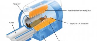
Progress of the procedure
Before the examination, the patient must remove metal clothing and accessories located in the abdominal area. The patient is positioned on the retractable tomograph table lying on his back. The patient must lie still throughout the entire procedure.
Special belts and pillows are used to secure the body. During diagnosis, to exclude displacement of the pancreas and obtain more accurate images, the specialist will ask the patient to hold his breath for a few seconds.
The patient with the table moves inside the device so that the area to be examined is located opposite the scanner. Before turning on the tomograph, medical personnel clear the room to protect from radiation.
On this topic
- Tomography
Indications and preparation for computed tomography of the abdominal cavity
- Olga Vladimirovna Khazova
- February 28, 2020
At the same time, using a special window, the doctor constantly monitors the patient’s condition, who, if side effects occur, is able to contact a specialist via voice or button.
First, cross-sectional images are taken, after which the data obtained are recorded, and the patient is given an intravenous contrast agent. After contrast is administered, repeat images are taken. Images of the organ are visualized on the monitor, and the doctor selects the most informative ones.
Computed tomography allows you to obtain a three-dimensional reconstruction of the organ under study, which is necessary for diagnosing the development of pathology. The patient receives CT results a few hours after the procedure.
How is a CT scan of the pancreas performed?
Before the procedure begins, the patient lies down on the mobile table of the tomograph; in order to keep him still, belts and bolsters can be used. During the examination, the scanning ring of the tomograph rotates around the table with the patient lying down.
High quality and accuracy of images can only be ensured if the patient is absolutely still and calm. During the procedure, the patient is periodically asked to hold his breath. The examination lasts about 15-20 minutes. The patient does not experience any discomfort or pain during the CT scan.
Possible complications
Computed tomography is performed using x-rays. Even the newest devices irradiate the body, so it is important to do such diagnostics no more than once a year. If a CT scan is performed with a contrast agent, the patient sometimes develops a normal reaction in the form of pain, heat flushes, and a sour, metallic taste in the mouth.
In some cases, the substance causes side effects such as swelling of the face, dry throat, itching of the skin, rashes, decreased blood pressure or bronchospasm.
During the procedure, you may experience a panic attack, nausea or dizziness, which should be reported to your doctor immediately. Some time after diagnosis, some patients develop an allergic reaction to the contrast in the form of hives, migraines or vomiting.
How is magnetic resonance imaging of the pancreas performed?
The duration of magnetic resonance imaging is, as a rule, no more than 30 minutes. Before the procedure, the patient does not need to perform any special measures. The day before the MRI, it is recommended to adhere to a gentle diet, avoid drinking alcohol, spicy and fried foods.
When performing an MRI with contrast, the patient must first be tested to ensure there is no allergic reaction. After the procedure, the patient does not require medical assistance and is allowed to go home.
Magnetic resonance imaging of the tail of the pancreas is performed if there is a suspicion of insulinoma, a malignant tumor that seriously disrupts the endocrine state of the body, which leads to excessive secretion of insulin. Sometimes this process can lead to the development of hypoglycemic coma.
MRI can detect all forms and types of pancreatic cancer. This study is the most accurate method with which the doctor has the opportunity to determine the nature of the tumor and its exact location at the earliest stage of development.
Using MRI, it is possible to identify cystadenocarcinoma and adenocarcinoma, the direction of development of these malignant neoplasms, thanks to which a specialist can develop a competent treatment regimen.
Magnetic resonance imaging can also detect metastases that have spread to the pancreas from other organs.
MRI, CT scan of the pancreas: price (Moscow)
The cost of a pancreatic CT scan may vary depending on whether a contrast agent is used. In addition, the price is influenced by such parameters as: the rating of the diagnostic center, the level of the tomograph machine, and the qualifications of doctors. As a rule, during a comprehensive examination of the pancreas, a CT scan of the liver and spleen is also performed.
The price of a CT scan in Moscow is slightly higher than in other regions of Russia. For example, if a CT scan of the pancreas with contrast is prescribed, the price is, on average, 6-10 thousand rubles, while a CT scan without the use of a contrast agent will cost 3.5-6 thousand rubles.
Treatment of pancreatic pathologies at the Yusupov Hospital includes consultations, diagnostic, therapeutic and rehabilitation measures. Thanks to the rich experience and competence of the doctors at the Yusupov Hospital, their use of the latest medical technologies and expensive high-tech diagnostic equipment, high treatment results are achieved. The clinic’s team of specialists - gastroenterologists, surgeons, endoscopists, radiologists and nutritionists develop individual therapeutic tactics for each patient, taking into account the characteristics of the patient’s body, the nature and stage of the pathological process.
Author
Tatyana Aleksandrovna Kosova
Head of the Department of Rehabilitation Medicine, Physiotherapy Physician, Neurologist, Reflexologist
Scanning with contrast
To visualize all organ structures as accurately as possible, a contrast agent is injected. It accumulates in tissues in a certain way and makes it possible to diagnose even the slightest disorders. Administration of the drug intravenously allows you to see blood vessels and tumors. In most cases, the administration is tolerated easily, without discomfort or side effects. If you still experience unpleasant sensations, itching, urticaria, or increased heart rate, you should immediately inform your doctor.