Briefly about radiography
X-ray is an effective technique for diagnosing pathologies in the body. The study was first done in 1895, when V.K. X-ray discovered the properties of rays, later called X-rays.
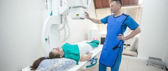
Using this study, the body is scanned with X-rays and an image is obtained on a light-sensitive film (more harmful) or a computer monitor. Diagnostics is carried out using both stationary and mobile, as well as portable devices. This makes it possible to take a picture even in the operating room, if necessary.
Today, the technique has undergone a number of positive changes, which has made it possible to reduce the dose of exposure that the patient receives by a hundred times. This was made possible thanks to the use of semiconductor linear detectors that convert the energy of the rays into light.
X-ray examination receives a full-format image and a reduced one. A striking example of a small format image is fluorography.
Radiography also takes overview and targeted photographs. By adjusting the device in the optimal projection, it is possible to identify disorders that are difficult to visualize, for example, pathologies of the lungs and chest.
Radiography became the first non-invasive diagnostic method, which later gave birth to modern research methods, for example, computed tomography.
LiveInternetLiveInternet
Quote from DejaVu57
Read in full In your quotation book or community!
November 8, 1895 120 years ago Physicist Wilhelm Roentgen discovered “X-rays”
It is in vain that they say that an elderly person is not capable of making an outstanding discovery. The famous German physicist Wilhelm Conrad Roentgen discovered mysterious rays, later named after his last name, when he was already over 50. Old age did not at all affect the scientist’s attentiveness, as well as his hard work and desire to establish the truth.
In the modern world, gripped by gerontophobia, we often hear that if a person did not do anything outstanding in his youth, then in old age he will no longer be able to do this. Unfortunately, this absolutely incorrect statement is becoming popular in the scientific community. Often, institutes prefer to hire inexperienced junior researchers, while refusing older professors - they say that they are not able to bring benefit to the institution, since they are already breathing their last.
However, those who talk like this for some reason forget many examples of outstanding discoveries that were made by famous scientists in old age. And the most telling example is the so-called X-rays, which in many countries are now called X-rays after their discoverer. The famous German physicist Wilhelm Conrad Roentgen discovered this interesting natural phenomenon in 1895, when he was already over 50. And he not only discovered, but also thoroughly studied the rays he discovered, not entrusting such an important work to his graduate students.
It should be noted that this phenomenon was discovered completely by accident (as often happens in science). In fact, Professor Roentgen, who at that time had already headed the department of physics at the University of Würzburg for several years, studied a completely different phenomenon - the so-called cathode rays. Now everyone knows that they are a stream of electrons emitted by the cathode when current is applied to it, but in those days they did not even know the word “electron”. Therefore, physicists were very interested in why a strange glow was observed when current was passed through a cathode tube, that is, how electricity could produce light.
And so on November 8, 1895, Professor Roentgen, as usual, stayed late in his laboratory. All his assistants had already left, but he continued to work - he turned the current in the cathode tube on and off, while taking measurements of various characteristics and recording the results. However, the years took their toll - around midnight, Roentgen felt tired and realized that he had to go home. The professor looked around the laboratory and, making sure that everything was in place, turned off the light. It was this action that led the professor to his outstanding discovery - X-ray suddenly noticed a luminous spot in the darkness.
The scientist approached the light source and discovered that it was a screen made of barium sulfide used in his experiments (which always reacts to electromagnetic waves, including visible light). But why was it glowing? After all, it had been dark outside for a long time, the cathode tube was turned off, and besides, it was covered with a black cardboard cover (the neat professor always did this when he finished work). And then Roentgen realized that he apparently forgot to turn off the cathode tube.
Reproaching himself for forgetfulness, the scientist groped for the switch in the dark and stopped the flow of current. But along with it, the screen instantly went dark. X-ray became interested in this phenomenon - he turned the tube on and off several more times, and the mysterious glow again appeared and disappeared. There was no doubt - it was the cathode tube that caused it.
But how could this happen? After all, the cathode rays had to be blocked by a paper cover, and in addition, the meter-long air gap between the tube and the screen was completely impenetrable to them. Wanting to understand the situation, Roentgen decided not to go home, but to continue the experiments. In vain that night Frau Roentgen waited for her husband - he, captivated by his accidental discovery, continued his experiments under the cover of the stormy Württemberg night.
So, leaving the case on the tube (so that the cathode rays were closed), the professor, with the screen in his hands, began to move around the laboratory. It turned out that even a distance of two meters is not a barrier for these unknown rays. During the study, Roentgen discovered that they easily penetrated books, glass and other objects. When the scientist’s hand was in the path of these unknown rays, he saw with horror the silhouette of her bones on the screen! That is, they passed through living flesh without hindrance.
However, there was no time to be surprised - as an experienced experimenter, Roentgen understood that he would need irrefutable evidence of his discovery. And so he took out the photographic plates lying in the closet in order to take the world's first x-ray. After this, a new series of experiments began, during which the researcher found out an interesting thing - not only do the rays illuminate the plate, but they also do not diverge spherically around the tube (as light would do), but have a certain direction.
Only in the morning, tired, but very satisfied, Roentgen showed up home. However, after resting a little, he again hurried to the university - he was attracted by the secret he had discovered. Realizing that at his age time is not just precious - it is actually priceless, he spent seven weeks hopelessly doing experiments, ordering food to be brought to his laboratory and a bed to be placed there. Everything was forgotten: students, friends, family, and even health. Only after 50 days did the scientist finally figure out what he had discovered.
It is curious that the first person to whom Roentgen demonstrated his discovery was his wife Bertha. A photograph of her hand with a wedding ring on her finger was attached to Roentgen’s article “On a new kind of rays,” which he sent to the chairman of the University Physico-Medical Society on December 28, 1895. The article was quickly published as a separate brochure, and Wilhelm Roentgen sent it to leading physicists in Europe. Thus began a new era in the history of medicine and other branches of science and technology - the era of studying the internal structure of an object using X-rays.
By the way, the outstanding physicist himself, who was simply a fantastically modest person, was against calling the radiation he discovered x-ray. Later, representatives of industrial companies approached Roentgen more than once with offers to profitably purchase the rights to use the invention. But each professor refused to patent his discovery, because he did not consider his research a source of income. And even when in 1901 the scientist became the first Nobel laureate in physics, he, having accepted the award, refused to attend the ceremony (because he could not stand congratulations, applause and other attributes of recognition, considering all this nonsense). The prize was sent to him by mail, but he did not use it himself - when the German government during the First World War asked the population to help the state with money and valuables, the modest and sympathetic scientist Wilhelm Roentgen gave away all his savings, including the Nobel Prize.
By the way, the fact that the greatest discovery in the field of medicine was made in Würzburg should be considered a kind of historical curiosity. The fact is that this ancient Bavarian city earned a reputation in the Middle Ages as the capital of the German “witch hunt.” Its ruler, prince and part-time bishop of Würzburg, Julius Echter, founded a university in 1582 to train Catholic theologians with the goal of “destroying all scum and infection in the city.” This was the same university where the aforementioned X-rays were later discovered.
However, in those dark times, this educational institution became famous as a hotbed of obscurantism - its graduates were famous for such cruel acts as the forced conversion of their flock to Catholicism, the expulsion from the city of Protestants who did not want to change their faith, the confiscation of the property of local Jews and witchcraft trials. During Echter's reign, more than 300 witches and wise men were burned - more than in any other German city. And only by the middle of the 17th century were the trials of witches and their executions stopped. Therefore, the fact that the discovery, which subsequently saved many human lives, was made at this university is a kind of symbol of the restoration of historical justice - this is how the University of Würzburg made amends for its guilt before humanity.
So, advanced age is not at all a hindrance to making discoveries, and Wilhelm Conrad Roentgen proved this with an example from his own life.
His half-century of life did not at all affect his attentiveness, hard work and desire to establish the truth. It should be noted that often it is young scientists who lack those qualities that the discoverer of X-rays possessed in full. On March 27, 1845, 170 years ago, Wilhelm Roentgen, German physicist, Nobel laureate Anton Evseev was born. Sources - https://www.calend.ru/, https://www.pravda.ru/science/useful/14-11-2012 /1133947-rentgen-0/
When is it necessary to take an x-ray, and to whom is it contraindicated?
Radiography is used in various fields of medical knowledge - in orthopedics, traumatology, pulmonology, gastroenterology and others. For each area, X-ray solves specific problems. Indications for radiographic examination:
- if necessary, assess the condition of internal organs;
- for the purpose of diagnosing injuries and damages;
- to find foreign bodies (for example, swallowed small objects);
- to identify tumor tumors;
- if necessary, identify specific changes in bone tissue and articular joints;
- to identify congenital developmental anomalies;
- when assessing the treatment provided;
- to determine degenerative-dystrophic processes in tissues;
- to assess the correctness of installed structures (for example, during dental implantation);
- in preparation for surgery.
When performing x-rays, there are both indications and contraindications. There are much fewer of them, but they make it difficult to diagnose some diseases. The influence of x-rays is contraindicated in the following cases:
- patient sensitivity to components containing iodine;
- open injuries;
- diabetes mellitus with decompensation;
- severe kidney and liver diseases;
- period of pregnancy and lactation in women;
- thyroid dysfunction;
- active pulmonary tuberculosis.
These are the main contraindications for which X-rays are not performed on patients. Some of them are relative, therefore, as soon as an examination is possible, such a diagnosis must be carried out.
The effect of x-rays on the body of children and adults
The dangers of X-ray radiation are one of the first questions that arose after the discovery of this type of research. Scientists have found that radiation is not as clear-cut as previously thought and has negative sides. Dangerous aspects of X-ray examination:
- medical radiation dosage is much higher than what a person receives daily from nature;
- the human body does not have mechanisms to adapt to artificial radiation;
- the effect of x-rays on a sick body weakens it even more;
- the examination is often carried out unevenly, when the same organs are examined repeatedly, which creates an inadequate radiation dose and puts these organs and systems at risk;
- a large dose of radiation provokes undesirable consequences, including malignant degeneration of cells.
These features indicate that x-ray diagnostics have an ambiguous effect on the body of both adults and children, and in many ways even have an unfavorable effect. The data obtained gave impetus to the study of the effect of x-rays on the child’s body, and this is what scientists were able to find out:
- children have increased sensitivity to radiation, and therefore any dose of radiation is undesirable for them;
- The developmental features of children's bodies lead to the fact that their internal organs receive a high dose of radiation compared to adults.
In view of these features, doctors are recommended to reduce the radiation dose in children to the minimum acceptable, and also to diagnose only according to indications, i.e., to exclude when other methods can be used.
Is it harmful to take an x-ray?
The maximum permitted dose for humans is 150 mSv per year. The usual standard procedures that have to be completed annually do not exceed 20 mSv (millisievert). However, in case of obesity and pregnancy, it is better to refuse X-ray diagnostics. Electromagnetic radiation can harm an unborn baby - affecting the normal formation of its internal organs and tissues. And an excess of fat will prevent you from taking a clear picture - dark spots appear on the film.
For children under 16 years of age, X-rays are recommended only in emergency cases - fractures, head injuries, hip dysplasia, pneumonia. If procedures have to be performed frequently, the maximum radiation dose to a child should not exceed 50 mSv.
Before X-rays are taken for a child under one year old, in order to avoid unnecessary movements, he is fixed using a special device.
The danger of X-ray examination lies only in the case of the patient receiving a large dose of radiation. The harmful effects of electromagnetic rays on the human body are manifested by:
- erythema - sunburn, characterized by deep and persistent damage to the skin;
- changes in blood composition - short-term with a small excess of radiation, with prolonged exposure to rays there may be irreversible changes;
- early aging of the body;
- formation of tumor-like formations;
- infertility;
- development of genetic abnormalities in offspring.
Of course, such consequences cannot but alert every modern person. However, if the influence of X-ray equipment poses such a danger to the patient’s condition, why is X-ray examination so widely used in medicine and can it be replaced?
How to “make friends” with x-rays
Some patients think about the effect of the procedure on the body and are concerned about their health after the study. Doctors rush to reassure such people - X-ray irradiation for diagnostic purposes is completely safe.
Firstly, people don't take X-rays often enough to tell about the cumulative effect of radiation on the body. X-rays are often harmful, but this does not apply to isolated cases. One-time exposures will not harm your health and will not cause serious pathologies in the future.
Secondly, X-ray doses are minimal and therefore safe. The volume of radiation is enough to get an image of a particular organ, however, such numbers are not enough to cause serious harm.
Thirdly, a person receives a certain dose of radiation every day, even when he walks down the street. The cumulative effect of rays does not appear at low doses, just as when irradiated by a device.
Fourthly, after an x-ray examination, you can help remove products from the body, which will reduce the load.
And the last “iron” argument in favor of x-rays is its high diagnostic value.
There are extremely few people who have been proven to have died from exposure to X-rays, but there are many more patients who were not correctly diagnosed because X-rays were not used.
Therefore, on this scale, the benefits of x-rays outweigh its negative features.
Types of modern X-ray machines
Different types of X-ray machines are needed to examine different areas. There are a lot of them. Here are the most common ones.
- An angiograph is a device used to study blood vessels.
- A fluorograph is a device designed to take x-rays of a person’s lungs. Usually gives data in two projections. The image is projected onto the film in a reduced scale.
- X-ray mammograph. From the name you can understand that the device helps to obtain data on the condition of the mammary glands. The test performed by a doctor is called a mammogram.
- A mobile ward unit, unlike a stationary X-ray diagnostic unit, is mobile. This multifunctional device is used in hospitals. Has a relatively compact size. The advantage of such a device is its transportability. There are mobile and portable units.
- Dental X-ray machine. The device is used in dentistry to clear the jaws. It allows you to identify damage and diseases of the teeth and their immediate location in the oral cavity.
- Operating X-ray machine. The device is used during surgical procedures, allowing timely detection of intracavitary bleeding and monitoring the progress of the operation. Such devices provide highly accurate results and have large dimensions.
- Digital X-ray. This is the most complex type of X-ray machine. All data is recorded not on film, but on an electronic matrix. They go directly to the workstation display in real time.
X-ray examination or Mantoux test: which is better?
It is not always correct to compare the harm of a tuberculin test and an x-ray, since the specifics of the research are different, as is the purpose of diagnosis. The Mantoux reaction is a mass test carried out to prevent the infection of children's groups with tuberculosis. If preventive tests reveal a carrier, then he is protected from the group, which saves a significant number of children from infection.
X-ray examination is done according to indications, and one of these is a positive Mantoux reaction. X-rays are not done en masse; they are a diagnostic procedure prescribed when a problem is suspected. Signs of tuberculosis are prolonged fever, cough, weakness, emaciation, decreased immunity and other signs indicating tuberculosis.
When considering whether x-rays are harmful, they are classified as unfavorable tests than the Mantoux test. However, you should not neglect it if the doctor sees the need to conduct research.
What is more harmful - Mantoux or X-ray?
Today, practicing medical specialists pay great attention to the issue of identifying infection of the population with Mycobacterium tuberculosis and diagnosing the disease in the early stages of development.
Children undergo the Mantoux test annually; in adults, diagnosis is carried out using the following studies:
- preventive fluorography;
- plain radiographic examination;
- bacteriological analysis of sputum;
- computed or magnetic resonance imaging.
X-ray diagnostics are prescribed for children only in special cases, therefore, to detect infection with Koch's bacillus, a tuberculin test is performed
The Mantoux reaction is the introduction into the child’s body of a small dose of waste products of mycobacterium tuberculosis, which causes an immune response. The degree of reaction of the child’s body corresponds to the presence of infection.
The tuberculin test has a number of negative aspects. The size of the post-injection papule depends on the reactivity of the child’s body. If a small patient has an allergy, a violent immune response is observed - the size of the spot exceeds the permissible 5 mm. A weakened immune system can cause the reaction to be negative even in the presence of infection.
Is fluorography harmful?
The diagnostic procedure must be carried out in an anti-tuberculosis dispensary. However, it is often carried out in children's institutions where the staff does not have a good command of the technique of performing the test.
In addition, water should not get into the tuberculin injection site, it should not be rubbed or injured, and children do not always comply with such requirements. This leads to a false positive reaction.
Despite the small dose of the diagnostic test, a child with a certain susceptibility may develop clinical manifestations of an allergic reaction:
- hives;
- bronchospasm;
- Quincke's edema.
Low specificity – the accuracy of the method does not exceed 50%. A positive test result is also observed after vaccination against tuberculosis (BCG), which stimulates the production of immune antibodies that provide protection for the body when it “encounters” the causative agent of the disease, infection with non-pathogenic forms of Tuberculosismycobacterium. However, this technique is still widely used by pediatric phthisiatricians because of its simplicity and accessibility.
Permitted radiation dosage
The safe amplitude of X-ray radiation is the range from 0.02 to 0.1 mSV. The critical dose at which radiation sickness develops is 0.7 pollutants. When performing fluorography on a digital device, a person receives a dose of 0.03 mSV, which corresponds to the norm. A person receives the same dose of radiation from the external environment.
Computed tomography is done at a dosage of 4-20 mSV; it will take two years to receive such a dose from the outside. The load is high compared to equivalents.
Therefore, there is no need to worry about conducting a short-term X-ray examination - it is safe, the devices operate according to sanitary standards and deliver the dosage that is permitted and does not cause harm to health.
Elimination of the consequences of radiotherapy
Radiation is a kind of poisoning when the body receives something that should not be in it. To avoid the complications of X-ray radiation, it is useful to drink more milk and eat fresh vegetables and fruits to remove radiation - they help cope with the consequences. The same recommendations are given to patients after radiotherapy.
X-ray examinations are associated with health risks. It is carried out exclusively according to indications, when there is a need for it. You should not be afraid of x-rays, because its health benefits are many times greater than its harm.