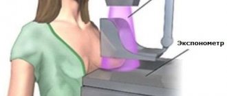
Dental treatment is often accompanied by an X-ray examination. This diagnostic method is fraught, according to many patients, with some danger.
Pregnant women are especially wary of X-ray examinations in dentistry, as they fear a negative impact on the development of the child.
But are these fears justified?
Is there a danger to mother and fetus?
Today in medicine there is no clear opinion about the dangers of exposure to x-rays . Some doctors argue that the use of this equipment during pregnancy is unacceptable.
European doctors analyzed statistical data, which revealed that more than 5% of women who underwent this examination had children born with underweight. But at the same time, they cannot prove that the reason for the child’s low weight was the x-ray.
© Kzenon/Fotolia
Most experts consider X-ray examination to be non-hazardous and a completely acceptable diagnostic method at certain stages of pregnancy.
It is only not recommended to use this type of examination in the early stages , during the formation of the child’s organs, since his cells at this time are most susceptible to radiation.
At other stages of gestation, if dental problems are detected, it is not only possible to take photographs, but also necessary. This will allow for timely and high-quality treatment, which will reduce the likelihood of the negative impact of pathological microorganisms on the development of the fetus.
During pregnancy, it is better not to take x-rays in public dental clinics; most of them still use old equipment that produces dangerous loads.
Modern equipment has low-frequency exposure with a radioactive load of up to 0.02 msv. For example, one flight by plane over a distance of about 2500 km produces a load equal to 0.01 msv.
A small dose of gamma radiation allows you to take pictures with modern equipment up to 5 times per visit. The average annual permissible dosage during the period of fetal development and growth is 3 mv, which allows for repeated treatment using this type of study.
During pregnancy, it is simply impossible to get such a dose.
What is the safety of a visiograph?
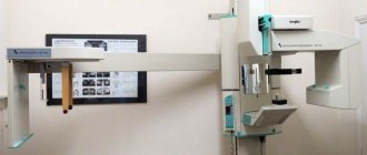
In modern dentistry, X-rays are taken using a radiovisiograph with a narrow beam. The flow passes only through the desired area, in this case the tooth, and does not reach other tissues. Those. the beam will not be able to influence the pelvic area of a pregnant woman in any way.
The microdoses emitted by the visiograph do not exceed the value of the natural background radiation that a person receives while living in modern conditions.
Is it possible to take x-rays during pregnancy?
We are exposed to radiation every day from various sources: food, water, air, flying from one city to another, solar and cosmic rays, in the end. But these are small doses. The average annual permissible level of radiation exposure is 3 millisieverts (mSv). One X-ray image carries a load of approximately 0.02-0.03 mSv.
So is it possible to do dental x-rays for pregnant women? Answer: yes, but only on modern equipment.
3 rules that pregnant women need to know
- The use of a film radiograph is not recommended. The safest would be a computer visiograph; its radiation beam acts locally. Any computer equipment provides approximately 10 times less radiation exposure compared to an outdated fluorograph.
- Before the procedure, you need to cover your chest and abdomen with a protective lead apron. This metal does not transmit x-rays. Without it, an examination cannot be carried out under any circumstances.
- Be sure to tell your doctor about your special situation and indicate the exact duration of your pregnancy.
Operating principle of a computer visiograph
Features of the event
The main role in diagnostics using gamma irradiation is played by its correct implementation. Despite the fact that the procedure is considered conditionally safe, it can only be prescribed if there are certain indications .
In this case, it is necessary to take into account the duration of pregnancy and ensure compliance with all safety measures for this procedure.
In what cases is the procedure necessary?
© Halfpoint / Fotolia
If you have a toothache, don’t worry in advance; dentists often treat and remove teeth without examining an x-ray. This approach is possible in case of obvious caries, in the absence of hidden, inexplicable problems.
It is important to illuminate the tooth and see what is happening inside when the carious process is advanced.
Indications for the study include:
- extensive focal infection. For example, deep caries of a group of teeth;
- hidden pathologies of the crown or root part of the tooth;
- inflammation of periodontal tissue;
- purulent pathologies: cyst, abscess;
- diseases that occur during the development and eruption of wisdom teeth;
- injury to the crown or root part;
- deciding whether to preserve or remove a tooth.
Any dental intervention must be based on a detailed picture of the condition of the units being examined, since even the most professional dentist cannot accurately determine the position of the roots, dental canals and the condition of the pulp chamber .
Only thanks to this, treatment can be carried out efficiently. In the absence of all the necessary data, therapy will not give the desired effect and will lead to an exacerbation of the disease, which will require re-treatment.
But if you take pregnancy planning seriously and monitor the condition of your oral cavity, then caries of such proportions will not have time to develop. And new, superficial caries, with a 99% probability, will be treated without x-rays.
What is dental physiotherapy and how is it performed.
This publication describes fissure caries.
Will garlic help with toothache? Details at the link.
Optimal timing
Dental treatment and x-ray examinations should be carried out in accordance with the timing of pregnancy. The most dangerous option would be a study conducted in the first trimester .
It is the most important for the normal development of the fetus and the most risky in terms of negative effects on its developing body. According to statistics, the largest number of miscarriages and the development of fetal pathologies occur during this period.
The first weeks of gestation are considered especially dangerous. At this time, diagnostics can be carried out in exceptional cases when the indications for the procedure do not allow for a delay.
But, as a rule, such situations arise in isolated cases and examination, as well as treatment, can be postponed until later in pregnancy.
© Halfpoint / Fotolia
If the x-ray was taken before the woman found out about her pregnancy, then in the future it is necessary to conduct genetic screening to identify abnormal fetal development in the early stages.
The most optimal period for carrying out such procedures is the second trimester . At this time, all the systems and organs of the child are already fully formed, and the influence of external negative factors has minimal impact on them.
Studies have shown that in women who underwent this type of diagnosis, the fetus developed without any abnormalities. Therefore, all existing pathologies that require medical attention must be examined and treated precisely at this time, since in the last trimester the risk of negative effects increases again.
Before going to the dentist, it is recommended that you also read this article, which tells you whether pregnant women can have their teeth treated.
Security measures
Even during the safest period of the second trimester, x-rays must be carried out in compliance with certain safety measures, which include:
- Use of special protective aprons and collars . They consist of lead plates that do not transmit gamma radiation. To prevent the metal from injuring the patient, it is placed in a cover made of thick fabric, which is shaped like an apron.
- Ensuring optimal distance . To obtain a detailed diagnostic picture, the maximum distance is selected. The farther the woman is from the beam tube of the equipment, the less radiation she will receive.
- Selection of the required exposure time to radiation . For one visit, no more than 5 photographs are allowed, with an interval of several minutes between them. In this case, the woman should not remove her protective apron for another 5 seconds after radiation exposure, since the decay time of gamma rays lasts up to 5 seconds.
Compliance with these rules for the safe conduct of x-rays will protect the woman and child from negative effects.
When can you do without an x-ray?
In some cases, x-rays are not necessary. Already during a visual examination, the doctor determines the disease and treatment tactics. Thus, with caries, it is possible to do without an x-ray; the affected areas are visible to the naked eye.
However, there are situations where diagnostics are extremely necessary and can affect the course of therapy:
- the need to seal canals located in such a way that there is a high risk of perforation;
- the presence of a neoplasm on the gum surface;
- injury to the subgingival zone;
- the doctor suspects the presence of an inflammatory process of soft tissues;
- with pathology of wisdom tooth eruption.
List of abnormalities in the fetus caused by radiation
If a woman had an x-ray in the first 12 weeks of pregnancy , this can cause serious deviations in the development of a number of systems and organs in the fetus:
- spine;
- bronchi;
- hearts;
- faces;
- jaws;
- reduction in the size of the skull and brain.
Radiation contributes to the development of health problems in the unborn child:
- dystrophy;
- blood diseases;
- incurable chronic diarrhea;
- predisposition to cancer.
Also possible:
- frozen pregnancy;
- miscarriage.
But all of the above is unlikely. To cause abnormalities in the fetus, a pregnant woman needs to receive a radiation dose of 3 mSv, and one procedure on modern dental equipment produces a load of 0.02 mSv, and on older devices 0.3 mSv.
The danger of exposure of women and men when planning conception
As explained above, to influence the development of the fetus, you need to receive a radiation dose of at least 3 msv. If, when planning a pregnancy, a woman underwent procedures that produced such a radiation load, then the cycle in which the examination or treatment was carried out should be skipped.
Scientists have proven that radiation affects not only the fetus, but also the egg.
The father of the unborn child should not receive such doses of radiation for 72 days (two and a half months), which is equal to the period of sperm renewal. There is a hypothesis according to which radiation accumulates in sperm and affects the formation of the fetus when the sperm enters the woman’s uterus.
How to perform an x-ray
In order not to harm the pregnant woman and the organism developing inside her, the exposure is selected depending on which tooth should be reflected in the picture. It is strictly prohibited to exceed this value.
In recent decades, X-ray machines have become much safer; new generation visiographs are often used, in which radiation exposure is several orders of magnitude lower. For pregnant women, a special class E film is mainly used, which allows you to work with reduced exposure. When using such equipment, the beam is strictly directed, which does not extend its influence to the entire body of the pregnant woman, but is concentrated only in the selected area.
Alternative Methods
Today in dentistry there is no alternative to x-ray examination. In their quality, we can only consider various types of equipment of this type, which are represented on the medical equipment market by a wide range of assortments.
The best option is a digital visiograph , characterized by a minimum radiation dose for a single exposure. It allows you to immediately receive images and store them on digital media.
The only disadvantage of a visiograph is that it can only be used to take targeted images covering no more than 3 teeth.
© Dan Race/Fotolia
To obtain a three-dimensional image of one or both jaws, an orthopantomograph , which, with a small dose of radiation, produces high-quality digital images.
an apex locator is used as an alternative , allowing one to measure the length and condition of the dental canal. Exposure to x-rays is completely eliminated.
The device is used for intermediate diagnostics, which avoids additional X-ray exposure.
How is a panoramic dental photo taken and is it harmful?
What is a caries marker and how is it used?
What to do if a temporary filling falls out, see the link.
Is it possible to get pregnant immediately after hysterosalpingography?
When planning a pregnancy, the doctor will definitely prescribe an X-ray if the long-awaited conception does not occur for a long time. The test is called hysterosalpingography (HSG). It must be carried out in order to determine the quality of patency of the fallopian tubes. If adhesions are found in the tubes, fertilization is impossible.
To check whether the tubes are patent, an HSG is performed, during which a contrast agent is used. It is with its help that you can see in the pictures the condition of the pelvic organs. Thanks to this procedure, the doctor can also detect formations that are dangerous to the woman’s health.
An interesting fact is that after completing this study, a woman who has not been able to get pregnant for a long time may discover two cherished stripes.
There is a completely scientific explanation for this “miraculous” healing. The same liquid used for X-rays is injected under pressure. The result of this procedure is the divergence of small adhesions and restoration of the patency of the fallopian tubes.
This important point must be taken into account after the x-ray, and your doctor will tell you when you can become pregnant. But in any case, during the menstrual cycle when the woman was examined, she needs to use protection, since the egg received a serious dose of radiation.
Prevention measures
Avoiding exposure to x-rays is possible only with timely detection of diseases and their prevention. The main preventive measures include the following:
- Attention to the quality of dental care .
It is necessary not only to follow the cleaning process, but also to choose the right products for oral hygiene. During pregnancy, dental and periodontal tissue become the most vulnerable, so additional cleaning products must be included for care. - Pregnancy planning should include complete oral hygiene.
- To avoid dental pathologies, you need to regularly conduct preventive examinations at the dentist .
As a rule, to maintain health, it is enough to visit the dentist 2 times a year. During pregnancy, a dental examination is routine and scheduled for the first trimester. If there are or periodically appear dental problems, then the woman should indicate them to the doctor for further diagnosis and treatment. - When planning a pregnancy, to make sure there are no dental problems, you can take an orthopantomogram of all teeth and heal those where there is the slightest suspicion of further development of caries.
- © Martinan / Fotolia
A woman needs to balance her diet so that it includes as many foods rich in vitamins and minerals as possible..
Vitamins help strengthen periodontal tissue, and minerals saturate tooth enamel, increasing its resistance to aggressive microorganisms.
Additionally, you should limit your consumption of foods with a lot of carbohydrates, as they promote the growth of bacteria.
The listed preventive measures are general. In order to get more detailed advice, the patient needs to visit a dentist, who will give detailed advice - see an example in the video:
How to prevent exposure
X-ray radiation will bypass a woman carrying a child if she does not develop caries and consults a doctor in a timely manner.
It's even better if she takes proper care of her oral cavity. Additional funds are used for care during pregnancy, as teeth and gums weaken during this period.
A woman planning a pregnancy must first take care of the health of her oral cavity and undergo a preventive dental examination.
It is better to heal incipient caries immediately. An orthopantomogram can serve as diagnostic equipment for all teeth.
A proper diet, including essential vitamins and minerals, which have a beneficial effect on strengthening tooth enamel and gums, will contribute to the preservation of teeth.
Confectionery products should be avoided. High carbohydrate foods like these stimulate the growth of bacteria in the mouth.
The video presents the opinion of one of the experts.
conclusions
Having studied world practice and research on the use of X-rays in dentistry during pregnancy, the following conclusions can be drawn:
- the harm of x-rays has not been scientifically proven;
- the safety of x-rays has not been scientifically proven;
- conclusions about the effect of x-rays on the egg and sperm during pregnancy planning are not based on anything and have no practical evidence;
- in theory, only a radiation dose of 3 msv affects fetal development;
- a radiation dose of 3 mSv is physically impossible to obtain even during the entire 12 weeks of pregnancy if images are taken every day;
- modern equipment delivers irradiation in a narrow direction, so the beam only affects the tooth specifically;
- It is not recommended to take x-rays in the first trimester;
- The recommended period from which x-rays can be taken is 16 weeks;
- if the degree of neglect of the dental problem is serious and there is a possibility of a negative impact on the health of the woman (and therefore the fetus), we can neglect the theory about the dangers of x-rays;
- The final decision should be made by the patient together with her doctor.
If you find an error, please select a piece of text and press Ctrl+Enter.
Tags: diagnostics during pregnancy
Did you like the article? stay tuned
Previous article
Small lower jaw: etiology and treatment methods
Next article
Infiltration anesthesia in dentistry: what it is and how it is carried out
Obstetrician-gynecologist answers
We asked obstetrician-gynecologist Elena Artemyeva to answer questions that concern expectant mothers.
“My husband and I have been planning a pregnancy for a long time. But he had to take an unscheduled photo of the tooth. Will radiation affect my ability to conceive? Is it dangerous to have a dental x-ray when planning a pregnancy? Should I stop planning?
— The amount of X-ray exposure in your case will not affect the male quality of conception.
— How long should it take after an X-ray of the lungs to conceive?
— You can plan a pregnancy already in the next cycle.
— I have a regular menstrual cycle. Sexual intercourse occurred two days before ovulation. And a day later I had to have fluorography and an x-ray of the hand in two projections. When the picture of the hand was taken, an apron was put on the stomach. And now I feel signs of pregnancy: weakness, drowsiness and cravings for salty foods, although it’s too early to take a test. If I'm pregnant, it turns out that I was irradiated before the egg was fertilized. Does this mean that I will give birth to a child with a pathology? Is it necessary to terminate the pregnancy?
— There is a theoretical basis for your fears. However, there is no need to worry ahead of time. Indeed, exposure to x-rays on the body on days when the likelihood of conception is high is undesirable. But this does not mean that the pregnancy must be terminated. First, make sure that you are pregnant.
To do this, you need not only to do a test, but also undergo an ultrasound examination.
In this case, the “all or nothing” principle applies. If a harmful effect on the fetus occurred, and it was strong enough, then pregnancy will not occur at all. If fertilization occurs and the fetus develops, then most likely a healthy baby will be born. Therefore, in your case there is no reason to panic. Start taking folic acid and wait quietly for the test results.
Using most medications during pregnancy can harm the baby. But even at the planning stage, a woman should be more careful in her actions.
If a married couple does not use contraception, then conception can occur at any time. All dental manipulations are recommended to be carried out long before conception. There are significant reasons for this.
Dental treatment before pregnancy
Planning a pregnancy involves visiting all doctors, including the dentist. The fact is that it is undesirable to treat teeth while carrying a child. The use of certain medications during this period is strictly prohibited. Microbes that spread throughout the body during the development of caries can cause intoxication. This negatively affects the development of the child , especially in the early stages of pregnancy, when the fetus is not yet protected by the placenta.
ON A NOTE! According to statistics, women who take vitamins when planning pregnancy are less likely to suffer from dental and gum diseases while pregnant.
Is it possible to take a dental x-ray when planning a pregnancy?
The main danger of dental treatment during pregnancy is that it is strictly forbidden to take x-rays at any stage. X-ray is a type of radiation exposure. It may affect the child's development.
Radiation has a particularly strong effect on dividing cells. They are in a vulnerable state. Radiation rays enter cells and change the structure of DNA. How this will turn out later is never known for certain. The most likely pathologies include:
- Microcephaly;
- Underdevelopment of the adrenal glands;
- Defects of the heart muscle;
- Immune deficiency;
- Anemia;
- Diseases of the gastrointestinal tract;
IMPORTANT! If an x-ray is unavoidable, then when planning a pregnancy it is better to do it in the first half of the cycle. During the procedure, it is advisable to cover the lower abdomen with a lead apron specially designed for this purpose.
Is it possible to remove teeth?
Tooth extraction before pregnancy is successfully practiced, and this is also possible during pregnancy. But there are certain risks, so it’s still not worth it to remove it. It is not possible to take an x-ray after removal. In this case, fragments of the tooth root . This is fraught with subsequent inflammation.
For pain relief during pregnancy, only those drugs that do not harm the baby are used. One of these is Ultracain . It acts in exactly the same way as other anesthesia, but does not harm the development of the fetus.
REFERENCE! You can relieve pain for a short time by rinsing with a solution of soda or salt.
Dental implantation before pregnancy
Doctors recommend inserting dentures first, and only then starting to plan a pregnancy. If it is possible to postpone the procedure until the postpartum period, then you can do so. Dental implantation is a long and complex process.
First, an implant is implanted into the gum. A certain amount of time must pass before it finally takes root. Typically this period ranges from 2 to 6 months. It all depends on the quality of the patient's bone. Only after this a crown is placed on the implant.
Dental prosthetics are not practiced during pregnancy. At the stage of pregnancy planning, this procedure can be carried out only in the most exceptional cases. Before the implant is implanted into the bone, the woman needs to take painkillers. Taking many of them is incompatible with maternity planning.
The situation is further complicated by the fact that the onset of pregnancy can affect the adaptation process . Hormonal changes characteristic of the pregnancy process provoke rejection of a foreign object. This will result in wasted time and money.
Reasons why dental treatment is necessary
During pregnancy, even women with healthy teeth may experience some difficulties. Very often there is a lack of calcium , which leads to tooth decay.
ON A NOTE! Daily oral care should include not only brushing, but also the use of dental floss and mouthwash.
The most common ailment during pregnancy is bleeding gums . The presence of caries or any inflammation in the oral area further complicates the current situation. The main reasons why you should treat your teeth before pregnancy include:
- The likelihood of rapid development of diseases in a pregnant woman increases significantly;
- Caries is a source of bacteria that harm the child's health;
- Toothaches during pregnancy affect its course;
- During the process of bearing a child, it is forbidden to take potent medications;
- There is no possibility to take an x-ray ;
- The healing process of the hole in the case of tooth extraction may be delayed;
A healthy oral cavity at the planning stage of motherhood provides a woman with confidence that nothing bad will happen to her child. A preventive examination is much less problematic than treating an existing disease. The earlier caries is detected, the easier it is to get rid of it.
A tooth can get sick, and the gums can become inflamed completely spontaneously - no one is immune from the problem. The female body is especially vulnerable to diseases, including those of the oral cavity, during pregnancy - hormonal changes aggravate the existing disease. Expectant mothers worry about their child and often wonder whether it is possible to do an X-ray on a pregnant woman?
The patient has concerns, because x-rays are radiation, which in large doses will harm even a healthy adult, but what will happen to a developing baby? Only a specialist assesses the risks, and radiography is carried out under the supervision of a doctor who is managing the pregnancy, if there is evidence for it.
Write a comment
Marina
February 21, 2020 at 03:24 pm
Although opinions differ about the harmful effects of x-rays on the fetus, I think the decision should be made by the doctor in a specific case and for a specific pregnant woman. For example, in the seventh month I had a toothache. The dentist removed the nerve and filled the tooth without checking with x-rays. And he said that it would be better to check it later and, if necessary, change it, rather than endanger the child.
Dasha
February 21, 2020 at 10:29 pm
I believe that the dental issue needs to be resolved before pregnancy. Dental treatment in any case involves the use of some medications, drugs, painkillers; all this is not advisable for a pregnant woman. But if this is already necessary, then you need to consume vitamins to the maximum, without them you can’t go anywhere. In general, I think there is nothing wrong with this.
Nastya
February 23, 2020 at 07:22 pm
During pregnancy I was going to get braces. Naturally, it was necessary to take two pictures... I didn’t risk it + I had an ultrasound scan 4 times. Although I remember reading on the Internet, many girls are not afraid - they do it. My friend, a dentist, told me that pregnant women often come with recently taken photographs... I don’t know... here, of course, everyone makes their own choice.
Inna
March 9, 2020 at 03:33 pm
When I was pregnant, I still tried to protect myself from x-rays, the older generation even prohibits the use of cosmetics, and this is an x-ray, no matter how radiation. And as per the law of meanness, it was when I was pregnant that I was diagnosed with a cyst, naturally, that’s why I needed an x-ray. Despite this, the pregnancy went well, the baby was born on time.
Lili
February 8, 2020 at 04:47 pm
This is all nonsense, they are just intimidating naive mothers about how much radiation is needed to affect the fetus... If you listen to the media, you can’t eat meat, drink milk or wash with tap water. They constantly make a big deal out of nothing. I know people who, even if they are not pregnant, are afraid to take an x-ray, and do not yet use a microwave and do not fly on airplanes.
Nastena
February 9, 2020 at 07:41 pm
I’m 5 weeks pregnant, I haven’t gone to the dentist and in fact I only recently found out that I’m pregnant. They sent me to the antenatal clinic to the dentist’s examination room. She scolded me and made me take an x-ray because I had a lot of bad, almost completely destroyed teeth. Well, I’m afraid of dentists; I haven’t been to the dentist for a long time. So I read that x-rays are not allowed, in case there is a miscarriage, what should I do?
Evgeniya A
February 10, 2020 at 09:17 pm
During my second pregnancy, I fell on ice and broke my leg. So at 10 weeks they did an x-ray of my legs! And nothing, ugh, ugh, happened, my son is 8 years old. And here are the teeth... hmm, girls, if you have problems with your teeth, don’t be afraid of this meager radiation exposure, the infection that spreads from a bad tooth throughout the body is much worse. If mommy’s tooth keeps rotting, then the child also comes under the influence of bacteria.
Prescriptions and contraindications
In dentistry, like any branch of medicine, there are a number of mandatory indications for radiographic examination. When treating teeth “by touch,” the doctor may miss one of the foci of inflammation, which, in turn, will lead to infection of the oral cavity, or even the entire body. Such consequences will cause much more harm to the child than minor x-ray exposure.
Indications for the procedure:
- acute inflammation of the connective tissue in the tooth cavity (acute pulpitis);
- inflammation of the root tissues of the tooth (periodontitis);
- diagnostics of the condition of dental tissue around the upper part of the tooth root (periapical region);
- jaw injuries;
- identification of possible purulent formations (abscesses) and pathological cavities (cysts);
- inflammation of the periosteum (periostitis);
- the process of decay in a filled tooth (hidden caries);
- abnormal position of the wisdom tooth (dystopia) or delay in its eruption (retention).
By taking an x-ray, the doctor will be able to assess the full scale of the disease and select the best methods to eliminate it. A pronounced contraindication to radiation diagnostics is a woman’s condition in which there is a risk of spontaneous termination of pregnancy (threat of miscarriage). Pregnant women with this diagnosis should postpone the examination.
A radiologist may refuse to perform an examination on the basis of the Sanitary Rules and Standards (clause 7.12), according to which x-rays of pregnant women are permissible from the second half of their term. If a woman only suspects that she is pregnant, the procedure is considered based on the assumption of pregnancy and an x-ray is not performed.


