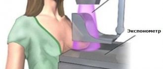
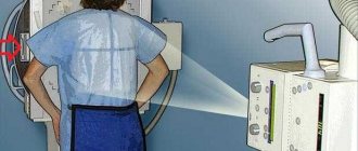
Modern technologies in medicine make it possible to diagnose various diseases even in the initial stages. The most popular methods nowadays include radiography and fluorography. It is believed that x-rays provide more accurate results. But fluorography is more accessible. These forms of diagnostics are actively used in various medical fields. However, there are cases when both studies are needed to determine an accurate diagnosis, clarify it, or if a specialist doubts the correctness of a previously made one.
Information that radiation is harmful to health is everywhere, everyone has heard about it. Therefore, doubts about the advisability of conducting two surveys alternately are clear. Patients want to know how safe it is to take an X-ray after fluorography, and what consequences are possible.
When should a chest x-ray be taken?
Plain radiography of the chest organs is carried out:
- During an annual preventive health examination to identify risk factors and life-threatening diseases such as cancer or tuberculosis.
- For pain in the chest and heart area.
- With painful or bubbling breathing, sensations of lack of air.
- With loss of appetite and a sharp decrease in body weight.
- For swelling and high blood pressure.
- For injuries, to identify foreign objects in the esophagus, lungs, and respiratory tract.
- If you suspect lung diseases: pneumonia, abscess, pleurisy, silicosis, etc.
What pathologies are detected by x-ray?
X-rays are quite widely used in medicine for diagnosing various diseases, pathologies and anomalies of almost all internal parts of the body and organs. The digestive system is examined to analyze diverticula, ulcers, gastritis, tumors and intestinal obstruction.
A chest x-ray is done to diagnose tumors and infectious diseases. This procedure is often prescribed as part of the study of the internal organs of the abdominal region and genitourinary area, various glands, and teeth. In addition, it remains one of the basic examination technologies in the process of diagnosing ailments of the skeletal and joint system of the body. Now we will find out when it is not advisable to use this method of analysis.
When is it necessary to do a plain radiography of the spine?
X-ray of the spine: coccyx, cervical, thoracic, lumbosacral regions is used:
- For pain, impaired mobility, and a feeling of stiffness in the back.
- For inflammatory diseases such as arthritis or arthrosis.
- For curvature of the spine: scoliosis, kyphosis, lordosis.
- For back injuries, vertebral displacements.
- For osteochondrosis and osteoporosis.
- Suspected tuberculosis, metastatic lesions and spinal tumors.
What do fluorographic images reveal?
This technique can be carried out as part of a routine examination. However, in case of some symptoms, the procedure is prescribed by a doctor. It is often given to those patients who complain of fever along with sweating, fatigue, weakness and a severe chest cough. The examination can detect tuberculosis or bronchitis, and, in addition, cancer and ailments in the bones of the chest.
How many times can fluorography be done? It is recommended no more than once a year.
When can an x-ray be prescribed?
A traumatologist or surgeon may prescribe digital radiography of bones and joints (skull, bones of the shoulder, forearm, foot, hand, femur, hip bone, etc.; temporomandibular, elbow, knee, wrist, hip joints, etc. ):
- If the patient experiences pain, crunching, movement blocking, or joint instability.
- In case of injury or suspected injury: dislocation, fracture, sprain.
- In order to make or exclude a diagnosis: longitudinal flatfoot.
- For the diagnosis of growths and calcified areas, metastatic lesions and tumors.
What is radiography?
This is the name of the method of studying the internal structure of a certain area of the human body using X-rays with further recording of images on photographic film or on photographic paper, and, in addition, in the memory of a digital medium. The technique does not involve receiving a significant dose of radiation to the body, it is relatively inexpensive and has great accuracy.
The fact is that its resolution reaches 0.5 mm or more, and the indicator increases along with a decrease in the area under study. Therefore, after fluorography, specialists sometimes send patients to undergo x-rays to obtain informative images.
How to prepare for an x-ray examination?
Before examining the lumbosacral spine, pelvic bones and hip joint, a cleansing enema is performed in the evening before the procedure. A light breakfast is allowed in the morning.
Before examining the abdominal organs, preparation may be required (do not take any food or medications 6-8 hours before the procedure, do not drink or smoke, etc.). People suffering from increased gas formation are recommended to exclude brown bread, baked goods, dairy products, cabbage, apples, carrots, etc. from their diet 2-3 days before the examination.
People suffering from increased gas formation are recommended to exclude brown bread, baked goods, dairy products, cabbage, apples, carrots, etc. from their diet 2-3 days before the examination.
Before examining the lumbosacral spine, pelvic bones and hip joint, a cleansing enema is performed in the evening before the procedure. A light breakfast is allowed in the morning.
In all other cases, no preparation is required.
Contraindications for radiography
Considering that this method is directly related to the body receiving a certain dose of ionizing radiation, people often wonder whether such a diagnosis can be done and what are the contraindications?
There are a number of absolute prohibitions that are worth remembering. X-rays are not done during pregnancy, especially in the early trimester. The fact is that during the period of formation the fetus is especially vulnerable, and the effects of radiation can harm its proper development.
Also, fluorography with x-rays on the same day is not recommended for patients in serious condition. A weakened body may simply not be able to withstand the load. They also try not to prescribe this in the presence of pneumothorax, scale pathologies, diabetes, pulmonary and pleural bleeding and some other diseases.
What device do you use for radiographic examination?
The Clinic on Komarova uses the latest generation universal X-ray complex - AGFA DX-D 300 (2016), the only one in the Far East. Modern digital technologies make it possible to obtain images of the highest quality with minimal radiation exposure, which have undeniable diagnostic effectiveness.
For comparison, when performing digital chest radiography AGFA DX-D 300, the radiation exposure is only 0.03 mSv (millisievert) per procedure, which is 17 times lower than with conventional fluorography (0.5 mSv), 10 times lower than with conventional radiography (0.3 mSv) and 2 times lower than with digital fluorography of the chest organs (0.05 mSv).
One of the unique features of the AGFA DX-D 300 is an individual approach to research. The German company has thought through everything to improve the level of patient comfort. The maximum flexibility and motorization of the AGFA DX-D 300 allow the equipment to independently take a position in space relative to the patient during the examination. This unique feature makes it easy to carry out a wide variety of examinations on any population, be it children, the elderly or patients with limited mobility.
Is it possible to do fluorography and x-rays on the same day?
The first question that interests many: “What is more harmful – x-rays or fluorography?”
Fluorographic examination has been widely used since 1936 for the diagnosis of tuberculosis and oncology. The method is based on obtaining an image on an X-ray fluorescence screen after passing a thin X-ray beam through the chest in a horizontal plane. The image is then transferred to film in a compressed scale. The reduced photo is the fluorogram itself. The radiation exposure on a film device reaches 1 mSv per procedure, depending on the technical characteristics of the device.
Today, radiation exposure during fluorography (FLG) is significantly reduced, since most clinics use digital devices. During one session, the patient receives a radiation load of 0.06 mSv. This is very little. According to the standards of the Ministry of Health, a single dose of radiation during a medical examination should not exceed 1.4 mSv.
Fluorography is exactly the test that everyone should undergo once a year. Thanks to it, doctors can make the following diagnoses:
- tuberculosis;
- cancer;
- pneumonia.
The image can reveal foci of fungal diseases and the presence of foreign bodies in the lungs.
X-ray differs from FLG in that it does not project the image onto a screen. Modern devices have a lower level of radiation exposure, and some of them have the ability to adjust the image quality. The radiation dose for radiography is 2-5 times lower than for fluorography.
Unlike FLG, x-rays are not done annually, but only when indicated. Using this method, you can identify pathologies such as:
- tumor;
- pneumonia;
- pleurisy;
- heart disease;
- changes in lung tissue of different nature;
- traumatic injuries to the respiratory system.
An X-ray image is clearer and of higher quality than a fluorogram. In some cases, a specialist needs to further examine the lungs to make an accurate diagnosis.
Many patients are surprised and do not understand when a doctor prescribes x-rays and FLG. Both methods involve radiation and, of course, a natural question arises whether these two studies can be done on the same day. Experts say that this is possible, but only if the danger from unknown pathologies exceeds the harm from the radiation received.
Is it possible to do fluorography after an x-ray?
Fluorography after an X-ray examination of the lungs does not make sense, since the X-ray image has greater resolution, and therefore higher accuracy. X-rays are usually counted as an annual FLG, the need for which is eliminated.
If the patient has an x-ray of other organs, for example, the bone tissue of the skull or limbs, and the patient received a minor radiation dose (up to 1 mSv), then FLG should be done. This is necessary if the survey was not carried out this year. If the total radiation dose is close to the maximum permissible, it is advisable to wait several months.
Is it possible to do an x-ray after fluorography?
It happens that during annual fluorography, strange changes were noted in the lungs, among them - similar to inflammation or neoplasms. To better examine the lesions, the doctor may order an x-ray, as this will provide more accurate information. Changes ranging in size from 4-5 mm are visible on a fluorogram, and from 3 mm on an x-ray image.
When a suspicious lesion is detected on FLG, the specialist needs to determine its characteristics and nosological affiliation. An x-ray will help with this. The procedure will be carried out not only in direct projection - most likely, targeted photographs will be taken from various angles. Thanks to a complete X-ray, the doctor will be able to accurately diagnose and determine treatment tactics.
How much time should pass between procedures
After an X-ray with a high radiation dose, it is recommended to wait several months so that the body has time to recover, because radiation tends to accumulate in the medium term. This is especially noticeable when taking several images over a large study area (for example, with an X-ray of the spine). The body is also exposed to stress when contrast enhancement is used.
If possible, it is better not to rush into x-rays after FLG and vice versa.
The doctor may order several x-ray examinations on the same day or with a minimum interval between them. However, he must assess all possible risks.
As for FLG, it is recommended to undergo this examination at least once every two years, but it is better to carry out the diagnosis annually. There are also categories of citizens who are prescribed fluorography more often - once every six months - due to high risks of infection, type of activity or other circumstances. These include:
- prisoners;
- conscripts;
- HIV-infected;
- members of families where the child was born;
- medical workers.
Unnecessarily, you should not often do X-rays of the lungs and fluorography, especially with a short interval between examinations. However, if there are serious indications, you should listen to the recommendations of the doctor, who will tell you exactly how long after FLG you can take x-rays.
Computed tomography and its advantages
So, computed tomography is one of the types of diagnostics using x-rays. It differs from a conventional X-ray machine in that the emitter (X-ray tube) rotates around the patient's body . The image is transferred to the laboratory assistant’s computer in the form of layer-by-layer sections. Modern devices take a photo every 2-3 millimeters . Thus, the device scans the organ or areas of interest being examined.
This diagnosis has its advantages:
- The tomograph takes images layer by layer, which improves visualization . Those. organs, tissues and vessels do not overlap each other. And you can accurately determine the location of the pathology.
- The tomograph step is 2-3 mm , which makes it possible not to miss even the smallest pathology and to identify it in time.
- Computer image processing . With the help of computer technology, it is possible to adjust images, if necessary, record video or generate a three-dimensional image. Which is sometimes more obvious, especially for surgeons.
- Fast research time (about a minute).
- An absolutely painless diagnostic method.
- High quality and contrast of images . This makes it possible to detect diseases in the early stages.
What can be seen on fluorography?
The image obtained after undergoing a fluorographic examination helps the doctor make an accurate diagnosis. Therefore, good or bad predictions can be made only after a specialist has analyzed the fluorography result.
Based on a fluorographic image, it is possible to identify:
- pathologies caused by previous injuries to the ribs and thoracic vertebrae;
- Petrification. Depending on where they were found and what size they are, the doctor can determine the type of tuberculosis;
- adhesions in pleural tissue;
- heart disease;
- tuberculosis processes;
- cancer diseases;
- bronchitis.
If the image shows changes in lung tissue, then based on this, the patient can be diagnosed with focal tuberculosis or cancer . But in any case, confirmation is necessary in the form of laboratory test results.
If the image shows an expansion of the mediastinal shadow, then most likely this is due to the onset of tuberculosis of the lymph nodes. If, in addition to this abnormality, there is effusion, this may mean that the patient begins to develop pneumonia or a relapse of earlier bronchitis occurs.
The presence of fibrosis of lung tissue in the image suggests that the person smokes too much. In this case, experts strongly recommend that such patients urgently give up their bad habit, otherwise they will soon develop cancer.
It is very easy to understand from a fluorographic image whether a person smokes or not . Unfortunately, it is difficult to determine bronchitis from an image. But identifying severe pneumonia, which often occurs with similar consequences, is not difficult.
The presence of a dark spot in the photograph, which is presented as an increase in transparency, allows you to identify emphysema. In some cases, it is mistaken for an extensive lesion due to bronchitis. However, these diseases differ in their symptoms.
Subsequently, emphysema, if not treated, can provoke the development of pulmonary edema, which very often happens to smokers.
You can recognize cancer in an image by a small spot. Changes in tissue structure will indicate metastasis from other organs .
An image showing changes in the edges of the diaphragmatic muscle indicates the presence of a disease not related to breathing. Most often, this can be explained by the accumulation of exudate or air in the abdominal cavity, or an inflammatory process in the liver, causing an increase in its size, or perforation of the diaphragm.
Is it possible to take x-rays at a fever?
This question arises often, especially among women with small children. Elevated body temperature is not a contraindication for x-rays, either for a child or an adult. On the contrary, temperature may indicate the presence of a serious illness, for example: pneumonia. In this case, an X-ray examination is simply necessary, and the sooner, the more favorable the prognosis.
Thermometer
If the temperature reaches high levels, which affects the well-being of a child or adult, you can take antipyretic drugs. This action will not in any way affect the results of the x-ray during decoding.
Temperature increase


