Radiography is a common diagnostic procedure that is used as a screening for certain diseases, and is also indispensable for confirming and clarifying the diagnosis. The fact that x-rays do not have the best effect on the body became known several years after its introduction into medical practice. Since then, radiation machines have undergone significant changes, making X-rays less dangerous. Nevertheless, there are still risks of negative consequences.
This material will discuss the main questions regarding whether x-rays are harmful and what dangers are hidden behind them. Readers will learn how often X-rays can be taken without harm to health, and what can be done to reduce the likelihood of consequences.
What are the dangers of x-rays and their effect on the human body?
To understand why x-rays are dangerous, it is important to know the essence and nature of this type of radiation. This type of ray belongs to the category of X-ray radiation, and the wavelength of such radiation is in the range between gamma and ultraviolet rays. Like other types of waves, x-rays have a certain energy potential - ionizing properties. When passing through tissue, an X-ray leaves a kind of trace: the structure of atoms and molecules changes due to a change in their “charge”.
Important! Even in small concentrations, X-rays always affect the body, and its effects have a cumulative effect - the longer contact with ionizing radiation continues, the greater the harm of X-rays.
When one-time exposure to large doses of this type of rays occurs, a person develops acute symptoms of X-ray exposure - radiation sickness. Internal organs are damaged (primarily the central nervous system and the hematopoietic system), a semblance of burns appears on the body, and multiple organ internal bleeding begins. Death can occur within the first hours after receiving a lethal dose. Regularly receiving non-life-threatening doses leads to chronic diseases.
The negative effects of X-rays are not limited to the body of the person exposed to the rays. The most dangerous consequences for the body are considered to be genetic changes that can be inherited. This is due to the fact that the gonads and reproductive cells - sperm and eggs - are most exposed to harmful effects. The damage to their DNA structure fully demonstrates how harmful X-rays are for humanity as a whole.
Is radiography considered dangerous?
The body of all people is characterized by individual resistance to radiation. But despite this, there are generally accepted indicators that medical workers adhere to. Answering the question of how many times a year an x-ray can be done, some doctors are of the opinion that the frequency of this procedure depends on how much the patient’s condition requires it.
Sometimes frequent monitoring is necessary for timely detection of pathologies. This opinion is not always rational, since a greater number of chest diseases can be detected using the safest methods, which include:
- general blood analysis;
- Ultrasound diagnostics;
- listening.
This judgment is rational if there is a suspicion of lung cancer or pneumonia. X-rays load the human body. X-rays are especially dangerous if you live in conditions of increased environmental pollution, which is acceptable in any large industrial city. Of course, if possible, it is better to avoid frequent examinations, but there are times when there is an urgent need for x-rays.
In addition, when answering the question of how harmful x-rays are, most doctors claim that serious radiation exposure is only possible when using an old device. Today there is a big difference between the X-ray equipment of the last century. A modern device significantly reduces the dosage of radiation that has a negative effect on the patient.
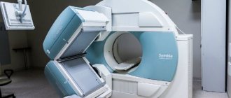
In addition, there is a non-destructive X-ray, in which the examination is carried out on a selected area. Patients undergoing CT and MRI are exposed to radiation, which is directed to a separate area.
How much radiation does a person receive during research?
Having realized how harmful x-rays are to humans, doctors had the opportunity to calculate what a safe dose of radiation should be. In medical practice, this concept is known as recommended radiation exposure.
In modern devices, the radiation dose from X-rays does not harm health, since its indicators are hundreds of times lower than the lethal dose, which is 1 Sv. It is this dose of radiation for a person that is fraught with the development of radiation sickness. It poses a danger in terms of long-term consequences and leads to various diseases of internal organs and systems. As for the concept of a lethal dose of radiation for humans, it implies a higher dose load:
- over 4 Sv - leads to death 1-2 months after irradiation due to damage to the bone marrow and dysfunction of the hematopoietic system;
- over 10 Sv - leads to death 1-2 weeks after exposure due to large-scale hemorrhages in the internal organs;
- over 100 Sv - causes enormous harm, causing death several hours (maximum 48 hours) after irradiation due to cessation of the functioning of the central nervous system.
Experts note that even modern x-rays are harmful if x-rays are taken too often. In this case, the ability of radiation to accumulate after the next procedure is affected.
Calculation of permissible radiation dose
According to WHO recommendations, the average annual X-ray dose for an adult should not exceed 0.5 Sv or 500 mSv per year. This level of radiation exposure is two times lower than that which provokes radiation sickness. However, in most cases, doctors ensure that the permissible dose received through X-rays per year is 10 times lower, that is, 50 mSv per year. This is due to the fact that a person is daily affected by background radiation even without medical procedures: solar, emanating from devices, etc. It does not cause direct harm to health, but also tends to accumulate.
Important! The permissible dose for children is 2-3 times lower than for adults, since it causes more harm to the growing body.
In order to correctly calculate the permissible number of rays for an individual patient, the background in the place of his permanent residence, other environmental factors and lifestyle are taken into account. For example, for people who frequently fly on airplanes, the rate of exposure during X-ray examinations can be reduced, since in the upper layers of the atmosphere there is stronger radiation than at the surface of the earth.
To determine how often a particular test can be done, the permissible annual dose of 50 mSv is written down throughout the year in the medical record. If at the beginning of the term it was necessary to frequently make diagnostics and the limit was exhausted, the adult will not be given an x-ray until the end of the billing period.
Received radiation doses for different types of x-rays
In modern facilities, radiation doses to patients are not much higher than background radiation. This made x-rays safer for repeated use. Even when taking a series of repeated images, the total X-ray exposure does not exceed 50% of the recommended annual load and is not harmful, but the final figures depend on the type of study.
Different procedures are characterized by different radiation exposure to the human body:
- analog fluorography (an outdated option for diagnosing lung diseases) - up to 0.2 mSv;
- digital fluorography - up to 0.06 mSv (in the latest generation devices up to 0.002 mSv);
- X-ray of the neck and cervical spine - up to 0.1 mSv;
- head examination - up to 0.4 mSv;
- image of the abdominal organs - up to 0.4 mSv;
- detailed radiography (includes X-rays of different parts of the body and joints) - up to 0.03 mSv;
- intraoral (dental) radiography - up to 0.1 mSv.
The greatest radiation exposure to the human body occurs during fluoroscopy of internal organs. Despite the insignificant radiation power indicators, they reach impressive figures due to the long duration of the procedure. On average, up to 3.5 mSv of radiation is transmitted to an adult in one session. Computed tomography has even greater indicators, in which the patient receives a dose of up to 11 mSv. Although such amounts of radiation are not harmful, such tests are not often done.
Acceptable frequency of conduct by type of research
The doctor determines how many times a year or month X-rays can be taken, depending on the individual characteristics of the body. Usually one photo is taken per day. It is not recommended to undergo x-rays twice in a row. If necessary, you may need to take the photo again at intervals of several days.
The recommended break for repeat X-rays is 3 weeks. Fluorography is recommended for people over 15 years of age once a year. This examination is not dangerous to health.
Using old equipment, you can do x-rays of organs without harm to your health:
- Zubov. Five times a year if the photo is taken from the side. Once a year if the rays pass through the brain or spine.
- Nose. For sinusitis, an x-ray is taken in the same way as an image of the brain, no more than once a year.
- Skulls. X-rays of the skull can be done no more than once a year.
- Spine. With caution, it is recommended to take an X-ray of the spine no more than once a year.
When using digital equipment, radiation exposure is reduced by several tens of times. Therefore, X-rays can be taken more often.
How X-rays work is discussed on the Jit zdorovo channel.
How to calculate the permissible radiation dose
When irradiated by an X-ray machine, the total radiation is measured in Roentgens, and the dose received by the patient is calculated in Sieverts. Microsievert (mSv) shows how much radiation the patient has received and how organs react to harmful radiation. Based on the total annual radiation exposure - up to 150 mSv / year, they calculate how often X-rays can be taken. If the patient has already exceeded the permissible radiation exposure limit, examinations must be limited or stopped.
Received radiation doses during procedures
Depending on the study, a person receives a different dose of radiation. For example, the radiation absorbed by the body during a chest x-ray is several times lower than during contrast fluoroscopy.
| Type of examination | Radiation exposure |
| X-ray of the sinuses | 0.6 mSv |
| Mammography | 0.7 mSv |
| X-ray of teeth | 0.35 mSv |
| CT scan of the spine | 6 mSv |
| Fluorography of the chest | 0.3 mSv |
X-ray during pregnancy
Before planning a pregnancy, it is recommended to refrain from x-rays. After the x-ray, you should refrain from planning a pregnancy for at least a month.
An adult can undergo several x-ray procedures per year without harm to health, while x-rays can cause serious harm to a baby in the womb. Therefore, doctors prescribe it during pregnancy in extreme cases. X-rays are especially dangerous in the first trimester. If there is an urgent need for it, laboratory technicians and doctors try to protect the fetus as much as possible.
To do this, x-rays are done using:
- protective plate;
- a lead apron covering the stomach;
- special screen.
X-rays may be prescribed for pregnant women if:
- fractures;
- inflammatory processes;
- suspected organ diseases.
In the second trimester, the procedure is no longer so dangerous. After the examination, mothers are required to undergo an ultrasound to see the condition of the child.
X-ray in childhood
For children, x-rays are prescribed to diagnose bone integrity. A child’s extremities can be x-rayed no more than five times a year. They try not to prescribe X-rays of the brain and organs for children. However, modern equipment can reduce the harmful effects on the child’s body several times.
Doctors prescribe x-rays for children when:
- serious dental problems;
- injuries;
- frequent attacks of bronchial asthma.
It is also recommended to refrain from performing fluorography on a child under 15 years of age. If doctors insist, it is necessary to find a clinic with the most modern equipment.
Is digital x-ray harmful?
Unlike the outdated analog X-ray, digital has less radiation exposure and causes less harm, but allows you to obtain higher-quality images. Considering that the radiation dose on digital X-rays is several times lower, specialists have the opportunity to do studies more often.
Good to know! Even when taking a series of images or repeated studies using digital installations, the resulting radiation dose is 2-3 times lower, so their harm is minimal.
When using a digital camera, photographs can be taken a second time within 24 hours. This may be necessary when you receive a blurry picture or find indistinguishable details in it. However, even here, radiologists take into account the potential harm of radiation, and try not to do diagnostics often, especially for children.
Where can I get an X-ray of my child’s lungs?
Such studies are carried out by medical institutions that have on staff a qualified specialist with access to this type of work, an equipped office with equipment equipped with the necessary documentation, and, of course, permission for this type of activity.
Most often, such rooms are equipped in clinics, emergency rooms or specialized TB medical institutions. According to sanitary standards, X-ray equipment must be located in a separate building. It is not allowed to set up such an office in residential buildings, the first floor of which is given over to a medical facility.
Who is allowed to carry out this procedure? To have access to x-ray examination, a specialist must have completed education in the field of medicine at a level not lower than therapeutic, in addition to undergo special training. This work is classified as hazardous, therefore employees of X-ray rooms have the appropriate category and preferential length of service.
How many times a year can you take an x-ray without harming your health?
To calculate how often an x-ray can be taken without harm to the body, it is necessary to take into account a number of factors. The main attention is paid to the total values of exposure per unit time. Taking X-rays too often is harmful, especially if large areas of the body are exposed to the rays. In addition, when calculating the period between studies, experts take into account the index of susceptibility of various tissues to radiation. The most pronounced harm is observed with irradiation of the brain and endocrine glands, including the gonads, so it is not recommended to diagnose them more than once a year.
Fluorography and x-rays of the abdominal cavity can be done 2 times a year. The average time between such diagnostic procedures can be reduced to 45 days. This is necessary so that the organs have time to partially recover after exposure to radiation. X-rays of peripheral parts of the body (limbs and joints) can be done more often - up to 6 times a year. However, here too the potential harm to health should be taken into account. You can do no more than three such procedures per month.
How long after can I do it again?
In some cases, patients need repeat x-rays:
- to clarify the diagnosis after fluorography;
- to track dynamics during treatment;
- to monitor the effectiveness of therapy;
- to clarify pathologies when receiving a low-quality image.
Only a specialist can determine the frequency of x-rays. This takes into account the ratio of the radiation load created by the device with the area of radiation exposure and individual harm to tissues. For example, when diagnosing a fracture of the hand, the image can be repeated after two days, while fluoroscopy of the intestine can be performed at intervals of at least two months. X-rays that affect the endocrine glands (neck, hip joints in women, etc.) are allowed no more than twice a year.
Important! An exception is for cancer patients who need regular monitoring of tumor dynamics. They can undergo up to 4 procedures per month, regardless of the area of study.
What will happen if you do it often?
There are different situations in medicine: some patients have to take x-rays 2 times in a row to establish an accurate clinical picture. At the same time, patients often worry whether it is dangerous to do x-rays so often. Experts say that if there are unconditional indications and it is impossible to use other diagnostic methods, an x-ray taken 2 times a day does not cause significant harm to the human body.
In situations where photographs need to be taken frequently, clinic staff use minimal dosages and try to protect the patient’s body from radiation as much as possible. This to a certain extent reduces the risk of receiving the maximum permissible doses of radiation. If the total radiation exposure is close to the maximum permissible standards, the doctor may refuse to take an image. But this rule also has exceptions: if the patient’s life is in danger due to the lack of important data, X-rays will often be taken even if the total dose did not significantly exceed the recommended values.
The main harm, which determined the rule why x-rays should not be taken frequently, is the gradual change in the functions of internal organs and systems. If the patient receives doses of radiation regularly, there is a risk of changes in the blood picture: leukopenia, erythrocytopenia, thrombocytopenia. The main sign of their appearance is excessive fatigue, weakness, bleeding gums, severe bleeding even from small wounds. Such conditions require special therapy and radical abolition of x-rays.
Conclusion
In general, there is no such thing as “permissible radiation exposure.” X-rays are performed strictly for medical reasons, and as a preventive measure - only for adults. Of course, when it comes to a person’s life and the extreme need for this diagnostic procedure, it is carried out in the quantity required, including for children.
Today, modern radiologists and equipment are somewhat encouraging, because they provide a minimal dose of ions, compared, for example, with equipment of past decades. Also, this radiation exposure now becomes more accurate and allows you to study very specific selected areas.
Does X-ray affect potency in men?
Among the male population, the effect of x-rays on potency is of particular importance. The question of what harm the procedure does to the male body interests male patients much more than the potential harm of x-rays for other areas of health. Radiologists reassure us that the radiation in modern installations is not enough to radically worsen the functioning of the reproductive system. Moreover, during each procedure, a man’s intimate organs are protected with a special lead apron in order to 100% eliminate the possibility of irradiation of the gonads.
Good to know! The male part of the population can have X-rays done as many times a year as women.
The only situation where x-rays can harm potency is the consequences of acute radiation sickness, that is, more than 1 Sv in one session, which is completely excluded if you do regular x-rays. In this case, deterioration of erectile function will be a secondary symptom. It will arise over time due to dysfunction of the gonads and a general deterioration in health.
Indications for x-rays
Indications for radiography:
- fractures;
- diseases of the chest organs;
- foreign objects in the respiratory system and gastrointestinal tract;
- dislocation of the hip joint;
- skull injury;
- suspicion of the formation of cancerous tumors;
- diagnosis of dental condition.
In newborns
A newborn is never given an x-ray for prevention. A doctor may prescribe this diagnostic method to children when the diagnosis needs to be made quickly and accurately.
Indications for x-ray of a newborn:
- injuries sustained during and after birth;
- pneumonia;
- ingestion of foreign objects into body organs;
- musculoskeletal pathologies;
- intestinal obstruction;
- preparation for surgery, if you need to find out more information.
Grudnichkov
X-rays are taken for infants under one year of age for the same reasons as for newborns. Complications after childbirth may not appear until several months later. Since X-rays are not taken just in case, after a difficult birth mothers need to closely monitor the health and behavior of the baby. If there are complaints about the child’s health, the doctor will send him for examination. Birth injuries are better visible on x-rays and may not appear externally.
Children from one year old
Children over one year of age undergo x-rays for the following indications:
- body injuries caused by falls;
- if the child has swallowed a foreign object or there is a suspicion that he has;
- diseases of the chest organs.
A child two years old and older may undergo a preventative X-ray of the head after a fall to rule out traumatic brain injury.
New X-ray machine on the channel "GTRK Mari El".
How to reduce stress and precautions
To reduce the harm from x-rays, you can do examinations no more often than your doctor recommends. In this case, it is worth giving preference to medical institutions that have the latest generation devices installed. They allow you to take health-safe images more frequently than older analogue X-ray machines.
To reduce the harmfulness of x-rays, clinics use special precautions. Most often they are expressed in limiting the area of radiation exposure with the help of special reflective devices: hats, sleeves, aprons and diapers made of lead rubber. They cover parts of the body that do not require diagnosis.
In order for the radiography to take place safely, the patient should follow the specialist’s recommendations for behavior during the procedure. Even minor disturbances (careless movement, uneven breathing, etc.) often lead to cloudy images, so doctors have to do a repeat session, that is, additionally irradiate the patient.
To track the total radiation exposure for each person, a special X-ray passport has been created, in which you need to make notes about the timing of the procedures and the doses received. Often the patient does not have access to them, so if it is necessary to do x-ray diagnostics in private clinics, you can take an extract from such a card. This will help reduce the likelihood of harm to health due to overexposure.
Video
About the pros and cons of the procedure, as well as how often you can do an x-ray for a child on the “Doctor Komarovsky” channel.
Do you have any questions? Specialists and readers of the HROMOSOMA website will help you ask a question
Was this article helpful?
Thank you for your opinion!
The article was useful. Please share the information with your friends.
Yes (100.00%)
No
X
Please write what is wrong and leave recommendations on the article
Cancel reply
Rate the benefit of the article: Rate the author ( 1 vote(s), average: 5.00 out of 5)
Discuss the article:
Duration of procedures
The safety of radiation emitted by X-ray and fluorography machines is also guaranteed by the fact that its effect on the body lasts a very short period of time.
X-ray
The entire procedure for obtaining an image of the lungs takes several minutes - from removing clothes to putting them on. Moreover, the shooting process itself lasts less than a minute.
Procedure:
- Remove clothing (to the waist) and jewelry (from the head and neck).
- If your hair is long, put it in a high ponytail or bun.
- The doctor covers the patient's abdomen and groin with a special protective apron.
- A person occupies a certain place and position between the emitter and the receiver.
- At the command of the radiologist, breathing is held.
- Upon completion, the protection is removed, the patient dresses and waits for pictures and descriptions.
Thus, preparation for the image takes much more time than the irradiation itself. The procedure time may be increased by the need to take photographs in several projections. In this case, the doctor will enter the treatment room between shots, reconfigure the equipment and change the patient’s position.
Fluorography
This examination is carried out in exactly the same way as an X-ray of the lungs, with the only difference being that the photograph of the lungs is taken in only one projection. This significantly reduces the procedure time, unlike radiography.
Many people wonder how long fluorography lasts. The examination result is valid for one year. And if a person took a photo, for example, in March, and gets a job in December, then the result of the procedure will be valid for admission to the staff. An exception may be the food industry and working with children.
Is it possible to go at home?
The latest technology makes it possible to perform chest x-rays at home. For this purpose, portable X-ray machines are used to examine a patient who is unable to leave the hospital bed. In public medical institutions, such an examination is possible only for patients in the intensive care unit or intensive care units, strictly according to the doctor’s indications.
To conduct an examination at home at your own request, even with a referral from a doctor, you will have to use a paid procedure at a private clinic that provides chest x-ray services. Where to do or, more precisely, order such a service is obviously on the websites of diagnostic and treatment centers.
The images obtained during the procedure at home, as well as the conclusion of the radiologist who performed the chest x-ray at home, can and should be used by doctors of government institutions at the level of official diagnostic studies. A government doctor does not have the right to require a patient to undergo an additional X-ray examination at a district clinic if the R-image data is not expired and is clearly readable.
True, the validity period of a chest x-ray is not indicated, since there are no documents regulating the “expiration date” of an x-ray. An image taken last year can be considered “overdue” if it relates to the diagnosis of tuberculosis. And when it comes to fractures, the dynamics of events develop faster and images need to be taken much more often to determine the correct fusion of bones. That is, determining the need to update X-ray data is again in the hands of the doctor.
Indications and contraindications for radiography
In each individual case, the decision on the method of technical examination is made by the attending physician. In what cases is each of these methods used? For a survey (complete) or targeted (fragmented) examination of the lungs, preference has been given to radiography for many years. The same method is mainly used to obtain information about the general condition of the respiratory system, as well as for prevention purposes.
Diseases that require a lung x-ray if suspected:
- low-quality tumors;
- problems with the bronchi;
- pleurisy;
- tuberculosis;
- pneumonia and some others.
Frequent colds may be a reason for this type of examination.
In case of prolonged cough, chest pain, pulmonary wheezing, severe shortness of breath, the patient will first be sent to the X-ray room. In addition, the legislation of our country provides for mandatory lung prevention. For some categories of citizens, for example, military personnel, some medical workers, people who have had tuberculosis and people in contact with such patients, undergoing fluorography or x-rays is mandatory twice a year. IDPs, migrants, refugees, carriers of severe chronic diseases, employees of institutions for children should undergo an annual examination, and all other categories should do this at least once every two years.
If we draw a parallel between radiography and fluorography, it turns out that there is a qualitative difference between these procedures. Being a more accessible procedure, fluorography has a low degree of accuracy due to the blurring of the resulting image, which prevents the identification of many problems with the lungs, leaving behind many questions. In addition, during the flux test, the condition of the tissues of the lungs and heart remains unclear.
As for contraindications, they apply to children under 15 years of age and to women who are pregnant. Exceptions are possible only if, when confirming the diagnosis, there is a real threat to their life that exceeds the harmful effects of radiation. At the same time, fluorography is absolutely contraindicated for persons under 18 years of age.
To reduce your radiation dose, go to clinics with modern equipment.
special instructions
Not every person can have an x-ray.
When x-rays are contraindicated:
- bleeding;
- multiple fractures of the spine and ribs;
- open form of pneumothorax;
- serious condition of the patient;
- pregnancy and breastfeeding (there are restrictions).
If the patient has no contraindications, he has been prescribed an examination, and he is worried about his safety, you can give some advice on how to mitigate the effect of the radiation received during the shooting.
How to reduce the negative impact:
- Take vitamins and Omega-3 regularly;
- before and after x-rays, it is recommended to consume more fermented milk products;
- oatmeal, prunes, grain bread will help remove harmful substances faster;
- drink more green tea;
- red fruits and vegetables will help cleanse the body;
- add garlic to your diet.
During the examination itself, the radiologist covers vulnerable areas of the body with a special apron - the groin, abdomen, neck and others (depending on the area being photographed).
At what age can you do it?
A child's developing body is much more susceptible to negative radiation, which can lead to more severe consequences than in an adult. Without a justifiable risk, the doctor will not prescribe such a procedure to the child, trying to replace x-rays with safer alternatives.
An exception may be cases where the danger to life from the disease is higher than the potential harm that X-rays can cause - these are severe injuries or pathologies.
Upon reaching 15 years of age, restrictions are lifted, fluorography and radiography are carried out in the prescribed manner.
X-ray during menstruation
Whether it is worth undergoing this examination if your period has begun is an ambiguous question, and the answer to it depends on several nuances.
The examination is not prohibited if:
- It is aimed at the organs of the upper body - chest, arms, teeth, and so on. In this case, the pelvic area is covered with a protective apron and the reproductive organs do not receive any negative effects.
- An urgent photo is needed when finding out the diagnosis using this method will help save a life. In this case, the consequences of delay can be much more dangerous than radiation, which means you need to “close your eyes” to the phase of the menstrual cycle.
- The woman feels well and is confident that the X-ray machine will not affect her well-being in any way.
In all other cases, it is better to wait until the end of your period and get examined after a couple of days.
Which is better: radiography or CT?
For some reason, some patients have the opinion that they have the right to choose the diagnostic method that seems to them the most modern, the best in terms of reviews and all other parameters.
Yes, a person has the right to invest in more effective diagnostics if it is medically indicated and makes some sense. But asking the question about a chest x-ray or a CT scan - which is better, we are simply showing our ignorance in this matter.
There are situations when x-rays, which are less “radioactive” than computed tomography, are quite sufficient to make a diagnosis and determine the extent of organ damage. Why not be satisfied with this simple and relatively safe method?
When performing a chest x-ray, radiation occurs within a fraction of a second. CT involves repeated scanning of the areas under study in different planes (“slices”), which creates an additional radiation load on the body (up to 12 mSv). It is, of course, also not very dangerous, but not particularly useful either. Therefore, this method of clarifying the diagnosis is resorted to only when other methods, including x-rays, turn out to be uninformative. Most often, this is necessary when carrying out differential diagnosis of tuberculosis and bronchopulmonary neoplasms.
If you think a chest x-ray is harmful to your child, isn't it harmful to give him an even more powerful CT scan? It is simply incorrect to compare these diagnostic methods. Only a doctor can determine which of them will be appropriate in each particular case.
How to resist radiation?
If the total annual radiation dose does not exceed one megasievert, the probability of negative consequences approaches zero. If this condition is not met, they can be severe and even catastrophic - up to the development of tumor diseases. In any case, you just need to follow some simple rules:
- before and after the start of the procedure, you should reinsure the body with antioxidants;
- when radiography, whenever possible, prevent healthy organs from being irradiated, using lead aprons and collars;
- to increase immunity, it is recommended to carry out vitamin (A, C, E) diversion from time to time;
- Various fermented milk products such as sour cream, cottage cheese, matsoni, kefir, as well as grainy breads, prunes, and oatmeal cope well with radiation.
Products containing calcium are excellent at removing radiation.