Myths about the dangers of MRI
Nowadays people often talk about the negative impact of geomagnetic storms, the dangers of living near power lines, and the dangers of radiation from mobile phones. Therefore, many patients worry that the MRI procedure is harmful to their health, because the tomograph creates an electromagnetic field.
The uniqueness of MRI is that the device creates magnetic fields of very high frequency, which do not have a negative effect on the body - both now and in the distant future.
This is confirmed by numerous studies that scientists have conducted on animals, and clinical experience using MRI to examine people.
In addition, when performing tomography, X-ray radiation is completely excluded, which in large doses is harmful to health. This is the main advantage of MRI, allowing for safer diagnosis than using X-rays or computed tomography.
Radiofrequency radiation is absolutely harmless to health, so the procedure is prescribed regardless of age, including small children.
But no matter what they say about the harmlessness and effectiveness of this procedure, MRI should only be prescribed by the attending physician. This is a complex and expensive study, so the patient must have compelling reasons and indications for the examination.
You absolutely cannot “prescribe” yourself an MRI, since the procedure, although safe, has a number of contraindications.
Only a doctor determines the advisability of conducting research for a specific disease. You should not perceive MRI as a panacea and perform a scan for any reason.
Many diagnostic procedures in some cases are considered more effective and informative, especially if a comprehensive examination is used.
Contraindications and disadvantages of the method
Relative contraindications to the study are:
- Pregnancy in the first trimester. The risk of interruption due to fear of the examination is much more harmful than the procedure itself.
- Claustrophobia is the fear of closed spaces, because this is how diagnostics are done.
- Decompensated heart failure. Intensification of symptoms due to activation of the sympathetic-adrenal system, stress during the examination.
- Metal implants because the magnet will attract them to itself.
- Obesity of the 3rd degree.
The disadvantage of this method is the immobility of the patient for quite a long time - 30-45 minutes. If there are abnormalities in the neurological status, diagnosis is carried out using a sedative.
To avoid unwanted reactions, it is necessary to remember that the MRI procedure may require the administration of a contrast agent. Therefore, there is a risk of developing neurogenic fibrosis in the presence of pathological changes in the kidneys . Contrast can be harmful.
How is the examination carried out?
The MRI machine looks like a large cylinder into which the patient is placed. During the examination, the patient is placed on a pull-out couch and the limbs are secured with straps, this allows for a clearer image (the pictures turn out blurry when moving).
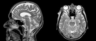
When examining the brain, a special device with wires is placed on the patient's head, which sends and receives radio waves.
After the patient is moved inside the capsule of the device, the diagnostic doctors go to another room, from where they observe the procedure, and a speakerphone is provided to check the patient’s condition.
The tomograph consistently produces a group of images of a layer-by-layer section of the examined tissues, with the help of which an accurate diagnosis can be made.
Usually the procedure takes no more than 30 minutes, with contrast – 50 minutes. The contrast agent is administered through a dropper during the examination, this allows you to obtain more accurate data.
The examination is painless, but to obtain high-quality images you have to remain completely still. The attending physician interprets the images.
Brain MRI algorithm
The MRI procedure takes place in several stages:
- Laying the patient on the table and fixing him
- Anesthesia, administration of a contrast agent (if necessary)
- Placing the subject in a machine to obtain a series of brain images
- Decoding the results, issuing examination findings to the patient
If a pathology of the nervous system is suspected, MRI diagnostics has an advantage as a modern, safe method of obtaining reliable information about the state of brain tissue, which is not harmful to the patient’s body. The examination results help to make an adequate diagnosis and prescribe comprehensive individual treatment.
What are the harms of MRI to health?
During the procedure, the patient’s body is exposed to radiofrequency radiation, which accompanies us every minute in everyday life. The high-frequency electromagnetic field is completely safe for our health, this is confirmed by many years of research by scientists.
Many people are interested in: is it dangerous to do an MRI of the brain and how can it affect mental activity?
In diagnostic practice, there is no evidence of harm caused to the patient’s health or deterioration in well-being after the examination.
If a doctor prescribes this test, it means that it is really necessary to undergo it in order to confirm the diagnosis or choose the optimal method of treatment.
Comparison of MRI, radiography and CT in relation to harm to the body
It is hardly possible to compare the procedure of magnetic resonance imaging and conventional radiographs. First of all, the final data from fluoroscopy are inferior to organ scanning in terms of information content. In some cases, computed tomography provides sufficient results; its cost is much lower than electromagnetic diagnostics, but the harm to the patient’s health is much higher.
Of course, exposure to ionizing radiation, which is used to obtain an X-ray, is strictly regulated, and radiation dose standards are calculated for different age categories and depend on the state of human health. Medical specialists who refer patients for CT or X-rays calculate the doses of radiation and determine how many times the diagnostic procedure can be performed so as not to harm the patient.
Under certain circumstances (for example, if the patient has loose ferromagnetic implants or endoprostheses), an MRI may be unsafe, and a CT scan will solve this problem.
Is MRI with contrast dangerous?
Contrast tomography is a more complex procedure, mainly used to diagnose tumors at an early stage. A special contrast agent is administered intravenously; the drug usually contains the metal gadolinium, which is safe for health.
In rare cases, the patient has an individual intolerance to the components of the contrast agent. Nausea, dizziness, mild itching of the skin, and decreased blood pressure may occur. These complications occur in only 2% of people.
An absolute contraindication for contrast MRI is renal failure. Patients with this diagnosis have difficulty removing the medication from the body.
For pregnant women and nursing mothers, contrast tomography is also contraindicated. Doctors fear that a strong magnetic field and the introduction of a chemical into the body may negatively affect the development of the fetus.
Principles of MRI of the brain
The use of magnetic resonance imaging is not harmful to the patient at all. It allows you to detect various types of tumors in the brain, diseases of the circulatory and nervous systems. In addition, MRI can be used to examine the working activity of the brain.
The study is performed using a contrast agent , which is not harmful to the patient, or without invasive interventions. When conducting a contrast examination, it is necessary to remember the patient’s possible allergic reactions, therefore this method is prescribed with extreme caution.
The procedure is performed from 18-20 levels of the skull and brain without causing any harm. The more intense the magnetic field and the more modern the device, the more accurate the images and the more reliable the results.
How often can an MRI be done without harm to health?
Since this high-tech technique does not pose any health hazard, MRI can be performed up to several times a year.
If there is a need to confirm the diagnosis, the doctor can almost immediately prescribe a repeat examination. But it is not advisable to carry out MRI too often; it is a complex and expensive procedure, which is prescribed after other examinations to finally clarify the diagnosis.
Carefully consider the need for contrast MRI
The bottom line is to avoid using MRI scans with contrast unless absolutely necessary. Often, doctors order these tests only to protect themselves from a legal point of view.
If this is your case, simply skip the contrast test. If necessary, consult with other doctors who can give you different advice.
This is especially important if you have a condition such as MS, which requires multiple MRI scans. Also remember that multiple MRIs with contrast will be especially dangerous if they are performed close in time.
Relative contraindications:
- heart failure;
- the presence of tattoos containing metals on the human body;
- pregnancy (first trimester);
- epilepsy;
- severe pain syndrome;
- involuntary movements (tremor);
- psychoneurological disorders.
People with claustrophobia are given sedatives before the procedure, and sometimes they have to resort to general anesthesia. Patients with large body weight are examined on an open tomograph.
If your doctor has prescribed an MRI for you, do not be alarmed. The key to a successful examination is the strict implementation of all the doctor’s recommendations. Before the procedure, it is important to notify the specialist about all health problems.
Prevention of complications during MRI
The person being studied needs:
- inform a specialist about problems with the urinary system;
- do not undergo examination if you have metal prostheses, pacemakers, or during pregnancy (especially in the first trimester);
- during the procedure, remove all jewelry;
- wear clothes without zippers, fasteners or other metal parts.
The effect of MRI on the body
There are scientific publications that indicate that the electromagnetic energy generated during the MRI procedure has a genotoxic effect. Some theories suggest a link between exposure to electromagnetic fields and early tumor formation, but there is no evidence to support this health risk. After all, even the mentioned genetic damage is reversible.
Static fields
Several studies have been conducted on volunteers exposed to MR. The purpose of the study was to evaluate the relationship between exposure to static magnetic fields and human health. We studied in detail changes in the activity of the central and peripheral nervous system, behavioral and cognitive functions, sensory perception, heart function, respiratory rate, and body temperature.
Some patients experienced dose- and time-dependent dizziness and nausea. No significant changes in several physiological parameters (heart rate, blood pressure, blood oxygenation, temperature, respiratory rate) were observed.
On the other hand, there have been reports in the scientific community of a statistically significant increase in the number of spontaneous abortions in women who underwent MRI examinations during pregnancy.
A 2006 World Health Organization document states that there is no evidence of short- or long-term adverse effects of static magnetic fields on human health.
Gradient magnetic fields
In 2000, patient safety in the effects of gradient fields associated with MR scanning was analyzed. Scientists have concluded that excessive stimulation of cardiac activity in modern systems is unlikely, but with sufficient amplitudes, the peripheral nervous system is excited, which can cause discomfort in the patient. Current safety standards, which were developed by the International Electrotechnical Commission, indicate that the lower threshold for cardiac pacing is significantly higher than that resulting from gradient fields. That is why the likelihood of ventricular fibrillation on MRI is extremely low.
Radio frequency fields
In 2000, a large-scale review summarized the physiological changes in visual, auditory, endocrine, nervous, cardiovascular, immune, and reproductive functions that were associated with radiofrequency exposure during MR procedures. It is believed that interactions between radiofrequency fields and biological tissue may be unsafe for patients. The majority of reported accidents are burns.
Don't miss: MRI of soft tissues of the chest
At the same time, it is believed that an MRI machine that generates a radiofrequency field is unlikely to be genotoxic, but to date there are no studies to assess the possible long-term consequences of exposure to MR fields on human health.
Find out more:
MRI of the cervical spine: preparation for the procedure, how it goes and how much it costs?
MRI of the hip joint: preparation for the procedure, how it goes and how much it costs?
How often and how many times a year can an MRI be done?
MRI of the joint and soft tissues of the shoulder
How is an MRI of the knee performed and how much does it cost? What does it show and how long does it last?
Difference between MRI and X-ray. Is it possible to do an MRI and X-ray on the same day?
MRI of the knee joint: indications, contraindications, features of the procedure
Ultrasound of the shoulder joint and its ligaments
Blood test for ACCP in rheumatoid arthritis
Choosing a clinic and device
Before the examination, you need to find out which device is best for doing an MRI of the spine. The magnetic field strength generated by the instrument should not be less than 0.3 Tesla to ensure optimal accuracy. Open type tomographs have such indicators.
Closed-type devices with a voltage of about 1–3 Tesla are more accurate. At the same time, the reliability of the images increases significantly. The quality also depends on the technical condition of the device. Its service life should not exceed 5 years. The best tomographs are made by the following companies: Siemens, General Electric, Philips, Toshiba.
The latest equipment is usually used in public medical institutions, since purchases are carried out using funds from the budget. Private clinics rarely have large sums of money and prefer used equipment. The main limiting factor is the cost of the procedure, because an image of one part of the spine in Russian hospitals costs about 8,000 rubles.
Read how to choose a bolster for your back: popular types of fillers, how to use a bolster correctly.
Find out how laser removal of a herniated disc is performed: preparation and rehabilitation after surgery.
Read what blood tests you need to take for osteoporosis and the rules for preparing for the test.
How many times can you do an MRI of the spine?
A spinal examination is prescribed for the following reasons:
- post-traumatic condition of the spinal column, mechanical curvature;
- hernias of various etiologies;
- malignant and benign neoplasms, cysts, tumors;
- osteochondrosis, osteoporosis and other diseases of the spine.
If, after an examination, the patient is diagnosed with an intervertebral hernia, then if the dynamics are static and there is no sharp deterioration, a repeat procedure is prescribed only once every 2-3 years.
An exceptional case is considered to be increased pain as a result of repeated injuries or excessive physical exertion. Sudden muscle apathy in the pelvic area also requires urgent examination.
An MRI of the spine will be needed if it is necessary to clarify the diagnosis and subsequent planning of treatment measures. The patient’s first step when sudden back pain occurs is to visit a specialist surgeon. He can refer the patient to a course of manual therapy, but first he will need to obtain visual confirmation of the problem. To do this you will have to undergo a tomography.