Why is fluorography of the lungs done and what is the procedure?
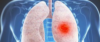
Fluorography is a type of x-ray examination in which photographs of the chest are taken in a special fluorescent screen. This is a fairly common diagnostic method, which is primarily aimed at screening (preventive) detection of tuberculosis and lung cancer.
Carrying out the procedure
What does chest fluorography show? Thanks to this study, it is possible to identify a number of diseases of the respiratory system. Radiologists analyze the images and issue a conclusion. These specialists study the fluorogram and, if pathological changes are present, make a presumptive diagnosis of a particular disease of the bronchopulmonary system.
The final verdict, as a rule, is made after additional radiography of the lungs.
Myths about the dangers of smoking
Myth 1 . A smoker's lungs simply take on a darker hue. They cannot have resin on them.
Disclosure : A person who smokes actually develops tar on their lungs as a result of the constant intake of soot. However, it is worth remembering that people who work in hazardous industries, drivers of diesel vehicles, and even those who heat the room with a stove are susceptible to such harmful influences. Of course, in these cases the harmful effects on the body are less noticeable.
Myth 2 . The relationship between smoking and lung cancer has not been officially proven.
Disclosure : Yes, this is true, but before you stop worrying about your own health, it's worth paying attention to the statistics. Thus, only 10% of those who contract cancer have nothing to do with this bad habit.
Myth 3 . People with a long history of smoking die from quitting.
Exposure : Unfortunately, the consequences of a harmful activity that a person has endured for many years (more than 30 years of experience) lead to death.
However, you shouldn’t “leave everything to chance”: quitting smoking now can give you several years of life.
Here lies additional motivation for young nicotine addicts: think about the consequences, because with prolonged smoking they will turn out to be much more serious than at first glance.
Types of fluorography
Today there are two main types of fluorography:
- Film , in which the image is only printed on the photograph;
- Digital is a more modern and convenient method. The research result can not only be printed, but also saved as a file on a computer or transmitted by e-mail. This method allows you to create an electronic database. In addition, when performing digital FLG of the lungs, the patient receives a lower dose of radiation exposure than during film FLG.
What diseases does it detect?
Fluorography of the lungs reflects characteristic changes inherent in the following diseases:
- Pulmonary tuberculosis;
- Malignant and benign tumors;
- COPD (chronic obstructive pulmonary disease), especially in the later stages;
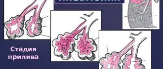
- Pneumonia;
- Lung abscess;
- Pleurisy.
Despite the fact that the main purpose of the study is to identify diseases of the respiratory system, other pathologies can be identified along with them in the image. Thus, an experienced radiologist can detect myocardial hypertrophy, indicating hypertension. Also, fluorography of the chest reveals some pathologies of the skeletal system, for example, fractures of the rib or collarbone in the past in case of improper fusion.
Important! It is not always easy to detect chronic bronchitis, which is characteristic of smokers, on fluorography, since in the early stages of development the disease practically does not change the structure of the lungs and bronchi. A typical fluorography picture for bronchitis appears already when the pathological process is quite advanced.
The appearance of shadows on a fluorogram
If additional shadows are found on a fluorographic image, this may not always indicate a person’s addiction to cigarettes. Some professions have specific working conditions that can lead to:
- diaphragm hernia;
- bronchitis;
- pulmonary tuberculosis;
- bronchial asthma and others.
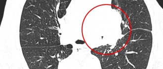
Round light spots on the image indicate the presence of inflammation. If the inflammatory process occurs constantly, the body becomes weaker, its protective functions deteriorate, which can cause the development of severe tuberculosis infection.
Can fluorography show that a person smokes?
Based on the above, we can conclude: the fluorography results of a smoker and a healthy person will have noticeable differences only if the former has a significant history of bad habit. If a person has recently smoked, then changes in the lungs may already begin, but for now they will not be so pronounced as to be reflected in the picture.
Snapshot
Features of the procedure
In order to answer your questions, you must first understand what fluorography is and what can be diagnosed with it.
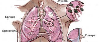
A fluorogram is a photograph of organs, which is obtained through the use of X-rays. Fabrics differ in their permeability to radiation. This makes it possible to see in the image organs of different densities and any changes that have occurred in their structure. A fluorogram helps show:
- foreign bodies and neoplasms;
- tissue sclerosis and fibrosis;
- abscesses;
- various pathologies;
- tuberculous cavities and others.
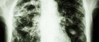
The lungs of a healthy person appear light gray in the photo. The organs have clear outlines, and the silhouette of the ribs is reflected against their background. Therefore, if smokers take a fluorographic photograph of the lungs, then in the absence of pathologies, the fluorography will not be able to show anything. But in an experienced smoker, you will notice changes that can lead to a conclusion about the presence of an addiction.
The difference between healthy lungs and smoker's lungs
Let's start with the fact that chronic bronchitis of a smoker is not an x-ray diagnosis. That is, it is impossible to identify this disease only on the basis of fluorography results of a smoker. In order to establish a diagnosis of COPD, an examination by a doctor and additional tests (in particular, spirometry) are required.
However, chronic obstructive bronchitis is distinguished by some radiological signs, which are detected as a result of fluorography of a smoker’s lungs. The first typical change is the strengthening of the bronchial pattern due to the formation of hilar and then diffuse fibrosis.
In the later stages of COPD, a complication such as emphysema often develops, which is characterized by the following symptoms:
- Lung fields have increased transparency;
- The intercostal spaces are widened;
- The lung pattern is depleted;
- The lungs of a smoker on fluorography are characterized by a relatively large volume, due to which the diaphragm, which usually protrudes into the chest cavity, flattens and its position becomes almost horizontal.
Effects of smoking on the lungs
How do the lungs of a smoker and an ordinary person differ at different stages of smoking? Let's look at this question:
With 1 year experience:
- The organ has already acquired a gray tint.
- According to recent studies, when consuming a standard pack of cigarettes, about a glass of tar accumulates in the human lungs.
- Immunity weakens: a person suffers from infectious diseases more often.
- A smoker gets tired much faster.
- At times you feel a slight tingling sensation in your chest.
What do lungs look like after 10 years of smoking?
- A colossal amount of harmful substances leads to spasms - the so-called smoker's bronchitis appears.
- Tobacco smoke has already had an impact on the organs of the digestive system - many people develop stomach ulcers.
- Not only the secondary pulmonary lobules are occupied, but also the functional tissue parts of the lungs.
- The skin looks much worse: a yellow tint and wrinkles appear.
- There are various tingling sensations and discomfort in the heart area.
15 years of experience:
- Now the smoky lungs are covered not only with tar, but also with green mucus.
- The brown color changed to black.
- The ability to conceive a child in women who smoke is reduced by 3-5 times compared to non-smokers.
- The risk of developing and developing a cancerous tumor is several times greater than that of a healthy person.
- 90% of tuberculosis patients are precisely this category of people.
Smokers with 20 years of experience or more:
- A noticeable deterioration in the blood circulation process, disruptions in the functioning of the heart.
- Pulmonary emphysema develops.
- Serious dental problems appear - deep caries, periodontal disease, tartar.
- The risk of diseases such as Buerger's disease, thrombosis, and stroke is extremely increased.
- The absorption of calcium deteriorates significantly, and as a result, diseases of the musculoskeletal system develop.
Is it possible to smoke before fluorography?
Many smokers wonder whether it is possible to smoke before fluorography and whether this will affect the result of the study. A smoke break before the procedure will not change the doctor’s conclusion, since the image reflects only those changes in the lungs that are already firmly formed.
Important! The fluorograph is not able to detect the remains of cigarette smoke in the bronchi, since it is not radiopaque. Therefore, a smoker can afford to smoke before the study.
Is it possible to cleanse the lungs before fluorography?
The process of cleansing the lungs of tar and other tobacco products is quite lengthy, it takes more than one month and requires a complete cessation of cigarettes. Therefore, you should not even try to clear your lungs in a short time before fluorography. It’s better to just undergo a study; a specialist will describe the fluorography image and give an objective assessment of the changes in the bronchi and lung tissue.
Smoker's lungs
How to check your lungs?
The initial examination can be carried out by a therapist. If necessary, he will refer the patient for an x-ray and consultation with a pulmonologist.
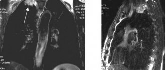
For the most complete diagnosis, it is recommended to undergo an R-study in 2 projections, or an in-depth study (MRI, CT, bronchoscopy).
You can also take a clinical and biochemical blood and sputum test.
- Many smokers (usually young people) are concerned about the question : is it possible to determine the fact of smoking using fluorography?
- Answer : no, fluorography cannot show changes in organs due to smoking, unless a tuberculosis or oncological process occurs. It will show the mesh structure of the organ. Using this study, you can trace white branches - the result of tissue inflammation.
Due to the fact that the internal organs increase in volume, the pictures show an abnormal expansion of the heart tissue.
An x-ray better demonstrates the pathologies of the bronchi and lungs - the image shows changes in the pulmonary pattern (vessels and interstitial tissue), which are especially noticeable in cancer, COPD, and emphysema.
What is the difference between fluorography and x-rays?
If fluorography is considered a preventive method for identifying pathologies of the respiratory system, then x-ray is a full-fledged diagnostic procedure. The benefits of x-rays are that they produce clearer images that help detect even small changes. In addition, X-rays can be performed with precision: in this case, the questionable area is photographed with the greatest magnification.
The lungs of a smoker on an x-ray have the same distinctive features as on a fluorogram. However, the first changes in developing bronchitis (increased hilar pulmonary pattern) can be detected at an earlier stage using this method.
Changes in the lungs on an X-ray during an advanced process can be very pronounced. But when COPD leads to such changes, the patient probably already knows about his problem. In this case, an x-ray of the smoker’s lungs only once again confirms the seriousness of this pathology.
Smoking before an x-ray, as well as before a fluorography, is not prohibited, since the very fact of a smoke break will in no way affect the result of the study.
Does fluorography show smoking and is it possible to identify a smoker in this way?
Doctors recommend undergoing a fluorographic examination once a year. This diagnostic procedure is included in the mandatory medical examination program. It helps to identify the initial signs of dangerous pathologies such as tuberculosis and cancer.
Does fluorography show smoking? This question is especially often asked by young smokers. They are afraid that, based on the results of the examination, their parents will guess about their bad habit.
What does a fluorographic image show? And is it possible to determine from it that the patient smokes? Let's try to figure it out.
The essence of the method
Does fluorography show cigarette smoking? To answer this question, it is necessary to understand the essence of this diagnostic method.
Fluorography is a type of x-ray examination. Nowadays, diagnostics are carried out using special digital devices, which allows the use of lower radiation doses.
However, the essence of the method remains the same. X-rays are passed through the patient's body and are unevenly absorbed by the lung tissue.
The image of the bronchi and lungs is displayed on a fluorescent screen and an image is taken.
Classic X-ray of the lungs and fluorography differ in the doses of X-rays required for the study. Conventional x-rays expose the patient to more intense radiation. For this reason, the image resolution is higher than with conventional fluorography.
Fluorography shows only gross and pronounced changes in the respiratory organs. This is a safer, but less reliable method. It is used during preventive examinations. As for classical x-rays, this examination is more often used to diagnose diseases. It shows a clear and accurate picture of pathological changes.
What does the photo show?
During fluorography, the condition of not only the lungs, but also other organs of the chest (bones, heart, blood vessels) is examined. In the picture you can see the following structural features of the tissues:
- structure changes;
- accumulation of gases and liquids;
- seals in organs.
Does fluorography show smoking? This study cannot establish the very fact that a person has a bad habit. It is impossible to determine from the image whether the patient smokes or not.
But, as you know, nicotine and tobacco tar have an extremely negative effect on the respiratory system.
If the patient has already developed pathologies of the lungs and bronchi due to smoking, then a fluorographic image will show these changes.
Is it possible to identify a smoker?
If a person actively and regularly smokes, sooner or later this affects the condition of the respiratory system. X-ray signs of a disease appear, which doctors call “smoker's chronic bronchitis.”
However, the cause of such bronchitis may not only be nicotine. The image can determine the presence of pathological changes, but it is impossible to establish their exact etiology. To suggest a possible cause of bronchitis, the doctor needs to take a medical history. The fact of smoking can only be established during a conversation with the patient.
Beginning tobacco lovers often ask the question: “Does fluorography show smoking if you’ve been smoking for a year?” At such an early stage, chronic bronchitis does not occur in all patients. However, much depends on the initial state of health and the number of cigarettes smoked. In some cases, heavy smoking can lead to bronchitis within 6-12 months.
Hookah and electronic cigarettes
Many people mistakenly believe that smoking hookah is practically harmless. However, hookah tobacco also contains toxic substances that irritate the respiratory system. Of course, the harm from regular cigarettes is much greater. However, it is impossible to talk about the complete safety of hookahs.
Does fluorography show hookah smoking? The study will only reveal the consequences of this habit. Heavy hookah tobacco smokers also develop chronic bronchitis over time. In addition, intense tightening leads to a decrease in lung volume.
Nowadays, many people use electronic cigarettes. You can even hear the opinion that with their help it is much easier to quit smoking. But is it really that harmless? The composition of liquid for refilling electronic cigarettes includes various flavors. These substances can trigger the development of bronchiolitis obliterans. This is inflammation and narrowing of the small bronchi (bronchioles).
Does fluorography show smoking of electronic cigarettes? If the patient has already developed bronchiolitis, then obstruction of the small bronchi will be visible on the image. In advanced cases, examination will show the presence of scar changes in the tissues. Over time, this pathology can lead to decreased respiratory function and oxygen deficiency in the body.
Signs of the consequences of smoking
As already mentioned, with prolonged and active smoking, changes occur in the respiratory organs. Let's take a closer look at the difference between a fluorographic image of an experienced smoker and a healthy person:
- Availability of seals. Normally, the image should not show dark areas in the lungs. Smoking reduces the elasticity of tissues in certain areas. These areas of compaction appear as shadows.
- Heart volume. In a healthy person, the size of this organ remains within normal limits. When smoking, respiratory function is severely impaired. This leads to expansion of the heart. The organ looks somewhat enlarged in the picture.
- Vascular drawing. In smokers, the network of blood vessels is more clearly expressed in the image than in healthy people. This is due to the fact that exposure to nicotine and combustion products leads to the formation of growths on the arteries and veins.
- Spots on the lungs. Tobacco tar clogs the pores of the lungs. Areas with reduced gas exchange are formed, which appear on the image as dark or light inclusions. Normally, no spots should be visible on the image.
- Pulmonary drawing. In smokers, the shadows from the blood vessels in the image are not clearly expressed. In this case, doctors talk about a weakening of the pulmonary pattern. This sign indicates the onset of fibrotic changes in tissues.
- Thickening of the walls of the bronchi. This is a consequence of constant irritation of the respiratory tract by tar and nicotine. This feature indicates the presence of chronic bronchitis.
However, even based on such signs, it is impossible to make an unambiguous conclusion that the patient smokes. After all, similar changes occur in people without bad habits. To establish the exact etiology of the abnormalities, doctors prescribe additional examinations.
Smoking before the procedure
Does fluorography show smoking if you smoke immediately before the procedure? This question often interests patients. The cigarette itself will not affect the results of the study in any way. The image will show only those changes in the lungs and bronchi that the smoker already has.
Many diagnostic procedures require quitting smoking some time before the examination. However, when preparing for fluorography, this rule is not mandatory.
Conclusion
It is impossible to give a definite answer to the question of whether fluorography shows smoking. This examination cannot determine the actual fact of tobacco addiction.
But it accurately diagnoses the negative consequences of nicotine use. Therefore, if pathological changes are detected in a smoker’s picture, then most likely they are caused by a bad habit.
This means that you need to reconsider your lifestyle and quit smoking immediately.
Source: https://FB.ru/article/464843/pokazyivaet-li-flyuorografiya-kurenie-i-mojno-li-vyiyavit-takim-obrazom-kurilschika
Errors in radiographic examination
Unfortunately, sometimes it turns out that fluorography does not show any pathological changes, but in fact they exist and the patient requires treatment. Why might this happen?
Firstly , as we have already noted, fluorography is a screening method, that is, it is not accurate enough to detect the slightest changes in the lungs.
Secondly , the equipment used, especially in small cities, is often outdated or incorrectly configured.
In this regard, the diagnosis of any bronchopulmonary disease is never made solely on the basis of an x-ray examination. The patient must be examined by a doctor, perform auscultation, prescribe the necessary tests and other studies, and only then, after analyzing the whole picture (including X-ray data), make the necessary diagnosis and prescribe treatment.
Indications for the examination and interpretation of the results obtained
The following are required to undergo fluorography:
- patients over 16 years of age once every 2 years;
- patients undergoing treatment in a medical institution in the absence of the necessary data on FLG;
- persons living with pregnant women and newborns;
- conscripts;
- persons applying for work or applicants;
- HIV infected.
Unscheduled FLG is carried out:
- if you suspect the development of pulmonary tuberculosis;
- in the presence of tumors in the lungs and heart;
- inflammatory processes in the lungs;
- pathologies of the heart and blood vessels.
The instructions are simple. No special preparation is required for the manipulation. In the office, the patient will be asked to remove any metal jewelry and clothing above the waist. After such preparation, the patient should lean tightly against the screen of the device with his chin on a special support. The picture (photo) is taken while inhaling while holding your breath.
The disadvantage of the method is the questionable quality of the image; if there is a suspicion of pathology, the doctor refers the patient to X-ray.
The table describes the symptom complexes that can be traced in the image after fluorography and x-ray:
| What changes can be detected with fluorography? | |
| Change | Description |
| Enhanced pulmonary pattern | It manifests itself in the presence of neoplasms, inflammation and sclerotic changes. |
| Expansion and compaction of the roots of the lungs | The natural picture changes in the presence of lung pathologies. |
| Fibrous changes | May appear after pneumonia and other lung lesions. |
| Focal shadows | May have different sizes. Manifested in tuberculosis and pneumonia. |
| Calcifications | They are formed from deposits of calcium salts at the site of infection. |
| Fluid accumulation | The picture allows you to assess the level. A similar symptom is characteristic of pleurisy. |
| Displacement of mediastinal organs | The result of the accumulation of fluid or air in the pleural cavity. |
| Changing the aperture position | After injuries, surgical interventions, illnesses. |
Such changes can occur not only in smokers, but patients with nicotine addiction experience them somewhat more often. This is due to the harmful effects of the composition of cigarettes on the human respiratory system.