- home
- X-ray
- X-ray of internal organs
22.07.2019
Oncological diseases are insidious due to their hidden development: according to statistics, more than a third of cancer tumors are discovered by chance in the later stages. In the chest organs, malignant neoplasms are most often discovered by chance, since this area of the body is regularly examined using x-rays. Screening allows for timely detection of cancer, even if it is asymptomatic.
Most patients undergoing regular preventive examinations believe that lung cancer is not visible clearly enough on X-rays. According to radiologists, the use of modern digital equipment makes it possible to see small tumors in the image and differentiate them from tuberculosis and other pathologies. This article will tell you in detail what a cancerous tumor and metastases can look like.
How to evaluate x-rays for lung cancer
Compaction syndrome.
Infiltrate. Cancer. Lung cancer gives obvious radiological signs that are easy to compare with pathology. High-quality x-rays and increased attention from the doctor help diagnose a formation larger than 5 mm in the image.
Unfortunately, at the very early stage, when the tumor is just forming, it is still indistinguishable on X-rays.
If the doctor suspects cancer even without a visible nodular neoplasm, he may send the patient for further examination. Using computed tomography, a malignant tumor measuring 2 mm in diameter can be diagnosed.
When receiving an X-ray with suspected lung cancer, doctors pay special attention to the following parameters:
- the presence of a peripheral shadow with a vague, bumpy contour - such signs can be caused by adenocarcinoma or squamous cell carcinoma;
- if dark grooves are found along the darkened contour, then this is a sign of growth of a carcinomatous node into the bronchus;
- “rising sun syndrome” is a typical manifestation of central lung cancer in the image, as evidenced by additional intense shading;
- the rise of the dome of the right lung indicates the presence of cicatricial adhesions on the pleura;
- if against the background of intense shadows there are clearing cavities, this means that the tumor has entered the stage of decay;
- the radiant contour around the tumor has smooth outlines (with rough and uneven shadows, tuberculosis is more likely to be suspected);
- with a pronounced pathological path to the right root of the lung, lymphangitis is suspected.
When examining an X-ray image of lung cancer, you need to take into account that there can be both metastasis and growth of the primary tumor into neighboring locations. The tumor most quickly grows into soft tissue, but there are cases of damage to the ribs and collarbone.
Clinical picture
Today, a large percentage of residents of megacities are at risk of developing lung cancer. This is due to the environmental situation. Severe gas pollution and air pollution lead to various pulmonary diseases and a decrease in local immune defense. Weakened tissues become a favorable environment for the appearance of cancer cells. The risk group also includes workers in heavy industries associated with intense emissions, as well as active smokers.
The onset of the disease can be detected in time by its clinical signs. A tumor in the lungs is manifested by the following symptoms:
- a prolonged incurable cough that gets worse over time;
- the appearance of shortness of breath, even with minimal exertion;
- the appearance of wheezing;
- the occurrence of painful attacks of prolonged coughing;
- moist cough with admixtures of blood clots in the bronchial secretions;
- unreasonable increase in body temperature;
- decreased or complete loss of appetite;
- chronic fatigue syndrome.
X-ray diagnostics makes it possible to detect malignant nodular neoplasms of lung cancer at an early stage. This greatly increases hope for the effectiveness of the treatment course and the restoration of the vital forces of the patient’s body.
How long does it take for a tumor to develop in lung tissue?
The rate of development of the pulmonary oncological process in each specific case is difficult to predict. The growth of cancer cells is influenced by various factors, the main one of which is a decrease in immune defense. Doctors roughly distinguish three stages of growth of a malignant tumor:
- biological (first stage) – the stage continues from the very beginning of the appearance of the neoplasm until it is identified on a chest x-ray; it is difficult to diagnose;
- preclinical (second stage) – signs of the oncological process are visible on x-ray, but there are no accompanying symptoms, difficult to diagnose due to the absence of complaints from the patient;
- clinical (third stage) - the X-ray results are accompanied by clear signs of the oncological process; most often, cancer is diagnosed in the third period, because the symptoms force the patient to undergo an examination to identify the cause of the ailment.
It is impossible to predict the formation of the tumor body precisely in time. The aggressive behavior and histology of cancer cells determine the rate of development of malignancy. Very often, the first two stages of the disease last for several years, without causing any suspicion in a person.
For oncological diagnostics, various forms of research are used in practice: X-rays, CT (computed tomography), endoscopy, puncture techniques and laboratory tests.
Along with X-ray diagnosis of a tumor in the lungs, laboratory tests are used when symptoms associated with oncology appear. They include a whole range of analyzes of the patient’s biomaterial: blood, urine, bronchial secretions. Every year, clinical results play an increasingly important role in the early diagnosis of cancer.
Detection of lung cancer on an image
It is not so easy to detect the presence of oncology with fluoroscopy, because a tumor half a centimeter in diameter is visualized, which is not obscured by the shadow of other pathological processes, for example, inflammatory. Natural shadows, for example, from the heart or from the sternum, can also cover the tumor.
Therefore, in patients suspected of having a malignant neoplasm, an x-ray examination is performed in two projections.
During the first projection (direct), the examination occurs as usual, but during the lateral projection, the patient will be asked to turn sideways and press against the screen. Such two-stage control will help to detect even those tumors that are hidden and cannot be detected with a regular x-ray.
To detect a tumor, competent differential diagnosis is necessary. This requires, first of all, good knowledge and skills from the radiologist.
Indeed, with peripheral cancer, shadows do not reveal the presence of a malignant process in any way, so they are easily confused with calcifications, deposits on the pleura or calcification of bone tissue.
If the doctor makes such a diagnosis, he will prescribe a dynamic x-ray and after a while the patient comes back to take a second x-ray, and there a large tumor is already detected.
But if you take a picture in a lateral projection at an early stage, you can detect a pathological formation in the mediastinum, which cannot be missed - the tumor is clearly visualized in the picture.
What is radiography?
Radiography is an image of a specific part of the body made using ionizing radiation directed from a machine.
Fluoroscopy is an X-ray examination lasting several minutes, when the doctor, through a special screen, visually monitors what is happening inside the patient’s body, occasionally photographing what is happening.
Recording the internal state of an organ on film in real time - an x-ray or radiograph.
You can take an x-ray of any part of the body and get a true image; in fact, it is a black and white “photograph” of all layers of the anatomical area of interest.
The reliability of the image can be increased by adjusting the shutter speed and power of the X-ray tube. Tissues and organs filled with air will be dark in the image, bones will be light, that is, the greater the density of the tissue, the lighter it will be on the x-ray.
Radiography provides an image fixed at a point in time; several images in different positions of the patient will help to get an idea of the deviations of processes from the norm in dynamics, but the idea will be somewhat incomplete without the introduction of contrasting solutions into the vascular bed, hollow organ or ducts of the studied area.
Modern X-ray machines have not become completely harmless to the subject, but the dose of radiation received during the study is minimal and is offset by the importance of the diagnostic information obtained. Analogue devices are gradually becoming a thing of the past, giving way to digital ones, where everything is calculated by a computer program, the image is displayed on the monitor without the need to develop the film, and the pictures taken during the process are stored on electronic media.
Additional cancer diagnostic methods
If a doctor suspects a cancerous tumor in the lung on an X-ray, he will not limit himself to one study, since this technique also has errors, and visualization of a suspicious tumor requires a thorough examination. For additional diagnosis of pathology, you can use the following methods:
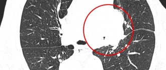
- Computed tomography is a basic examination for suspected cancer, since the technique obtains an image in layers. And with a minimum step of 2 mm set, doctors can see even the smallest nodes;
- bronchography - this technique helps to identify the connection between malignant neoplasms in the lungs and bronchial pathologies, for example, if the tumor has grown into the bronchi. As doctors point out, more than half of tumors can be diagnosed using bronchoscopy, even at a time when they are not yet visible on an x-ray.
- Tomosynthesis is an increasingly popular technique in the assessment of pathological changes in the chest organs.
Bronchography
All research methods are valuable and provide the doctor with important information for diagnosis.
What do lung metastases look like in the final stages?
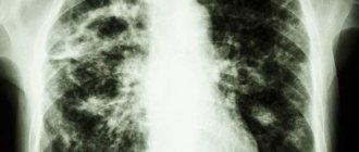
The visibility of metastases in the lungs on X-ray is quite good. Unlike primary tumors, such neoplasms are always multiple. In appearance, darkening is characterized as inhomogeneous structures on the surface or in the thickness of organs, with an unclear edge and irregular shape.
At the initial stage, metastases are defined as multiple nodules. As the metastatic process progresses, an X-ray of the lungs in cancer reveals widespread tissue damage, similar to a disseminated form of tuberculosis - spots can merge, forming extensive darkening. In the final stages, necrotic foci with disintegrating tissue and cavities filled with exudate become visible in the photographs.
In addition to damaged lung tissue, x-rays can reveal metastases in the bones of the chest. These may be secondary tumors of the ribs or metastases in the spine. The latter are detected less frequently due to the layering of images of the anterior chest, lungs and mediastinum. Metastases in bone tissue look like dotted shadows with a sparse bone structure.
CT scan for lung oncology
The best diagnostic data for cancer tumors is provided by lung tomography. This study also helps to detect accompanying signs that may, to one degree or another, illustrate the pathological process. According to the results of tomography, you can find:
- narrowing of the bronchi;
- complete obstruction of the bronchial lumen;
- problems filling the lungs with air;
- blurred contour of the bronchi due to damage by the tumor process;
- shadow of a tumor in the area of bifurcation of the trachea;
- increase in the angle between the bronchi;
- abnormal cavities;
- compression by bronchial metastases.
Lung cancer is not always visualized on x-rays, and if darkening is visible, the doctor still needs to differentiate it. That is why tomograms receive such important attention in diagnosing respiratory oncology.
Bronchogenic carcinoma
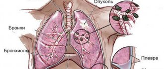
Lung cancer is best known among doctors under this name. Why bronchogenic? Yes, because this pathology arises from the mucous membrane of the bronchi. The walls of the bronchi are the most vulnerable part of the internal organs, because only they have direct contact with the environment and the toxic substances contained in it.
As a result, the tumor grows rapidly with the help of inhaled carcinogens. And it is growing by leaps and bounds.
Therefore, the question: “Does a fluorogram show lung cancer” can be answered in the affirmative – it does. This diagnostic method is convincing; it can show the disease, as well as determine what stage it is at.
Author of the article:
Bykov Evgeny Pavlovich |
Oncologist, surgeon Education: completed residency at the Russian Scientific Oncology Center named after. N. N. Blokhin" and received a diploma in the specialty "Oncologist" Our authors
X-ray for lung cancer: advantages and disadvantages of the procedure
The study has positive and negative aspects in terms of diagnosing oncology. The advantage is its accessibility, because X-ray machines are available in almost all clinics and hospitals.
The study can be carried out with high clarity using a contrast agent - this simplifies the correct diagnosis.
With a thorough examination of the patient using the X-ray method, it is possible to differentiate cancerous tumors from tuberculosis, knowing what lung cancer looks like in the picture and what tuberculosis looks like.
Among the negative features of X-ray diagnostics are the radiation exposure to which the patient is exposed during the examination.
The downside is the fact that tiny tumors are not visible on the image, and it is even more difficult to see the tumor on darkened X-rays. This contributes to delaying treatment and activating the growth of pathological neoplasms. For these reasons, doctors consider x-rays insufficient for oncology and prescribe additional screenings.
Types of radiography
The types of research are determined by the characteristics of the anatomical zone being studied and the process of taking the image.
Standard radiography is a survey, that is, a picture that gives an idea of the condition of the entire organ, or rather pictures in two mutually perpendicular projections - anterior and lateral.
In dentistry and for oral tumors, a version of the overview image is known as orthopantomography, but in radiography technology it is a panoramic image, during which the beam emanating from the apparatus passes along a wide arc, appearing on film in the form of the upper and lower jaw, and not an individual tooth.
The opposite of the survey view, targeted radiography allows you to “photograph” a specific area, for example, the upper mediastinum or the root of the lung altered by a tumor. This type is performed only after a survey x-ray.
Essentially the same targeted contact radiography is used in dentistry, when a film is placed in the mouth and only the diseased tooth is removed. In oncology, intraoral technique is used for cancer of the oral mucosa and oropharynx to assess the involvement of bone in the tumor conglomerate.
Similar to sighting is close-focus radiography, usually of small structures and at a close distance from the beam tube. In a small focus, the pathology is seen more clearly.
The introduction or intake of a contrast agent visualizes the cavitary organs of the gastrointestinal tract, urinary system and vascular network - contrast study of the intestine - irrigoscopy, bile ducts - cholecystography, urinary tract - urography, fistulas - fistulography.
X-ray according to Vogt is not used in oncology; a photograph of the eye without skull bones is necessary for injuries. For malignant eye processes, CT is used.
There is also rarely a need for x-rays with functional tests when the patient is filmed in a certain position, for example, with spinal tumors.
Soft tissue radiography is also not a widely used method, but may be useful in sarcomas.
How is a CT scan performed?
Before starting the study, the patient must provide the doctor with all the necessary data about his condition, as well as information about previous diagnostic methods.
Pregnant women are not recommended to have a CT scan due to possible risks to the baby. Tomography is performed only in emergency, life-threatening conditions.
Children under 5 years of age are usually given sedatives or anesthesia, which is directly performed by an anesthesiologist.
A computed tomography scan takes about 30 minutes, in rare cases – up to one hour.
You must remove all metal items and wear loose clothing.
Preparation for the examination depends on the area and the contrast agent used. In the latter case, it is necessary to refrain from eating food for 4 hours before the procedure, and, on the contrary, you need to drink as much as possible in order to quickly remove the contrast from the body.
If a CT scan is performed under anesthesia, it is prohibited to consume food and water for 4 hours.
The patient must lie still throughout the entire time to ensure reliable images.
The Yusupov Hospital provides patients with many services, including a complete diagnosis of cancer patients using a modern computed tomography scanner. The oncology clinic employs the best doctors who constantly confirm their qualifications and use European methods of diagnosis and treatment. The hospital has many awards and a huge number of grateful patients. Yusupov Hospital is one of the best medical institutions in the Russian Federation. The Yusupov Hospital operates around the clock, seven days a week, and therefore can receive you at any convenient time.
Author
Dmitry Yuryevich Frantsev
Doctor for X-ray endovascular diagnostics and treatment, Ph.D.
Classification
There are several principles for classifying this type of malignant tumor. According to histological structure, central lung cancer can be:
- Squamous cell (spindle cell).
- Small cell.
- Glandular (adenocarcinoma).
- Large cell.
- Glandular-squamous (dimorphic).
- Malignant tumor of the bronchial glands.
Based on the TNM classification, common to all malignant neoplasms, there are 4 stages of development of central lung cancer:
- Stage I corresponds to a tumor without spreading to neighboring organs or metastasizing.
- The malignant process of stage II is characterized by the presence of regional metastases or the transition of the tumor to the pleura, chest wall, or diaphragm.
- Stage III is assigned to cancer with metastasis to the lymph nodes (bifurcation, tracheobronchial, paratracheal).
- Stage IV neoplasm is characterized by the presence of distant metastases and extends beyond the lung.
According to the characteristics of clinical and radiological manifestations, its anatomical growth forms were determined:
- An endobronchial tumor characterized by exophytic growth and a polyp-like appearance. A clinical feature of this form of central cancer is early disturbances in ventilation of the affected area of the lung.
- Peribronchial form. The neoplasm in this case grows inside the bronchial wall. The formation of a narrowing of the lumen is possible due to its compression from the outside.
- Branched cancer also grows endophytically (towards the surrounding tissues) and takes the form of a bronchial tree.
Cases of detection of mixed forms of the disease are not uncommon. The formation of such neoplasms occurs due to the attachment of other components to the tumor during the development process.
Book a consultation 24 hours a day
+7+7+78