Since recently the number of exacerbations of bladder ailments (cystitis, sliding hernia, etc.) has been rapidly increasing, ultrasound examination is becoming a more popular diagnosis among the population. The medical procedure is based on the impact of sound waves that can penetrate the skin and scan the condition of internal organs.
When the “reflected” ultrasonic elements return to the receiver built into the sensor, a dark-light image appears on the computer monitor, from which key indicators are taken. Preparing for an ultrasound of the bladder does not involve any particular difficulties, but nevertheless, everyone who will subsequently undergo diagnostics should familiarize themselves with the basic rules of the preparatory stage for the procedure.
What abnormalities are detected using ultrasound?
The use of an echography apparatus allows professional sonologists to detect a certain range of ailments, among which the following can be identified:
- organ prolapse;
- atony;
- ureteral obstruction;
- sliding hernia;
- tumor formation;
- cystitis;
- increased tone;
- cystolithiasis;
- diverticulosis of the walls, etc.
If one of the diseases is identified, doctors issue a medical report to patients or their treating physicians, which serves as the basis for the treatment being developed.
What is being researched
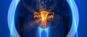
Using ultrasound of the internal organs of the small pelvis in women, the following structures of the genitourinary sphere are examined:
- Uterus - you can determine its position (anteflexio, retroflexio, etc.), shape, and take basic measurements. And also ultrasonography in M-mode allows you to evaluate the cavity and structure of the walls of the uterus, in particular, the endometrium (inner epithelial layer) and myometrium (muscular wall, in which benign tumors are often localized).
- Ovaries - the doctor evaluates their number, shape and location relative to the uterus, takes measurements of each ovary separately, their follicles and corpus luteum; folliculometry may be necessary.
- Uterine or fallopian tubes - allows you to determine their size, condition of the cavity and walls.
- The bladder is also located in the lower floor of the abdominal cavity and is visualized during ultrasonography. You can evaluate its shape, echostructure of the walls, size and calculate the volume of residual urine.
To determine the intensity of blood supply and differentiation of tumor formations, color Doppler mapping is used.
Drinking fluids before diagnosis
Since liquid is a good conductor of ultrasonic waves, the patient is recommended to fill the bladder 1–1.5 hours before the ultrasound, resorting to safe drinks: compote, weak tea, clean water, fruit drink or juice. Adults will need approximately 1–1.5 liters of fluid to keep their bladder sufficiently full; very young children (up to 3–4 years old) can get by with half the volume.
You should not drink coffee before an ultrasound scan, as its main components inhibit kidney function.
If a person does not want to drink immediately before the procedure, then he can refrain from going to the toilet for 4-6 hours after the last drink. It is important to remember that you are not allowed to drink alcohol for 1–3 days before the test.
Preparation
Preparatory measures require strict adherence to the doctor's instructions.
What can you eat on the eve of a bladder ultrasound and how much water should you drink before the procedure? What foods are undesirable to eat?
Diagnosticians regularly hear all these questions.
Video 1. Preparing for an ultrasound of the bladder.
Food
The examination procedure is not carried out on an empty stomach, so there is no complete ban on eating, but it is better to avoid dishes that cause flatulence.
Important! A few days before an ultrasound scan of the bladder, the patient needs to remove brown bread, milk, fresh fruits and legumes in any form, as well as raw vegetables, alcohol and soda from the diet.
Drink
Immediately before the examination (one and a half to two hours before the ultrasound), the right decision would be to drink plenty of fluids. More precisely, the patient needs to drink more than 2 liters of liquid without gas:
- water;
- tea;
- compote;
- juice
It is important not to go to the toilet for about 3-4 hours before the examination, because the key factor in high-quality visualization of the bladder is its fullness.
Reference! The standards for preparing for ultrasound are the same for all categories of patients, that is, men and women (including during pregnancy with minor nuances), as well as children, are subject to the same requirements for preparation for manipulation.
If you have a very strong, unbearable urge to urinate, you can partially empty your bladder. But then be sure to drink some fluid so that the loss is replenished.
If fluid intake is not possible
If, for a number of reasons, it is not possible to replenish the volume of fluid, then the physiological method can be used. It consists of holding urination for 5-6 hours before the ultrasound so that the procedure is performed on a full bladder .
Reference! If you urgently need to examine the bladder with ultrasound, and it is not possible to prepare for the procedure for several hours, then taking diuretics is recommended.
Diuretics have a pronounced diuretic effect and will help make ultrasound more effective.
If the procedure is scheduled for the morning?
There are also recommendations for those patients who have an appointment at the diagnostic doctor’s office in the early morning - they should not urinate after sleep; this can only be done after the procedure . If it is too difficult to endure, then it is recommended to set the alarm clock for 2.30-3.30 am and go to the toilet, but remember that in the morning, before the ultrasound, you can no longer urinate.
Why does the bladder need to be filled?
The bladder is a hollow organ, and in order for it to be clearly visible on the screen of an ultrasound machine, it needs to be filled, thereby forcing it to take its final shape.
Important! The fullness of the bladder is a chance to clearly see the organs located behind it (in women, this is the uterus and appendages, in men, the prostate gland).
Only if the organ is full will the doctor be able to examine in detail the shape, contours and dimensions of the organ, as well as effectively check the necessary parameters.
Preparation for the study, taking into account the type of procedure
At the moment, specialists use 4 main types of ultrasound, each of which is slightly different in the principle of preparation. If the patient has such an opportunity, he should discuss in advance with the doctor a method that is more suitable for his individual characteristics.
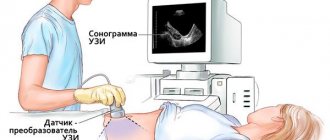
Abdominal method
This type of procedure is the most common type of ultrasound, which is performed superficially through the wall of the abdominal cavity. During the examination, the skin in the lower abdomen is lubricated with a transparent gel, which greatly facilitates the conduction of sound waves through a natural barrier. This is necessary to eliminate the so-called air cushion or layer between the sensor and the body. During an abdominal ultrasound, you do not need to prepare in any special way: all you need is a full bladder and a good psychological attitude.
Transrectal method
How is an ultrasound of the bladder performed in women?
It is prescribed, as a rule, to men to study the condition of the prostate gland. During ultrasound, an oblong sensor is inserted into the rectum, thereby reducing the distance between the organ being examined and the scanning machine - this key factor often allows the specialist to timely determine whether the patient has a malignant tumor.
It is extremely rare that a referral for TRUS is given to female representatives: the basis for this is the suspicion of any pathology in the rectum or pouch of Douglas. To make the procedure easier, it is recommended to cleanse the intestines the day before using a laxative or enema.
In case of chronic flatulence, the patient must adhere to a diet for 2-3 days, which involves the temporary exclusion of milk, raw vegetables and fruits, beans, peas, baked goods, smoked and fried foods. In addition, instant food (crackers and chips, including) also falls under sanctions.
Transvaginal method
This type of diagnosis is characterized by careful insertion of a transducer into the vagina with further scanning of organs of the genitourinary system such as the ovaries and uterus. Since a special condom is placed on the sensor before the study, if you are allergic to latex particles, you should inform either your doctor or sonologist about this.
There are several important aspects that must be studied in order to better prepare for the upcoming event:
- The procedure should not be performed during menstruation: days 5–9 of the cycle are considered a more favorable time for examination.
- The bladder must be empty during diagnosis; if this condition is not met, the doctor may direct the patient to the toilet to empty the organ.
- Immediately before the ultrasound, you should take a shower or bath.
Excess weight is one of the main indications for transvaginal ultrasound
Transurethral method
TUS involves inserting a thin sensor device into the urethra. The main goal of rare diagnostics is to detect direct connections between diseases of the bladder and the urethra. Since this type of ultrasound is performed with anesthesia, the patient must:
- stop drinking any alcoholic beverages one day, or preferably 2–3 days before the examination;
- do not smoke 2–4 hours before the procedure;
- Eat a fairly light breakfast or give it up altogether.
Under no circumstances should the study be carried out on those who have:
- severe disease of the cardiovascular system;
- allergy to the components of the anesthetic drug;
- inflammatory process in the urethral area.
The bladder, as in most cases, should be filled with liquid, preferably plain water.
Indications
Despite the safety of the method, ultrasound of the pelvic organs should be performed only when indicated:
- A woman has complaints of nagging pain in the lower and inferolateral parts of the abdomen.
- Inflammatory diseases of the internal organs of the reproductive system (salpingoophoritis, endocervicitis, endometritis, etc.).
- Suspicion of a cystic neoplasm of the ovary (isolated dermoid, edometrioid or functional cysts).
- Various types of menstrual disorders (algomenorrhea, opso- or polymenorrhea, dysfunctional uterine bleeding and amenorrhea).
- Acute and chronic inflammatory processes in the organs of the urinary system (in particular, if cystitis is suspected).
- Confirmation and determination of pregnancy duration as mandatory routine examinations. To exclude ectopic pregnancy.
- Female infertility of any origin.
- Suspicion of a malignant or benign tumor of the uterus, ovary or carcinomatosis of the pelvic peritoneum.
- Any complications resulting from childbirth or abortion.
- Recovery period after surgical interventions on the pelvic organs.
- As a control for the installation of an intrauterine device.
How to properly prepare a child for an ultrasound of the bladder?
There are no special differences in preparing children for the procedure. The only thing worth remembering is that children under 4–5 years old need to reduce the amount of liquid consumed to approximately 600–800 ml.
You should also take with you disposable wipes to remove medical gel from the surface of the skin and a special oilcloth that is placed under your back. If the patient has a number of questions related to the implementation of ultrasound, you need to come to an appointment with a specialist and discuss controversial or unclear issues with him.
How do they do it?
What algorithms exist for performing an ultrasound examination of the bladder?
Transrectal method
It happens that the doctor prescribes a transrectal examination of the bladder, that is, the sensor will be inserted into the patient’s rectum.
Reference! Transrectal scanning is used to examine virgins and representatives of the stronger sex.
Preparation for such a diagnosis has its own characteristics: in the evening, before the ultrasound, you need to visit the toilet, using a laxative (you can choose any form of release: from suppositories to tablets and microenemas). After waking up, a cleansing enema is recommended .
Transvaginal
Scanning the bladder through the vagina does not require patients to fill it first. The advantage of the study is the possibility of a comprehensive examination of the pelvic organs within one procedure.
Transurethral
With this scanning option, the doctor inserts a sensor into the urethra. This type of ultrasound is often performed with anesthesia and is used only for significant pathologies . The specifics of anesthesia and preparation for such a serious intervention should definitely be discussed with your doctor after prescribing an ultrasound.
Residual urine test
If an examination is prescribed, the purpose of which is to determine the volume of residual urine, then in this case it is necessary to fill the bladder . During the procedure, a standard examination is performed, then the patient goes to the toilet. And then the doctor uses a sensor to measure the volume of sediment, which is the residual amount of liquid.
When is the examination scheduled?
Indications for ultrasound of the bladder:
- suspicion of inflammation (frequent urination with pain);
- symptoms of stone formation (persistent dull pain in the lower abdomen when urinating);
- assessment of the condition of the bladder after surgery on any pelvic organ;
- confirmation / refutation of urinary oncology in patients with hematuria (presence of red blood cells in the urine).
The sensitivity of ultrasound (the ability to give the correct result) in diagnosing tumors is 87.1%. Specificity (absence of false positive results) - 98.1%.
Preparing for an abdominal ultrasound
During an ultrasound examination, internal organs must be open. They should not be obscured by gases and products that are in the intestines. To do this, the intestines need to be prepared.
First of all, within a few days of the planned study, exclude from daily consumption foods that cause bloating. These include beans, peas, brown bread, bananas, and sweets. Fresh vegetables and fruits, especially cabbage, can cause excessive gas formation.
It is also necessary to take espumizan for three days (as prescribed by the doctor). On the day of the study, you will need to increase the dosage to four capsules. They need to be taken once a day, and should not be washed down with water. Abdominal ultrasound is performed on an empty stomach. Before starting this procedure, you should not eat for 6 hours.
A patient suffering from diabetes is allowed a light breakfast.
What you can and shouldn't drink
It is best to drink water before the bladder ultrasound procedure. Also allowed:
- fruit drink,
- tea,
- juice.
Drinks prohibited by doctors are soda and milk, as they contribute to the formation of gases in the intestines, which complicates the examination.
Why is an ultrasound performed on a full bladder?
For diseases of the urinary system, studies are prescribed using modern diagnostic methods. Worsening urine test results, changes in the color of the excreted fluid, pain in the lower abdomen are reasons for undergoing an ultrasound of the bladder in women.
How to prepare for the procedure? What foods reduce gas formation in the intestines? Are there any contraindications? The answers are in the article.
Have you been fighting CYSTITIS for many years without success?
Head of the Institute: “You will be amazed at how easy it is to cure cystitis by taking it every day...
Read more "
- General information about the procedure
- Benefits of Ultrasound
- Indications for use
- Contraindications
- How is the procedure performed?
- What does a bladder examination show?
- How to prepare: rules and recommendations
- Decoding the results
General information about the procedure
Examination of internal organs using ultrasound waves is a highly informative non-invasive diagnostic method. The procedure has virtually no restrictions and does not require lengthy preparation.
The doctor moves the nozzle over the abdomen or performs the procedure on women transvaginally (through the vagina). A special scanner emits waves, reflecting from the walls of the bladder; ultrasound transmits the image using a transducer to an ultrasound machine, creating a certain picture on the monitor.
Based on the results of the study, the patient receives a black and white image, which shows all formations (hyper- and hypoechoic, anechoic) identified during the procedure. Doppler sonography is performed to assess blood flow and vascular condition.
The sonologist issues a transcript with a preliminary diagnosis, a description of the size, condition of the bladder, and other organs (during an ultrasound, it is possible to examine the ovaries, uterus, and appendages).
With the results obtained, the specialist refers you to a urologist or nephrologist to make a final diagnosis and prescribe treatment if there are deviations from the norm.
Find out how cystoscopy of the bladder is done in women and what the study shows.
The list and rules for the use of uroseptic drugs for pyelonephritis in adults and children can be found at this address.
OUR READERS RECOMMEND!
Our readers successfully use CystoBlock to treat cystitis. Seeing how popular this product is, we decided to bring it to your attention. Read more here...
Advantages of ultrasound
Preparing for a pelvic examination
This ultrasound can be performed on any day of the cycle. The exception is the days of menstruation. To clarify the diagnosis, a repeat ultrasound is performed on the day of the cycle, which is prescribed by a specialist. The location of the pelvic organs is the abdominal cavity. They are located deep, making them difficult to explore.
In order to examine the pelvis (uterus, ovaries, fallopian tubes), a special vaginal sensor is used. This research method is called transvaginal. If the girl is a virgin, then this method is not acceptable for her. Transvaginal ultrasound does not require preliminary preparation and is performed through the anterior abdominal wall. Before starting the study, you need to go to the toilet and empty your bladder.
An hour before the appointed time, virgins drink 3-4 glasses of non-carbonated liquid.
How to quickly fill the bladder for ultrasound - diet, signs, manifestations, herbs
How to quickly fill your bladder before an ultrasound
Menu
There are situations when people wonder how to quickly fill their bladder. Of course, this organ of the excretory system usually fills up on its own, and the speed of this process depends on the amount of fluid drunk and the functioning of the kidneys.
But sometimes you just need to fill your bladder as quickly as possible. For example, for performing an ultrasound of the bladder.
The essence of the research method
Ultrasound is a modern, minimally invasive procedure that is comfortable for the patient and at the same time very informative. The advantages of this method are the absence of age restrictions and the absolute absence of harm.
Thanks to this diagnostic procedure, it is possible to differentiate various diseases affecting a specific organ and monitor the effectiveness of the therapy.
Ultrasound of the bladder is always performed transabdominally (the sensor is placed by the doctor on the anterior abdominal wall).
conclusions
Three approaches are used for ultrasound examination of the bladder: through the abdominal wall (transabdominal), through the rectum (transrectal), through the urethra (transurethral). Each of these methods requires special patient preparation.
Proper patient preparation ensures a quality examination. Each method of examining the bladder has its own advantages and disadvantages.
For this reason, each method has its own indications for use and one method cannot be fully replaced by another.
Previous article - Ultrasound of the kidneys
Next article - What can be seen with an ultrasound of the bladder?
Indications
The bladder is examined in the case of:
- pathological processes in various parts of the urinary system;
- determination of the source of erythrocyturia;
- complaints characteristic of prostatic hyperplasia, cystitis, kidney stones and urolithiasis;
- establishing the cause of acute urinary retention and other dysuric symptoms;
- suspicion of the presence of benign or malignant neoplasms;
- suspicion of diverticulosis;
- traumatic lesions of the bladder;
- control of therapy of various types of therapy.
Indications for ultrasound of the bladder
Ultrasound examination is used to diagnose bladder diseases. Using this research method, the presence of various diseases is detected. Indications for ultrasound examination are:
- suspicion of cystitis, kidney stones or bladder stones;
- presence of blood in the urine;
- possible tumors in the urinary organs;
- sharp pain when urinating in pregnant women;
- urinary retention.
There may also be other problems with the organs of the urinary system that require specialist supervision.
Why is a full bladder necessary?
The bladder must be filled in order, firstly, to better visualize the walls of this organ, since it is in a straightened state and has its usual shape.
Secondly, in a full state, it pushes the intestinal loops apart and gets as close as possible to the anterior abdominal wall.
It may be necessary to quickly fill this organ if you forgot or did not know about this subtlety of the preparatory stage for the study.
How long does it take for water to enter the bladder?
Before we talk about how to drink and how to eat, let’s look at the maximum and minimum activity of organs.
How much water should you drink?
20 ml per 1 kg of weight in winter
25 ml per 1 kg of weight in spring and autumn
30-35 ml per 1 kg of weight in the summer.
How long does it take for 0.5 liters of drunk water to leave the stomach?
“How long does it take for food to be digested in the stomach?
average digestion time (this is mainly protein food)
long-term digestion food (this includes fatty foods and a combination of fatty and protein foods)
food takes too long to digest and is practically indigestible.
The first category includes: almost all fruits (with the exception of bananas, avocados and the like), vegetable and fruit juices (not mixed), berries, kefir.
Avoid or eat to a minimum those foods that belong to category 4.
It is not recommended to combine foods and foods that have different digestion times in the stomach.
Rope park
How and how much fluid to drink while hiking and climbing.
The water-salt balance is disturbed. And it’s not that there’s not enough water, but the salt content in the blood has increased,
the blood needs to be diluted. Drinking water enters the stomach through the esophagus and absorption into the blood begins. Once in the blood, water dilutes it to the desired concentration. We got drunk. oh soon we'll be thirsty again.
WHERE DOES THE WATER GO?
From the blood, water enters the skin and evaporates through the sweat glands. Sweat, like blood, contains water and salts, but still, mostly water is lost with sweat, and excess salt remains in the blood. Another consumer of water is the kidneys: they need to dissolve in something everything that the body has intended to be released. Then the water is sent to the bladder, and at the first opportunity to the grass.
WHEN THERE IS NOT ENOUGH WATER.
Source: https://xn—-7sbbabkwnixgwr4a5a.xn--p1ai/uzi/skolko-nuzhno-vypit-vody-pered-mochevogo.html
Why do you need to fill an organ for an ultrasound, and how to do it?
In most cases, the bladder is examined using ultrasound along with nearby organs. This is done in order to make an accurate diagnosis. For example, signs of inflammation of the fallopian tubes are similar to the symptoms of cystitis. It can also be confused with signs and symptoms of a number of sexually transmitted diseases.
A comprehensive examination of all pelvic organs will help the doctor draw specific conclusions. In children, ultrasound of the bladder and kidneys is usually performed simultaneously. The examination is absolutely safe for both children and adults.
Ultrasound of the bladder can be done in different ways, each of which requires specific preparation:
- when applying the sensor down the abdomen (transabdominal);
- when placing the sensor inside the vagina in women (transvaginal);
- when administered through the urethra in men (transurethral);
- when a sensor is inserted through the anus (TRUS, usually used in men).
add to favorites link thank
My bladder filled up the fastest when I was given a drip in the hospital, as far as I remember, about 1 liter of saline is poured into your vein within about 15 minutes. solution, and even if the bladder had been emptied before, I could barely wait for the procedure to end) I can’t imagine anything faster than 15 minutes)))
Also, when you swim in the pool for 1 hour, at the end of the swim you are already ready, since the skin, being in the water, absorbs it very actively)))
The bladder must be filled before an ultrasound of the pelvic organs, and filled by a certain time. I did the following: I started drinking mineral water two hours before the appointed time. In the amount of one and a half to two liters. For an hour and a half. Twenty minutes before the examination I drank furosemide tablet.
Also, when you swim in the pool for 1 hour, at the end of the swim you are already ready, since the skin, being in the water, absorbs it very actively)))
The kidneys produce urine, which then drains into the bladder. When you go to the bathroom, urine leaves the storage organ through an opening at the bottom and flows out through a tube called the urethra. In women, the opening of the urethra is located directly above the vagina, in men - at the tip of the head of the penis.
As the bladder fills, signals from nerves are sent to the brain, which eventually trigger the need to stool. When urinating, nerve signals coordinate the relaxation of the pelvic floor and urethral muscles (the latter are called the urinary sphincter muscles). The muscles of the organ tense (contract), pushing urine out.
Decoding the results
To make a preliminary diagnosis, the doctor only needs to study the ultrasound results. Important information for a specialist is the shape, size and other parameters of the bubble.
Norms
Normally, this organ of the urinary system has the following parameters:
- shape - pear-shaped when filled, saucer-shaped - after emptying;
- sizes and volume (one and the same) - the capacity of the female bladder reaches 250-550 ml, the male one - 350-750 ml;
- wall thickness is the same everywhere (2-4 mm when the bladder is full);
- filling speed - 50 ml/hour;
- residual urine - up to 50 ml.
What do changes in normal values indicate?
Uniform and uneven thickening of the walls of the bladder is observed in the following conditions:
- stones in the kidneys and urinary tract;
- papillomatosis - benign growths that provoke a precancerous condition;
- tumor neoplasm - malignant in 30-50% of cases;
- chronic cystitis is an inflammation of the walls, manifested as flakes in the urine sediment.
A pathological change in shape and a decrease in urea volume can be caused by functional and organic factors. In the first case, this is organ hyperactivity (frequent involuntary muscle contractions). In the second, there are a number of diseases, including tuberculosis of the urinary system, which provokes cicatricial wrinkling of the organ.
Research results
Deciphering an ultrasound of the bladder based on the results of the examination sometimes requires the additional involvement of a specialist in order to compare the ultrasound picture and the medical history.
What does the diagnosis show? Ultrasound examination of a hollow organ allows you to evaluate and measure the following indicators:
- volume and shape;
- filling speed;
- amount of residual urine;
- wall thickness;
- structure.
The bladder on ultrasound is defined as an anechoic cavity protruding from the pelvic cavity in a filled state. If the indicators are normal, the boundaries of the organ will be smooth, with symmetrical cross sections. The thickness of the walls varies depending on the level of filling (about 4 mm), but should be uniform and the same in all zones.
After urination, the organ is examined again for the presence of residual urine - normally its amount should not exceed 50 ml, so if it is present, the doctor records its volume. After this, the examination protocol includes examination of the ureters and kidneys.
Treatment for a shriveled bladder
Treatment of a wrinkled bladder must be timely, which avoids and prevents the development of serious and life-threatening complications for the patient. In addition, the treatment of a wrinkled bladder, which is carried out by specialists at our professional clinic, is always high-quality, reliable, effective and as safe as possible for each patient who comes to us.
Treatment of a shriveled bladder can be conservative or surgical. Conservative treatment involves the use of techniques and medications whose action is aimed at reducing the frequency of urination and increasing the storage function of the bladder. Surgical treatment methods are used when the isolated use of conservative therapy is expected to be ineffective (if there are certain reasons), or its long-term use has not led to positive results. Let us note that our doctors only proven, unique, effective and reliable methods surgical treatment of a wrinkled bladder
In any case, treatment of a wrinkled bladder in our highly specialized clinic is carried out only after consulting a doctor and carrying out a series of diagnostic measures to accurately determine the diagnosis and establish the scope and nature of treatment.