Is it possible to perform an ultrasound of the kidneys during pregnancy?
The longer the pregnancy, the higher the load on the kidneys. During this period, the organs of the urinary system are forced to work for two, that is, to remove waste products of both the mother and the growing fetus with urine. In addition, in the second half of pregnancy, the pressure of the enlarged uterus on the ureters and bladder increases, which can obstruct the free outflow of urine. The latter circumstance creates favorable conditions for the proliferation of pathogenic microflora and the development of inflammatory processes in the urinary tract. And if we add to this the inevitable weakening of the pregnant woman’s body, decreased immunity and sharp fluctuations in hormone levels during this period, then the risk of nephrological diseases in such women is extremely high.
In the case where the expectant mother suffered from a urological disease even before conception, during this difficult period of life it can worsen, seriously endangering her health and the life of the unborn child. Therefore, an ultrasound of the kidneys, if there is a reason for it, can and should be performed during pregnancy.
Kidney ultrasound can be prescribed at any stage of pregnancy
The question of the safety of ultrasound exposure on the body of a pregnant woman worries almost all patients who are prescribed this procedure. It should be truthfully noted that to date, the effect of ultrasound waves on the fetus has not been studied in detail. But regarding this, doctors say that the expectant mother need not worry: the study will not harm the unborn child at any time. As you know, all women routinely undergo ultrasound examinations of the pelvic organs and fetus three times during a normal pregnancy, and even more often if indicated. This study can be carried out during pregnancy as many times as the doctor deems necessary.
If even the slightest negative biological effect of ultrasound waves used in diagnostics on a child had been scientifically confirmed, then doctors would not have prescribed this procedure.
Measures to maintain kidney health in a pregnant woman
The paired filtering organ of the urinary system often fails during pregnancy. More than half of all possible problems can be avoided if you follow these tips:
- Drink more cranberry juice and reduce your intake of junk food.
- Drink at least two liters of water daily.
- Do not endure when the urge to urinate appears, but immediately go to the toilet.
- Do not wear tight underwear, tights or pants.
- Use clothes made from natural materials designed for pregnant women.
- Refuse to take a bath not only in the first weeks after conception, but until the birth of the child. It is better to give preference to the soul.
- Perform a special exercise. To do this, stand on all fours for 10 minutes several times a day. If this is done regularly, the kidneys will rest, since this position reduces the pressure on them. This position will allow you to cope with pain in the lower back, which especially intensifies in the second and third trimester.
If, despite all preventive measures, the expectant mother is prescribed a test, do not be nervous. To make sure that the technique is absolutely safe, you need to watch a video about how an ultrasound of the kidneys is performed on pregnant women.
Advantages and disadvantages
An ultrasound examination can promptly detect kidney disease, which gives the expectant mother a chance for early treatment and the opportunity to safely carry the pregnancy to the end. At the same time, the information content of the procedure and the accuracy of the diagnosis are close to 100%. Knowing the type and degree of development of the pathology, the obstetrician-gynecologist observing the woman will either independently monitor the condition of the expectant mother, or, if necessary, involve a nephrologist or urologist in the management of this pregnancy. Increased attention from doctors will help prevent the development of complications and toxicosis, and minimize the threat of premature birth.
This diagnosis is also good because it does not require introducing any chemicals or medical instruments into the body. It is painless and harmless, does not require special preparation or placement of the patient in a hospital, as it is performed quickly and on an outpatient basis. If indicated and referred by a doctor, kidney ultrasound is performed free of charge for pregnant women.
The advantages of ultrasound echography include its capabilities, allowing:
- differentiate a solitary renal cyst from a tumor;
- distinguish the renal origin of the neoplasm from the extrarenal one;
- detect X-ray negative (urate) stones that were not “seen” by the x-ray taken before pregnancy.
A tumor differs from a cyst in the presence of internal echo structures and uneven contours
There are practically no disadvantages to ultrasound examination. The only drawback of the diagnosis is the insufficient reliability of detecting small (up to 30 mm in diameter) parenchymal tumors - they are not visualized during ultrasound scanning. And also the impossibility of recording neoplasms of the hollow organ system during echography. In addition, ultrasound cannot determine the malignancy of the tumor. This requires more in-depth studies (for example, a kidney biopsy), which are most often contraindicated during pregnancy.
How much does the procedure cost in medical centers?
Ultrasound of the kidneys during pregnancy involves examining the pelvic organs through ultrasound. On average, the price of the procedure does not exceed 1,500 rubles in medical centers. The cost depends on the region.
Thus, an ultrasound examination of the kidneys during pregnancy in the Moscow region costs 700–800 rubles. If analysis of nearby organs, ureters and adrenal glands is required, the price will rise to 1000 rubles.
Ultrasound of the kidneys is performed for pregnant women according to indications. The condition of the child depends on the health of the mother. Therefore, it is important to undergo diagnostics at the slightest suspicion of a disease of this organ.
Have you undergone such a study? Tell us about the procedure in the comments. Share the article on social networks with your friends. Be healthy.
What is ultrasound diagnostics?
Ultrasound scanning of the kidneys is based on the registration by electronic equipment of ultrasonic signals sent by living tissues through a special sensor. In different parts of the kidney, different speeds of propagation of ultrasound radiation are observed. The coefficients of its absorption and reflection by healthy and diseased tissues are also different. Currently, pregnant women, depending on the suspected pathology, undergo one of two or both types of kidney tests:
- two-dimensional ultrasound sonography, which allows, due to the different densities of pathologically affected and healthy kidney tissues, to obtain information about the presence of inclusions in the organ that are different from normal, their size and location;
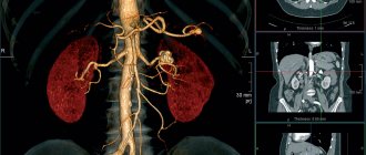
- Dopplerography (echography based on the Doppler effect), with the help of which information is obtained about the state of the circulatory system of the kidney and hemodynamic disorders in the organ are detected.
During Doppler sonography, a color image appears on the screen
The Doppler method is based on tracking changes in the frequency of ultrasonic waves reflected from the moving boundaries of media that are different from each other.
Video: an obstetrician-gynecologist talks about the dangers and benefits of ultrasound for pregnant women
Interpretation of research results
If the organ is in normal condition, there should be no stones in it. In addition, ultrasound on a healthy kidney does not detect cysts, new formations or abscesses.
If the organ is in normal condition, there should be no stones in it. In addition, ultrasound on a healthy kidney does not detect cysts, new formations or abscesses.
If the organ is in normal condition, there should be no stones in it. In addition, ultrasound on a healthy kidney does not detect cysts, new formations or abscesses.
If the organ is in normal condition, there should be no stones in it. In addition, ultrasound on a healthy kidney does not detect cysts, new formations or abscesses.
What features of the kidneys does ultrasound reveal?
During the examination, the ultrasound diagnostician takes several photographs of the kidneys in longitudinal, oblique and transverse sections. Examining the organs, the specialist determines:
- their contours (smooth or uneven);
- the thickness and condition of the capsules covering the outside of the kidneys;
- location and size of the kidneys;
- the presence of dilation of the renal pelvis and calyces (pyelectasis), its degree;
- volume and weight of each kidney;
- condition of the renal vessels, the presence of blood clots in them;
- the structure of the organ parenchyma, the thickness of its medulla and cortex in various places;
- the presence of neoplasms or cysts, their size and prevalence;
- congenital anomalies of the structure and development of the kidneys;
- the presence of solid inclusions (stones or sand), their shape and parameters.
With the help of ultrasound, diseases such as pyelonephritis, urolithiasis, tumor processes, hydronephrosis, nephroptosis and many others are easily detected. etc.
Photo gallery: what the most common kidney diseases look like on ultrasound sonograms
A solitary (simple) kidney cyst in the images looks like a volumetric dark formation
With polycystic disease, ultrasound images visualize many dark round areas
Ultrasound images show tuberculous cavities (marked with arrows): on the left (a) - in the upper and lower edges of the kidney; on the right (b) - multiple kidney cavities
The kidney abscess is indicated by cross-shaped markers against the background of unchanged light parenchyma
In the photo, number 1 indicates a carbuncle, and number 2 indicates unchanged parenchyma.
In acute pyelonephritis, hypoechogenicity, focal and diffuse heterogeneity of the parenchyma, as well as a decrease in the tone of the pyelocaliceal system are noted
The last stage of hydronephrosis is characterized by atrophy of the parenchyma and complete deformation of the pyelocaliceal system
A tumor thrombus in the inferior vena cava is indicated by crosses on an ultrasound image.
In the photo the arrows indicate: a - stone in the right pelvis, b - stone in the lower cup, c - stone in the middle cup, d - stone in the neck of the upper cup
Data decryption
Diagnostics is aimed at identifying deviations from the norm, which can manifest themselves in the form of:
- Acute pyelonephritis, in which the echo picture of the kidneys is practically no different from that normally. Occasionally, thickening of the walls of the renal pelvis, less often of the calyces, blurring of the contour of the kidney and swelling of the perinephric tissue in the sinus area can be detected.
- Urolithiasis. In this case, hyperechoic round formations with an echo-negative track are detected. The doctor must count them and indicate their location.
- Hydronephrosis, which occurs due to disruption of the normal passage of urine. But do not forget that during pregnancy the renal pelvis slightly increases in size and reaches 25-27 mm. This diagnosis is made on the basis of dilated calyces, pelvis and thinned parenchyma.
- Chronic glomerulonephritis. When performing an ultrasound of the kidneys during pregnancy, this disease is characterized by hyperechogenicity of the parenchyma and a decrease in the size of the kidneys.
- Nephroptosis – prolapse of the kidney below the established limit. When such a pathology is detected, a dynamic observation is carried out, recording the degree of prolapse.
For whom is ultrasound diagnostics of the kidneys mandatory?
Ultrasound scanning is absolutely indicated for suspected focal pathological process in the kidneys in pregnant women:
- carbuncle or abscess;
- nephrolithiasis;
- tuberculosis;
- cyst;
- tumor.
Indirect reasons for sending a pregnant woman for a kidney ultrasound are:
- the patient complains of pain in the lumbar region;
- unreasonably elevated temperature for several days;
- the appearance of blood in the urine, other sudden changes in its color and smell;
- changes in previously normal urine test results;
- kidney diseases diagnosed before pregnancy;
- back bruise;
- persistent increase in blood pressure;
- weakness, sweating, loss of strength;
- the appearance of swelling on the face;
- problems with urine excretion (rare, too frequent or painful urination, a sharp decrease or increase in the one-time volume of urine excreted).
The symptoms listed above can occur either in isolation or in combination with each other. At the urgent request of a woman, a referral for an ultrasound scan of the kidneys in some cases can be issued by an obstetrician-gynecologist and without any special indications. The desire of a pregnant woman to control her health within reasonable limits is always welcomed by doctors.
Some urologists even believe that ultrasound scanning of the urinary organs should be performed on all expectant mothers without exception, since the asymptomatic course of chronic pyelonephritis or urolithiasis is a common and frequent occurrence.
Video: all about ultrasound of kidneys in women
How to prepare
If the patient requires any special preparation, the doctor will warn her about this. But most often, preparing for an ultrasound of the bladder and kidneys is quite simple:
- if you are prone to gas formation, you should stick to a diet excluding legumes, brown bread, dairy products and cabbage for 2-3 days before the procedure;
- 1 hour before the examination you need to drink about 0.5 liters of water to fill the bladder;
- During the ultrasound, they take a clean diaper or sheet with them to lay on the couch; you can also bring sanitary napkins.
Before the procedure itself, the specialist will ask you to undress to the waist and remove your jewelry.
Preparation and conduct of ultrasound examination
There is no need to prepare specially for an ultrasound of the kidneys and urinary tract. On the eve of the procedure, the woman can adhere to her usual diet. However, an overview of the kidneys, adrenal glands and ureters will be better if the pregnant woman, for three days before the procedure, removes from her diet foods that increase gas formation: legumes, peas, cabbage, rye bread, as well as milk and meat. You can take Espumisan, activated charcoal, Pancreatin or chamomile infusion. This bowel preparation is especially useful before Doppler ultrasound of the renal vessels. On the day of the procedure, you can take a light breakfast.
During the examination, you should wear something comfortable to quickly expose the lumbar region. For example, these could be wide sweatpants and a spacious knitted pullover. The dress will not fit: it will have to be removed completely, which is not comfortable for all women.
In some budget clinics, the patient is warned to take a towel with her to wipe off the gel and a sheet to lay on the couch.
An hour before the examination, it is recommended to drink 0.5–1 liters of water to ensure that the collecting system and bladder are full of urine. This way, the ultrasound picture of all organs of interest to the doctor will be clearer.
In preparation for a kidney ultrasound, drink 1 liter of pure still water.
Video: do you need to prepare for an ultrasound of the kidneys and how to do it
How is an ultrasound examination of the kidneys performed?
Ultrasonography is performed in a slightly darkened room, since in bright sunlight or electric lighting, the eyes of the ultrasound technician will not be able to distinguish the entire spectrum of shades of gray on the monitor. During the examination, the doctor moves a sensor over the pregnant woman’s torso and looks at the screen on which the device displays an image of the kidneys. He dictates the parameters and digital data to the nurse, who writes them down in the patient’s card or on the results sheet. In order to prevent air from getting between the device and the woman’s skin, a special gel is applied to her lumbar region.
The gel is applied to the skin for tight contact with the sensor
Kidney examinations are usually performed with the pregnant woman lying on her side. However, during the procedure, the doctor may ask the patient to turn over on her stomach (in the early stages) or on her back for a better view of the abdominal cavity.
The doctor may ask the patient to turn on her stomach, back or side
If kidney prolapse (nephroptosis) is suspected, the examination is repeated in a standing or sitting position. To assess the degree of physiological mobility of the organ, ultrasound images are taken at the height of deep inspiration and at maximum exhalation.
The ultrasound procedure does not cause patients the slightest discomfort, so it is rightfully considered the most favorite type of diagnosis among pregnant women. The examination lasts no longer than half an hour.
The ultrasound machine sensor moves easily and painlessly across the skin, without causing any discomfort to the patient.
Preparing for the examination
After ordering an ultrasound examination of the urinary organs, the doctor will tell you how to prepare for the procedure. Preparing for a study for a pregnant woman is no different from other categories of patients. A few days before the screening, it is recommended to follow a diet that excludes foods that cause increased gas formation.
Eight hours before the ultrasound examination, it is recommended not to eat, and to fill the bladder you need to drink unsweetened tea, fruit juice or compote, and clean water without gas. If discomfort occurs in the intestines, they are relieved with several tablets of activated charcoal.
Interpretation of results and normal indicators in healthy pregnant women
The specialist conducting the ultrasound examination does not make a diagnosis. He only describes in detail the condition of each kidney. The results sheet is given to the patient for subsequent transmission to the doctor who prescribed the procedure. By comparing the ultrasound data with the indicators of tests and other studies, the doctor confirms or denies the presence of a suspected disease in a given pregnant woman.
This is what the kidney ultrasound results sheet given to the patient looks like:
Normally, the kidneys of a healthy expectant mother should have the characteristics described below.
Dimensions and shape
The length of the organ is 100–120 mm, thickness – 50–60 mm, width – 35–45 mm. Dimensions are among the most important parameters, because their change “screams” about the disease. For example, with pyelonephritis the kidney is enlarged, and with hydronephrosis it is reduced and wrinkled.
The organ has smooth, even contours and is surrounded by a clearly visible fibrous membrane. The shape of the bud is bean-shaped. In the cross-sectional photo it approaches round, and in the longitudinal photo it approaches oval.
Photo 1 shows a normal right kidney in a longitudinal section, and photo 2 shows a transverse section
Topography
The kidneys are located in the retroperitoneal space symmetrically relative to the spinal column, at the level of the 11–12th thoracic vertebra and/or second lumbar vertebra. The right organ is located slightly lower than the left, which is due to its displacement by the liver.
Mobility
The kidneys have limited mobility. Moreover, the amplitude of their physiological displacement should not exceed 2–3 cm. Pathological mobility of the organ, especially in combination with an increase in its size, is a sure sign of pyelonephritis. A significant downward displacement of the kidney in the patient’s vertical position indicates nephroptosis.
Properties of parenchyma
The structure of the parenchyma is homogeneous. Kidney tissue has different thicknesses in different areas: from 10–20 mm in the middle segment to 20–25 mm at the poles. Thinning of the parenchyma may indicate chronic latent pyelonephritis. The fabric does not contain any foreign matter. The medullary (pyramidal) and cortical layers of the parenchyma are clearly visualized and differ from each other on ultrasound images. The pyramids appear dark against the background of light cortical tissue.
Hollow kidney system
Normally, the pyelocaliceal system is not expanded and does not contain foreign inclusions or impurities. Its appearance on ultrasound images depends on the scanning direction. The renal cavities on sonograms appear light in comparison with the darker parenchyma. Expansion of the pelvis and cups is observed in case of obstruction of the urinary tract, when urine, not finding a way out, rises through the ureters back to the kidney (reflux phenomenon).
Renal vessels
Large vessels of the kidneys of a healthy pregnant woman appear as dark transverse stripes on ultrasound sonograms. Unlike a vein, an artery has:
- pronounced pulsation;
- smaller diameter;
- thicker walls.
Normally, the vessels are clean, not narrowed, and do not contain blood clots. The speed of renal blood flow should be 50 - 150 cm/sec.
An ultrasound image of a normal kidney, taken in cross section, shows the main renal vessels


