Vaginal ultrasound (transvaginal ultrasound) is a procedure performed to examine the condition of the pelvic organs in women. This diagnostic method is carried out using a special ultrasonic sensor. The main task of such an ultrasound examination is to evaluate the functioning of the woman’s internal genital organs (uterus, ovaries, cervix, fallopian tubes), as well as the genitourinary system. Vaginal ultrasound is significantly more informative than studies conducted on the external part of the body, since in this case the sensor is located in close proximity to the organs being examined. Therefore, transvaginal ultrasound allows you to obtain more accurate and objective information about the work and condition of internal organs and their systems.
What does diagnostics with a vaginal sensor show?
Let's figure out what transvaginal ultrasound is. The word “transvaginal” is translated from Latin as “through the vagina.” From the name it is clear that the sensor is inserted into the woman’s vagina.
The vaginal sensor has a length of 12 cm and a thickness of no more than 3 cm, so it does not cause pain. There may be some discomfort if a woman is hypersensitive. The exception is pain that increases with pressure during inflammatory processes, injuries, and late stages of cancer.
There is a puncture needle at the end of the vaginal probe. It is used when ultrasound-guided puncture biopsy is necessary.
During a transvaginal ultrasound of the female organs, the doctor sees on the monitor:
- condition and size of the pelvic organs (uterus, ovaries, fallopian tubes);
- location and mobility of organs;
- presence of neoplasms;
- inflammatory signs;
- pregnancy.
Transvaginal scanning can detect pregnancy and gynecological pathologies. The vaginal sensor transmits echo signals through the vaginal wall, which helps to better examine the female organs. This is the main advantage of the transvaginal method compared to the abdominal one.
Watch a video about vaginal ultrasound of the pelvic organs in women:
Is the study related to the menstrual cycle?
There is a direct relationship between the results obtained and the critical days when performing diagnostics, so in order to decide when it is best to do a transvaginal ultrasound, you should know exactly your menstrual cycle. The reliability of the sensor readings will depend on how much time has passed since the last ovulation, which is observed on days 12-14 of the cycle, after which changes occur in the uterus.
The first few days after the end of your period are best for performing a routine transvaginal pelvic ultrasound. To determine endometriosis, it is better to schedule a diagnosis in the second phase of the menstrual cycle.
If emergency conditions develop, the study is performed at any time. If it is necessary to monitor the maturation of the follicle or observe the progression of inflammatory processes in the pelvic organs, then ultrasound is performed several times a month.
Indications
Transvaginal examination is indicated under the following circumstances:
- screening in the first trimester of pregnancy;
- preventive examination of women who are sexually active, as well as during menopause;
- irregularity or absence of menstruation;
- violation of the volume of menstruation (heavy or scanty);
- intermenstrual bleeding;
- pain in the lower abdomen during and outside of menstruation;
- control of treatment of gynecological diseases;
- infertility;
- diseases of the mammary glands;
- urological diseases.
Indications and prohibitions for ultrasound
Ultrasound in the field of gynecology is recommended to be performed at least once every 2 years, and after 40 years - annually. However, if alarming symptoms occur, you should consult a doctor earlier:
- very short or too long periods;
- at the first signs of conception;
- pain during sexual intercourse;
- discomfort and disturbances after intrauterine devices;
- bleeding;
- cycle disorders;
- purulent or mucous discharge (including bloody discharge);
- pain in the appendages or uterus, lower abdomen;
- very painful or heavy periods.
Ultrasound examination is also performed in case of suspicion (or presence) of diseases:
- infertility;
- endometriosis;
- myoma;
- salpingitis;
- polycystic disease;
- cancer;
- polyps;
- cysts;
- inflammation of the ovaries;
- endometritis.
Ultrasound of the uterus and appendages is mandatory before planning pregnancy, IVF. The examination is performed at least three times during pregnancy. An ultrasound is done if inflammation of the female genital organs is suspected, to monitor the treatment of uterine fibroids.
Contraindications
A rectal examination is not carried out in case of intestinal obstruction or wounds in the anus. Transabdominal is not done if ulcers, erosions, or skin rashes appear on the abdomen. Transvaginal is not performed in case of bleeding. From the 2nd trimester, scanning is prohibited by any means other than abdominal. In other cases, ultrasound research has no prohibitions.
How to prepare for a transvaginal examination?
The most common questions that worry women: is it possible to have sex before a transvaginal ultrasound, is it possible to eat and drink before the examination, is it possible to do an ultrasound during menstruation.
This procedure does not require special preparation. It can be performed during menstruation, but preferably on days 5–7 of the cycle. You can eat and drink, but it is better to avoid foods that cause gas. For flatulence, it is recommended to take Espumisan or Smecta first.
Sex before an ultrasound is allowed only with a condom to avoid the accumulation of male discharge in the vagina.
Immediately before the ultrasound, it is enough to empty the bladder and carry out hygiene procedures for the external genitalia.
Rules for the procedure
In order for a transvaginal ultrasound to be as informative as possible, you need to properly prepare for it. It won't be difficult. Transvaginal ultrasound is performed only on women living an intimate life. For girls who have not had sexual intercourse, the procedure is not performed due to the risk of damage to the hymen.
For them, ultrasound diagnostics is used using a transabdominal sensor, i.e., studying the structure and pathologies of the pelvic organs in the usual way, when moving the sensor across the abdomen. There are cases when excess fat deposits in the abdominal area or excessive bloating of the intestines do not allow this procedure to be fully carried out. Then girls who are not sexually active are prescribed a transrectal ultrasound examination, i.e., a special sensor is inserted into the rectum.
Vaginal ultrasound does not require special preparation, but there are a number of recommendations and wishes that should be followed. Unlike classical (abdominal) ultrasound diagnostics, the bladder must be empty. If the last trip to the toilet was more than an hour ago, the sonographer will ask the patient to go to the toilet and empty the bladder. A very important point is intestinal flatulence. It is very important to make every effort to eliminate or reduce it. Medications aimed at reducing the concentration of gases in the intestines will come to the rescue. The doctor prescribing the procedure will help you decide on their choice and dosage.
Therefore, it is very important to inform your doctor about this problem if it is present. The next point is personal hygiene. It is very important to thoroughly wash your external genitalia and wear clean underwear before the procedure. If you need to go to the toilet before the procedure, do not forget to use wet sanitary napkins. This has no effect on the results, but from an aesthetic point of view, this point is very important. If an abdominal diagnosis is first performed before a vaginal examination, then you need to come with a full bladder and go to the toilet before using the vaginal sensor.
How is TVUS performed?
A transvaginal ultrasound method for examining female organs is prescribed on certain days of the menstrual cycle. Depending on the indications for diagnosis, you should take into account the days of the cycle when it is better to do a transvaginal ultrasound.
As a rule, transvaginal ultrasound is performed on days 5–7 of the cycle, i.e. immediately after menstruation. These days, female organs are visualized best. For some pathologies (endometriosis, assessment of the condition of the corpus luteum), ultrasound is performed in the second half of the cycle.
Follicle growth and number are monitored using a series of transvaginal examinations performed several times during one menstrual cycle. Folliculometry is recommended at the stage of preparation for pregnancy or if it is impossible to become pregnant.
In case of constant absence of menstruation, as well as in case of sudden symptoms of gynecological pathology, an ultrasound scan is performed any day.
On what day of the cycle and how often to do an ultrasound is determined by the attending physician. This allows you to get the most accurate results if you suspect a particular pathology.
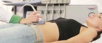
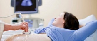
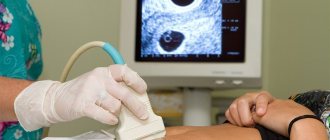
Method of performing transvaginal ultrasound:
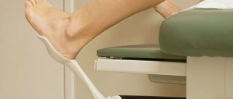
- The woman undresses from the waist down and sits on the couch. The ideal position for the examination is lying on your back with your knees bent and apart (butterfly position).
- A condom is placed on the sensor to prevent infection. Ultrasound gel is then applied. It improves penetration into the vagina and also improves visualization by eliminating the air gap.
- The woman is asked to relax the muscles of the perineum. The sensor is inserted into the vagina, after spreading the labia.
- The probe is gently rotated to ensure a thorough examination of all pelvic organs.
- The doctor records the received data, saves important images, and issues a conclusion.
A condom is a must when conducting a transvaginal ultrasound examination. Even if you did not bring it with you, do not worry - the doctor always has a means of protection against infections.
With a transvaginal ultrasound, you can get a lot of things only if the ultrasound is performed without a condom. But now this is not practiced anywhere: you are more likely to be refused research than to be exposed to the risk of infection. Protective caps for transvaginal ultrasound are affordable and sold in all pharmacies.
For the procedure for performing an intravaginal ultrasound, watch the video:
Features of transvaginal ultrasound
Vaginal ultrasound can be performed not only to study the female reproductive system. At the same time, the procedure helps to evaluate other pelvic organs. The study is also carried out for pregnant women, but only in the 1st trimester - up to 12 weeks. It is safe, the fetus is reliably protected from any influences by the uterine cervix, amniotic fluid and mucous membrane.
Transvaginal ultrasound is one of the most highly informative methods. Modern equipment allows you to obtain instant 3-dimensional images, assess blood flow and soft tissue structures, determine the general condition of the reproductive organs and identify even the slightest disorders.
Relationship between screening and menstrual cycle
For transvaginal ultrasound, a specific period of the cycle is selected. A routine examination is scheduled at the beginning, on the next day after the end of menstruation, but can be carried out on days 5-12. If a woman is suspected of having endometriosis, then the study is indicated in the 2nd half of the cycle. If it is necessary to monitor the process of follicle maturation, transvaginal ultrasound is performed repeatedly at periods (in days) - at 8-10, 15-16 and 22-24.
If bleeding or smears appear, but they are not associated with menstruation, then the examination is done immediately after the occurrence of alarming symptoms, regardless of the period of the cycle. During pregnancy, transvaginal ultrasound is performed mainly in the 1st trimester. Otherwise, penetration of the sensor may cause a miscarriage.
After week 12, only transabdominal examination is performed, which is carried out on any day of the cycle. However, in some emergency cases, transvaginal ultrasound can be performed in the 2nd trimester. Indications are the need to evaluate the uterine cervix or scar, abnormal location of the placenta. An ultrasound is also done if during a previous birth there was a cesarean section or placenta previa.
What does an ultrasound show?
Ultrasound waves can be used to visualize disruption or changes in the shape of the bladder. Endometrial nodes sometimes appear on the uterine walls, which are signs of a pathological process. Vaginal ultrasound can detect a number of diseases:
- endometriosis;
- hydatidiform mole;
- fluid in the lower abdominal cavity, fallopian tubes;
- pathologies of the genitourinary system;
- cysts;
- cancerous tumors;
- uterine fibroids;
- endometrial polyposis;
- benign tumors;
- cyst ruptures;
- chorionepithelioma.
Ectopic conception is characterized by improper attachment of the egg, which is clearly visible in the image. Transvaginal ultrasound also helps to determine premature menopause. It is characterized by an untimely decrease in the size of the ovaries.
TVUS during pregnancy
Transvaginal ultrasound in pregnant women is performed during the first screening examination in the first trimester. Diagnosis requires moderate filling of the bladder. To do this, you need to drink 0.5 liters of water 1 hour before the examination. The methodology does not differ from the usual one.
In the early stages of pregnancy, the doctor determines the presence of a fertilized egg, its place of attachment, the tone of the uterus, the condition of the cervix, the thickness of the chorion (the precursor of the placenta), and the threat of its detachment.
In subsequent trimesters of pregnancy, the examination is carried out through the anterior abdominal wall so as not to provoke the threat of miscarriage.
Preparing to Scan
Preparation for an ultrasound of the uterus depends on its type. Before transabdominal, it is necessary to fill the bladder with fluid. To do this, drink 1 liter of water 60 minutes before the procedure. With a full bladder, the uterus will be more clearly visible.
Also, 2-3 days before the scan, you need to abstain from foods and drinks that cause gas and take remedies to improve digestion. The last meal before the examination should be 12 hours before the scan (it is done on an empty stomach). For constipation, it is advisable to perform an evening bowel cleansing with an enema.
During transvaginal and intrauterine ultrasound, the bladder for collecting urine should be empty so as not to obscure the organs in the pelvic area. You need to take a condom with you that fits over the sensor. If there is an allergic reaction to latex, the doctor should be warned about this in advance.
Before a rectal ultrasound of the uterus and ovaries, the intestines must be clean. It is emptied in the evening and six hours before the scan (for example, using a Microlax microenema). Before it, you can take laxatives or put glycerin suppositories.
Normal indicators during the study
The conclusion of a transvaginal ultrasound indicates the ultrasound signs of the identified pathology, and the final diagnosis is established by an obstetrician-gynecologist. Normal results are usually designated as follows: “No ultrasound signs of pathology of the pelvic organs were identified.”
Pregnancy is a separate issue. During a normal pregnancy, the uterus is in normal tone, enlarged in accordance with the term, and there are no signs of chorionic detachment. At 5–6 weeks, the heartbeat of the embryo becomes audible.
A decrease in the size of the organs of the reproductive system after the onset of menopause is considered normal. These are natural age-related changes in the body.
Uterus
A healthy uterus has smooth and clear contours, uniform echogenicity, tilted anteriorly (anteflexio) or posteriorly (retroflexio). The second option of uterine inclination creates difficulties in conceiving and carrying a pregnancy. Painful menstruation and a tendency to constipation are possible.
Dimensions of the non-pregnant uterus in women of reproductive age:
- length: 70 mm;
- width: 60 mm;
- anterior-posterior size: 40–42 mm.
The uterine cavity is normally homogeneous with clear, even edges, without pathological neoplasms. The thickness of the endometrium (inner layer of the uterus) depends on the menstrual cycle:
- 3–4 days of the cycle: less than 3 mm;
- on days 5–7: within 3–6 mm;
- on days 11–14: about 8–15 mm;
- on days 15–19: up to 10–16 mm;
- on the eve of menstruation: no more than 10–20 mm;
- decidualization of the endometrium – possible pregnancy.
Cervix
A healthy cervix has a homogeneous echostructure without pathological inclusions. The cervical canal is up to 3 mm in diameter and contains mucus of a homogeneous echostructure. Both pharynx of the cervix (external, internal) are closed.
Dimensions of the cervix during transvaginal examination:
- length: 35–40 mm;
- anterior-posterior size: 2.5–3 mm.
Ovaries
The ovaries are located on the sides of the uterus, slightly behind it, connected to it by the fallopian tubes. The ovaries have a homogeneous echostructure with small areas of fibrosis, clear contours, and a bumpy surface due to the presence of follicles. A slight difference in the size of the right and left ovaries on ultrasound is considered normal.
Dimensions:
- length: 20–37 mm;
- width: 18–30 mm;
- thickness: 16–22 mm;
- volume: 4–10 cu.m. cm
Depending on the day of the cycle, the number and size of follicles vary. In the first half of the cycle, there are up to 10 follicles with a diameter of less than 6 mm and 1 dominant one. The maximum size of the dominant follicle before ovulation is 20 mm.
In the second phase of the cycle, a corpus luteum is detected at the site of the burst follicle, which decreases from 25–27 mm until it completely disappears by the time of menstruation.
Fallopian tubes
Unchanged uterine (fallopian) tubes are not visualized during transvaginal ultrasound.
Free liquid
In the first days after the rupture of the dominant follicle, a small volume of free fluid accumulates in the retrouterine space. This is considered the norm. There should be no free fluid on other days.
Decoding the results
The ultrasound result is deciphered by a specialist, comparing the average statistical indicators with the data obtained:
| Ultrasound parameter | Normal echo pattern | Signs of pathology |
| Size and position of the uterus | Length: 16-62 mm. Width: 15-60 mm. Thickness: 12-48 mm. In virgins and menopausal women, the organ is smaller. The more a woman gives birth, the larger the size of the uterine body. The contours of the organ are smooth and clear. The uterus is bent anteriorly or slightly posteriorly | An enlarged uterus and blurred contours can appear with inflammatory diseases, endometriosis. Fuzzy contours, lumpiness, compactions are a sign of oncological processes. Incorrect position of the uterus in the pelvic cavity can cause infertility and spontaneous abortion |
| Surface of the cervix | The contours are clearly visible, the surface is smooth | An uneven surface and blurred contours may indicate inflammation or congenital anomalies |
| The fallopian tubes | Not rendered | Thickening of the walls, increase in diameter, accumulation of fluid indicates the development of an inflammatory process, ectopic pregnancy |
| Free liquid | It is visible behind the uterus during the period of ovulation in a small amount | A large volume of fluid is a symptom of the inflammatory process in OMT |
| Ovarian size | Length: 20-25 mm. Width: 12-15 mm. Thickness: up to 12 mm. Volume up to 4 cubic meters cm. Contours are smooth and clear | An increase in the size of the ovaries, uneven contours may be a sign of polycystic disease, oncological processes, cysts, congenital pathologies of the organ structure |
Separately, it is necessary to consider the indicator of double endometrial thickness - M-echo, median echo.
The endometrium is the functional layer of the uterus, most susceptible to the influence of hormones, as a result of which the structure of the epithelium changes throughout the menstrual cycle.
Standard indicators of endometrial thickness with normal cycle duration (28 days):
| Phase of the menstrual cycle | Standard range M-echo, mm | Possible pathologies |
| Desquamation (1-7 days) | 3,0-8,0 | Hyper- and hypoplasia of the endometrium may indicate endometriosis, an inflammatory process. Serious deviations in endometrial thickness from normal are the cause of infertility |
| Proliferative (8-14 days) | 5,0-12,0 | |
| Periovulatory (15-21 days) | 6,0-16,0 | |
| Secretory (22-28 days) | 6,5-20,0 |
Vaginal ultrasound is only one of the methods for diagnosing pathological processes in the reproductive organs.
Deviations from the norm
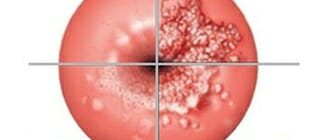
If there are deviations from the norm, a transvaginal ultrasound interpretation will be required. Changes in echogenicity, size, contours, and the appearance of neoplasms indicate the presence of female diseases. Ultrasound signs of gynecological pathology:
- An ovarian cyst is a round formation with the presence of fluid.
- Salpingo-oophoritis (inflammation of the appendages) – enlarged ovaries, fallopian tubes are visualized and thickened. The contours of the appendages are blurred, mobility is limited, the area of study is painful when pressed with a sensor.
- Uterine fibroids – enlargement of the organ, the contours are disturbed, a hyperechoic node is visualized.
- Endometriosis of the uterus (adenomyosis) – uneven thickness of the endometrium, jagged contours, heterogeneous echostructure.
- Endometriosis of other female organs – heterogeneous echostructure, ultrasound signs of “bubbles”.
- Endometritis (inflammation of the endometrium) - thickening, swelling, heterogeneous structure, blurred contours of the endometrium, hyperechoic inclusions.
- Neoplasms (polyps, cancer) - volumetric formations with increased echogenicity, deformation of contours and enlargement of the organ. Malignant tumors are often accompanied by tissue swelling.
- Pathological fluid in the pelvis - ultrasound signs of accumulation of fluid (blood, pus) in the retrouterine space.
- Pathological pregnancy - the location of the fertilized egg outside the body of the uterus (ovaries, cervix, tubes), detachment of the normally located chorion.
- Trophoblastic disease (chorionepithelioma, hydatidiform mole) is a homogeneous fine-grained mass (a “snow storm” symptom), visualization of an embryo with a damaged chorion is possible. Determined against the background of signs of pregnancy.
If we summarize the echo signs, it turns out that:
- signs of inflammation are a slight increase, heterogeneous structure of the organ, increased echogenicity, blurred contours, the presence of fluid behind the uterus;
- signs of benign or malignant neoplasms are a significant increase in the organ, an uneven surface, hyperechogenicity, the presence of fluid behind the uterus;
- signs of a normal pregnancy are increased size of the uterine body with the presence of a fertilized egg or embryo, the contours of the uterus are smooth and clear.
If the results are not normal
Pelvic ultrasound, which is performed transvaginally, is the most effective method for early diagnosis of pathological processes in this area.
What does a transvaginal ultrasound show:
- malignant tumors of the uterus and appendages;
- curvature of the fallopian tubes;
- infectious and inflammatory processes of the genitourinary system;
- cancer of other pelvic structures;
- neoplasms in the vagina.
If diseases and pathological conditions are detected, immediate treatment by a gynecologist or oncologist is required.
Ultrasound with a transvaginal sensor allows you to quickly and accurately detect the presence of abnormalities in the functioning of the pelvic organs, as well as identify developing pathological conditions in the early stages, which will help begin timely therapy.
In addition, gynecological ultrasound performed transvaginally makes it possible to determine the presence of pregnancy in the first weeks, as well as to detect the ectopic location of the embryo before the fallopian tube ruptures.
Author: Violeta Kudryavtseva, doctor, especially for Mama66.ru
TVUS cost
Transvaginal ultrasound diagnosis of diseases of the female organs costs an average of 1000–1500 rubles. The cost of transvaginal ultrasound depends on the region and clinic.
Average prices for ultrasound of the pelvic organs using the intravaginal method:
Since transvaginal ultrasound is an accessible, safe and informative examination, it can be performed several times during one menstrual cycle. This allows you to track the dynamics of changes in the pelvic organs (monitoring the effectiveness of treatment, tracking the growth pattern of cysts, the dynamics of growth of the ovum during pregnancy).
Share the article with your friends on social networks, it may be relevant to them right now. Tell us about your experience with transvaginal ultrasound. Be healthy. All the best.
Contraindications
Contraindications to ultrasound of the bladder depend on the method of diagnosis.
Transabdominal method (through the abdominal wall):
- urinary incontinence (ultrasound is performed only on a full bladder) - however, it is possible to catheterize the bladder immediately before the study;
- excess weight (a thick subcutaneous fat layer makes scanning difficult and reduces the diagnostic information);
- skin lesions in the lower abdomen (pyoderma, herpes, wounds, burns, infectious lesions due to syphilis and HIV);
- bladder defects (sutures and scars on the bladder wall).
Transrectal method (through the rectum):
- inflammatory bowel diseases in the acute stage (fissures, hemorrhoids, dysentery, Crohn's disease, etc.);
- absence of the rectum (as a result of surgery and replacement of this organ with an artificial anostomy to remove feces);
- narrowing (stricture) and obstruction of the rectum;
- intolerance, allergy to latex (medical rubber used to make gloves and condoms).
Transvaginal method (through the vagina):
- allergy to latex;
- the presence of a hymen;
- pregnancy more than 12 weeks;
- genital infections.
Transurethral method (through the urethra)
- intolerance to medicinal painkillers;
- inflammatory diseases of the urethra.
What does the sensor look like?
This special tube with a camera at the end. The diameter of the transvaginal sensor is only 3 cm, and the total length is 12 centimeters. Often there is a channel inside such a device where a biopsy needle can be placed.
Due to the peculiarities of the anatomical structure of the body and the specific location of the uterus, the sensor is designed with an oblique view relative to its axis. Thanks to this, ultrasound with a transvaginal probe is more convenient.
There are several types of device. Some clinics prefer a straight-handle probe to perform routine gynecological procedures. Reproductive health centers almost always use a sensor that has a beveled handle, thanks to which in vitro fertilization or biopsy can be performed.
A transvaginal sensor with a beveled handle is more convenient and ergonomic for examinations in the gynecologist's chair.
What is the frequency range of the sensor?
The frequency range of the sensors used for transvaginal scanning is in most cases 4-7 MHz. Higher frequencies, as a rule, are not used to study the uterine cavity.
The fact is that the depth of the uterus is easily detected by a gynecologist, so there is no need to purchase sensors with a higher frequency.
The scanning angle of such sensors varies from 120 to 140 degrees. This angle is sufficient to completely examine the uterus. There are also special sensors, thanks to which they receive a 4D image and simultaneously display the image on the screen.
Such equipment is capable of studying the structure of small parts of the fetus and its cardiovascular system, as a result of which doctors can identify abnormalities at an early stage.