Radiography remains the most popular method for diagnosing many diseases to this day. It is especially widely used in traumatology and orthopedics. This is a fast, reliable and relatively inexpensive way to detect pathological changes in bones and joints. However, exposure to X-rays is not always safe, so this examination has limitations. To ensure that the diagnostic procedure does not harm the body, you need to familiarize yourself with general information about it, and if there is a need to take an X-ray of the heel, it is best to consult a specialized specialist for advice.
What does a foot x-ray reveal?
Immediately after the shooting, the images are developed, and the radiologist begins decoding. An x-ray allows you to find out the cause of pain and determine the nature of the pathological process. To accurately interpret the result, the doctor can compare images of healthy and diseased heels.
If plantar fasciitis (the so-called heel spur) is detected, the specialist gives a detailed description of the image, where he notes the presence of an osteophyte, indicating its length and location. Typically, osteophytes are visible in the image in the projection of the upper edge of the posterior tuberosity of the talus.
The first thing a patient complains about when a calcaneal bone is fractured is pain. It can occur during movement or persist at rest. Upon examination, swelling may be detected that extends to the Achilles tendon. Also among the symptoms of a fracture are: flattening and widening of the heel, hematoma in the center of the sole.
When palpating the site of injury, the patient feels sharp pain. While standing, he cannot support his injured foot.
The images obtained during the study are interpreted by a radiologist immediately after the procedure. When conducting a study, you can clearly see the presence of defects indicating traumatic injuries.
An X-ray of the heel bone helps to determine the cause of pain and inflammation and allows you to see changes in bone tissue, indicating the development of a heel spur or malignant neoplasms.
During the procedure, you can clarify the diagnosis if tuberculosis, osteomyelitis, osteoporosis and other bone diseases are suspected.
Literature:
- Korzh N.A., Prozorovsky D.V. “Modern aspects of diagnosis and treatment of some foot pain syndromes in adults”
- Dryagin V.G. "Diagnostics and treatment of unstable calcaneal fractures"
The doctor receives X-ray images immediately after the examination and, upon completion of studying the image, makes the correct diagnosis. The results of a stress x-ray can be considered correct only if the study was carried out in compliance with all the rules.
For flat feet, such diagnostics will allow you to record the length and degree of curvature. With the final examination data, the patient should be sent to the attending physician, who will draw up a method for further treatment.
Other diagnostic methods
A spur on the heel in its development stage can be recognized by palpation and examination. The orthopedist probes the area of the heel tubercle and sole. With posterior localization, the spine is easily detected.
With a lower position, you need to apply light pressure on the heel. This causes the patient to feel acute pain. The doctor also examines the feet for inflammation of the fascia and tendons.
On a note!
Corns and calluses in the affected area can signal the imminent formation of a growth.
Computer examination
Often, when making a diagnosis, a CT or MRI of the legs is prescribed. These studies are indicated when it is difficult to identify spurs using x-rays when the spine is located inside a tendon or ligament.
Computer diagnostics are highly sensitive studies and give 100% results. The method is based on a sequential examination of all layers of tissue, which allows you to accurately determine the nature and cause of heel growths.
Analyzes
A spur on the foot manifests itself not only externally and symptomatically. The disease also causes changes in clinical tests. Usually, with spurs, the ESR in the blood increases. The C-reactive protein may increase. With concomitant joint damage, rheumatoid factor is detected in the blood.
Ultrasound
A spur on the foot is detected using ultrasound in a few minutes. And even pregnant women and small children can do the procedure. Ultrasound determines foci of inflammation on the sole and the size of the growth using high-frequency waves.
What can reveal
The principle of radiography of the heel bone is based on the ability of X-ray rays to penetrate tissues that are opaque to visible light. An X-ray of the heel shows a negative imprint, that is, the dense heel bone of the foot appears white in the image (the fracture area is clearly visible on the black background of the film), the cartilage tissue appears darker.
An X-ray of the heel bone can reveal the following diseases and conditions:
- fracture or crack of the heel bone;
- heel spurs;
- changes in the structure of the heel and foot.
Using x-rays of the calcaneus, flat feet and related problems, arthrosis, and degenerative processes in the foot and heel are detected. X-rays of the heel bone are performed to assess the condition of the bone tissue in gout.
Depending on the suspected pathology, x-rays are done in two ways:
- Lying on your back with your knees bent and your feet resting on the table.
- During weight-bearing x-rays, the patient stands on one leg and rests his or her weight on the leg being examined.
The projection of the heel bone is chosen by the doctor depending on the suspected pathology. No special preparation is required for an x-ray, but there should be no metal objects on the leg that could distort the quality of the x-ray.
The largest bone in the foot is the talus. When standing upright and walking, it bears the heaviest load. On a bone x-ray you can see:
- flat feet;
- heel spur;
- malignant and benign osteoarticular neoplasms;
- fractures;
Heel bone fracture on x-ray
- osteomyelitis;
- cracks;
- dislocations, subluxations;
- arthrosis;
- osteoporosis;
- arthritis;
- tuberculosis;
- gout
X-ray allows you to identify the disease and carry out differential diagnosis with other diseases.
The images obtained during the study are interpreted by the radiologist immediately after the procedure. An x-ray allows you to find out the cause of pain and determine the nature of the pathological process. If plantar fasciitis is detected, the radiologist gives a description of the image.
Orthopedic doctors examine the image
In conclusion, the doctor notes the presence of an osteophyte, indicating its length and location. Typically, osteophytes are visible in the image in the projection of the upper edge of the posterior tuberosity of the talus.
How is the examination carried out?
X-ray photos of the heel bone can be taken in various projections.
Direct radiography
To take a frontal view, the patient needs to lie on his back on the X-ray machine table. Legs bend at the knees and feet rest on the table.
X-ray with stress
The load on the heel bone occurs when a person stands. Accordingly, to perform weight-bearing x-rays, the patient must stand and place weight on the foot.
Treatment of plantar fasciitis
The plantar aponeurosis attaches to the calcaneal tubercle and phalanges, providing the longitudinal arch of the foot. The function of the plantar fascia is to reduce the load on the foot. Thus, the fascia provides shock absorption when walking. The plantar aponeurosis consists of collagen fibers. In an upright position, half of a person's body weight puts pressure on the fascia. In this case, the maximum stress is experienced by the place of attachment of the aponeurosis to the heel.
The causes of plantar fasciitis and heel spurs lie in disruption of the biomechanics of the foot and inflammatory and degenerative changes in the fascia. With each step, the aponeurosis is stretched. When the fascia loses its elasticity, it cannot withstand loads and tears occur. In this case, the person feels a sharp pain in the sole from the heel.
Over the course of several months, osteophytes grow at the site where the fascia attaches to the heel. They are called heel spurs. Osteophytes are detected on x-ray in half of cases of plantar fasciitis.
10% of people have spurs, but only 5% of people experience heel pain. Heel spurs do not cause any symptoms, but they can be complicated by fractures.
Plantar fasciitis is one of the most common causes of heel pain in adults.
Magnetic resonance imaging (MRI) is used to diagnose heel spurs. This method examines the condition of the bones, ligaments, muscles and fascia of the foot. It is MRI that reveals thickening of the fascia in the disease up to 6–10 mm. Normally, the aponeurosis is no thicker than 4 mm.
MRI is used to differentiate between cartilage, Achilles tendon, and heel spur injuries. Ultrasound of the foot is used for examination, but it is used more often for monitoring during treatment.
Answers on questions
Radiotherapy of the plantar fascia raises a lot of interest and many questions.
Which doctor should I contact to prescribe treatment?
If you are worried about pain in the heels, you need to visit a traumatologist or orthopedist. After collecting the analysis, examination and laboratory examination, the doctor will decide on the advisability of using radiation waves. Usually, medication and physiotherapeutic treatment of the disease are first used. If all methods of therapy are unsuccessful, before consulting a surgeon to decide on surgery, specialists prescribe a course of radiotherapy.
Research methodology
The largest bone in the human foot is the heel. It can withstand enormous loads. But even the heel bone can be injured. Fractures cause a lot of discomfort and can cause serious complications. Often, when falling from a height or unsuccessful jumps, people do not pay attention to pain, and during X-rays fractures are revealed.
It is imperative to treat a calcaneal injury, otherwise negative consequences are inevitable. However, it is not easy to diagnose this problem. It is impossible to do without a comprehensive X-ray examination. Only it provides comprehensive information about the condition of the bone.
The Bellaire angle data provides more information. Many traumatologists use this as a starting point when assessing the results of treatment. If everything is in order, then the degree will be no less than 20 and no more than 40. For serious fractures, the Bellaire angle data may give a negative result. But such information can only be obtained after radiography.
The best specialists in this field accept patients. Experienced radiologists accurately diagnose the problem in the shortest possible time. It is possible to perform the procedure on an outpatient basis.
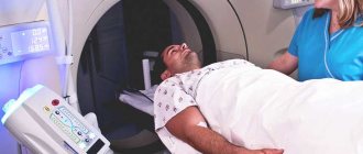
X-rays are performed in a supine position. The procedure is carried out in a relaxed state. The legs are bent at the knees. Your heels should rest on the table.
If the procedure is performed in an oblique projection, then the patient needs to lie on his side, bending his legs and holding a pillow between his knees. For radiography, the angle of the feet is important. The process lasts from 5 to 10 minutes; it all depends on the complexity of the suspected disease.
During the x-ray, the patient is given a special protective apron. It is necessary to protect yourself from radiation. Our center has such equipment even for the youngest patients. The aprons are made with lead material: this is what protects against the adverse effects of X-rays.
High-quality equipment and qualified specialists are the pride of the medical industry! Due to this, we carry out any radiography. In a short time, the examination result is given, a diagnosis is made and treatment is prescribed.
Features of the procedure
X-ray diagnostics are carried out in specially equipped and protected rooms strictly as prescribed by the doctor. In case of injury or other acute emergency conditions, the study is carried out immediately, in addition, in the vast majority of medical institutions this procedure is free. It does not require special preparation and does not take much time.
To increase the information content of the images, it is necessary to remove all metal elements: jewelry, clothes with rivets or zippers, mobile phones and other electronic devices, bank cards, etc. The metal delays and reflects X-rays, so the images are illuminated, which distorts the result.
It is extremely important to place lead protective aprons on those parts of the body that are not being examined immediately before the procedure.
X-Ray Protective Clothing
X-ray signs of a spur
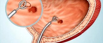
Heel spurs are bone growths on the surface of the talus, usually in the area of the tubercle. They grow due to flat feet, physical activity, poor footwear or inflammatory foot diseases. On an x-ray, a heel spur appears as a hook-shaped or awl-shaped growth in the area of the posterior or lower surface of the tubercle.
The main symptom of plantar fasciitis is pain when taking the first steps after getting out of bed.
Clinically, a change in gait is determined, in which the center of the body shifts to the forefoot and toes. In the projection of the spur, calluses are visible on the skin. The main complaint of patients is severe pain in the heel in the morning when supporting the foot. After some time of walking, the pain decreases.
A fracture of the talus occurs during a fall from a height or an accident. An x-ray image shows the displacement of bones and fragments. The length of the fracture is determined. For diagnostic purposes, radiography is done in two projections - lateral and axial.
During the examination, a hematoma and pain when pressing on the heel are noted. Foot mobility is limited.
Fracture of the talus is rare
A cyst of the talus does not show any clinical signs for a long time and is detected by chance on an x-ray. More often, a cyst develops as a result of constant foot injuries in athletes, including children. When pain occurs in the heel during physical activity or when walking, swelling of the foot appears.
The cyst is detected in the axial projection. On an x-ray it appears as an oval formation. As a result of the cyst, pathological fractures of the talus occur, which limit the mobility of the foot.
Localization
The location of the spurs has a significant influence on the appearance of the spurs on the legs. More often, the growth forms in the plantar part of the heel bone and is called the lower one. This localization is characterized by intense symptoms, but is hardly noticeable externally. The thorn penetrates the soft tissue of the heels and is impossible to see with the naked eye. X-ray examination methods help diagnose the disease.
In 10-15% of cases, the spur grows under the Achilles tendon and is called lateral or posterior. With such localization, the disease is easy to recognize independently. A wedge-shaped tumor with a sharp apex forms in the area of the heel tubercle. The clinical manifestations of this form have a less aggressive course. But signs of the disease are clearly visible in the photo of spurs on the heels, especially in a side view.
X-ray therapy for heel spurs
Heel spur (plantar fasciitis) is a fairly common foot disease, which is a bony growth at the bottom of the heel bone, leading to sharp, excruciating pain that intensifies when walking. If left untreated, the disease can cause complications such as heel bursitis (inflammation of the mucous bursae) and fasciitis (inflammation of the muscle tissue of the foot).
One of the methods of treating plantar fasciitis today is radiotherapy. The essence of the method is to expose the heel spur to ionizing X-rays, which split and destroy the resulting pathological bone growths.
X-ray therapy is a fairly effective and safe method. After just a few sessions, as a result of blocking the nerve endings, pain and inflammation are significantly reduced. In addition, the impact of radiation in this case is focused, its doses are minimal, and there is no negative effect on the body.
Positioning the patient
To take a high-quality photo, you need to ensure proper styling. The patient lies on his back. The foot is bent. A cassette is placed under the heel. The installation can be seen in the presented photo.
Hardware examination of the heel
You can take a photo while standing. The cassette is placed under the patient's sole. The leg is bent at the shin at a certain angle. The patient leans against the back of the chair.
X-ray allows you to study the volume and integrity of the bone, possible pathological abnormalities in relation to the norm.
X-ray is a non-invasive examination method that has no contraindications. Radiation exposure is minimal. There are relative contraindications during pregnancy, but if necessary, wear a special apron to protect the fetus from radiation.
The resulting images are interpreted on a special screen by an experienced radiologist, after which a conclusion is made. If there are any doubts or to clarify the diagnosis, a consultation of experienced doctors is held, who make a joint verdict in especially severe cases.