Cystography of the bladder is prescribed only for special indications, when other diagnostic methods (cystoscopy, ultrasound) did not provide a complete picture of the possible pathology. Cystography is not recommended for preventive purposes or as part of screening. The procedure is unpleasant and accompanied by painful sensations.
Cystography is an x-ray procedure for studying the anatomical and physiological features of the bladder. To obtain an x-ray image, a contrast agent is used, which is injected in a descending or ascending manner.
Why is the research being conducted?
Cystoscopy for a child can be performed to achieve the following diagnostic and therapeutic purposes:
- Examination of the bladder, taking a biopsy.
- Bougienage and catheterization of the ureter.
- Cauterization of erosion.
- Removal of polyps.
During cystourethroscopy, it is possible to remove a polyp
- Administration of drugs intravesically.
- Elimination of prostatic hyperplasia or removal of neoplasms.
- Crushing and removing stones.
- Stop bleeding.
What is cystoscopy?
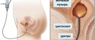
Pediatric cystoscopy is a method of endoscopic examination of the cavity and mucous membrane of the bladder (MB) using a cystoscope.
According to indications, the child may be prescribed urethroscopy - a method that allows you to examine the condition of the urethra (urethra) using a urethroscope.
The procedures are performed under the visual supervision of a doctor.
Diagnostic procedures are performed using pediatric cystoscopes and urethroscopes of various sizes. For each young patient, including newborn babies, a device is selected in accordance with his age and anatomical features.
Cystoscopy and urethroscopy are recognized as the safest and most informative diagnostic methods for examining the urinary tract.
Purposes of cystoscopy
Depending on the goals that guide the doctor when prescribing the study, survey, catheterization or resection (operative) cystoscopy can be performed.
During survey cystoscopy, the doctor examines and evaluates the cavity of the bladder, the color and condition of its mucous membrane and the orifices of the ureters.
Detects pathologies such as:
- congenital defects of the urinary system;
- the presence of neoplasms, stones or polyps;
- diverticulosis of the bladder (as well as pseudodiverticula of the bladder);
- bladder reflux in children;
- neurogenic dysfunction of the bladder (more about the causes and treatment of neurogenic bladder dysfunction in children here);
- MP damage, etc.
If congenital or acquired pathological changes are detected, the doctor has the opportunity to accurately determine them:
- location;
- boundaries and shapes;
- sizes and quantities, as well as other parameters.
In addition to the viewing function, cystoscopy allows you to perform some diagnostic and therapeutic procedures, including:
- catheterization and bougienage of the ureters;
- biopsy of the mucous membrane of the organ and neoplasms (which is very important for determining treatment tactics);
- administer drugs intravesically;
- crush and remove stones;
- remove polyps;
- cauterize erosion;
- stop bleeding.
Preparation and carrying out the procedure
The day before the scheduled cystoscopy, a broad-spectrum antibiotic is started, which is recommended to be continued for the next 7 days to prevent infection.
1-2 hours before the examination, it is necessary to empty the intestines (either on your own or with the help of a laxative).
If the procedure will be performed under local anesthesia, then no other preparation is required. If the study is carried out under anesthesia, then doctors recommend not taking food for at least 8 hours, and water for 6 hours.
If urethrocystoscopy is performed under anesthesia, the child must come for the examination on an empty stomach.
Next ─ about what and how to do during cystoscopy.
During the entire study, the patient lies on his back with his legs spread. If a decision has been made to undergo general anesthesia, the patient is put under anesthesia. If the procedure is carried out under local anesthesia, then a solution of novocaine is injected into the urethra. The doctor inserts the cystoscope into the urethra, past it, into the cavity of the bladder and fills it with a small amount of sterile saline for better visibility. The doctor receives all the necessary information about the condition of the mucous membrane and carries out the necessary manipulations (for example, cauterization of erosion, biopsy, bougienage or catheterization of the ureter, etc.). Cystoscopy is often done with chromocystoscopy ─ during this study, a contrast agent is injected into the patient’s vein and it is observed from which ureter and at what speed urine enters the bladder. Thus, with the help of cystoscopy, one can indirectly judge the condition of the kidneys in children.
The duration of the study depends on the qualifications of the doctor, the device used for the examination, the individual characteristics and diseases of the patient, as well as the purpose of the study. It can take from 5-10 minutes to an hour.
Everything that the endoscopist sees and does during the procedure is recorded in the protocol, and the results of the examination are explained to the patient by his attending physician (therapist or urologist).
How is cystography done in children?
For a newborn or older child, cystoscopic examination is prescribed in extreme cases. The volume of contrast agent for a child is 50-100 ml. Children over 13 years old are given 200 ml of contrast.
When the doctor takes pictures, parents or medical staff must fix the child in a stationary position (the image will be blurred when moving). After the bladder has emptied, final images are taken. In this case, the shoulders and legs must be raised at a right angle.
After the procedure is completed, the doctor analyzes the resulting images. If necessary, additional tests are prescribed, which together makes it possible to detect the presence of oxalates or other deposits in the urine. An X-ray image shows the contours and shape of the bladder, thickening of its walls, the presence of tumors, cysts and other structural changes in the organ.
Based on the information received, appropriate treatment is prescribed. This may be a course of drug therapy, dietary nutrition, hospital treatment or surgery (if ruptures or tumors are detected).
Diagnosed pathologies
The following types of pathologies are diagnosed using cystoscopy:
- Chronic cystitis.
- Bladder stones.
Bladder stones are a fairly common pathology.
- Urethral obstruction.
- Foreign body in the urethra or in the lumen of the bladder.
- Neoplasms of the lower urinary tract.
- Polyposis of the lower urinary tract.
- Bladder injuries.
Doctor's actions
Before prescribing the procedure, the doctor must establish all the indications and contraindications for its implementation.
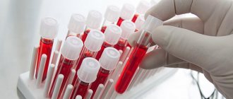
The patient is prescribed a standard set of tests to exclude inflammatory processes in the genitourinary system and bladder. The doctor is obliged to explain in detail the stages of preparation.
With ascending cystography, the urologist inserts a catheter and contrast, evaluates the images, identifying pathological changes.
In the descending version, the contrast solution is administered by the nurse. The doctor is obliged to promptly detect an allergic reaction as it develops and prescribe symptomatic therapy.
After diagnosis, the doctor should give recommendations regarding lifestyle for the coming days.
Possible complications
Cystoscopy, like many medical interventions, can cause the following complications:
- Lower urinary tract infection (cystitis, urethritis).
- Violations of the integrity of the urethral wall.
- Violation of the integrity of the wall or rupture of the bladder.
Carrying out cystoscopy in boys is somewhat more difficult than in girls due to the structure of the urethra and its length. Much depends on the skills of the endoscopist. In boys, in addition to cystitis and urethritis, prostatitis, vesiculitis, and erectile dysfunction may occur.
Compliance with the cystoscopy technique allows you to minimize the likelihood of possible complications
There are some conditions under which the doctor’s task during the research process becomes significantly more complicated. This occurs in the presence of congenital anomalies of the urinary tract, genitourinary fistulas, etc. Under such circumstances, complications may develop somewhat more often than usual.
With the right approach to treatment, most complications are reversible and can be dealt with within a few weeks.
Reviews
Reviews about the procedure make it clear how serious and unsafe it is.
Maria My son was prescribed such an examination last year (vesicoureteral reflux was suspected), but I kept putting it off until the last minute. Then the urine test results began to deteriorate significantly. My child is small; he was then one year and 3 months old. The doctor told us everything in detail and gave us recommendations for preparation. He said that anesthesia should be given, because it is difficult to force a small child to lie still. Lesha tolerated the anesthesia well and there were practically no tears. After the cystography, the doctor watched him for another two hours, then I watched myself (so that there was no blood in the urine). They recommended drinking Furamag for three days. After the procedure, the suspected diagnosis was removed and we were sent for an additional ultrasound examination. We had no consequences or complications. So there is absolutely nothing to be afraid of.
Elena My daughter had cystography done last year, when she was 2 years and 4 months old. I was very afraid of this, I read on forums about various complications. I can say one thing: it’s not that scary. The procedure itself went relatively well. Sveta cried a little, but this was while inserting the catheter. Then it was a bit difficult, I saw blood in my urine for about a day, it was painful to urinate. But everything returned to normal. The diagnosis was removed after an examination (reverse reflux of urine was suspected).
Indications
The pediatrician refers the child for ultrasound diagnostics in cases where certain symptoms are present:
- patient complaints of frequent urination, accompanied by pain;
- clinical urine analysis reveals the presence of oxalates or leukocyturia;
- in the absence of characteristic symptoms, hyperthermia is noted;
- a feeling of discomfort or pain in the lumbar or suprapubic areas.
Drinking fluids before diagnosis
The liquid consumed should not be carbonated.
Before the procedure you can drink:
- water;
- weak tea;
- juice;
- compote.
Coffee, milk, fruit drinks (cranberries, lingonberries) and alcohol are prohibited for consumption before ultrasound diagnostics.
The amount of fluid directly depends on the child’s weight. For 1 kg of body weight, you should consume 5-10 ml of liquid.
Infants are examined under the same conditions. The newborn needs to drink water, breast milk or formula at the same rate.
Older girls can take another 0.3-0.5 liters of still water half an hour before diagnosis.
Minimum fluid intake:
- children under 2 years old - at least 0.1 l;
- up to 7 years – at least 0.2 l;
- 8-11 years – at least 0.3 l;
- over 11 years old - at least 0.4 liters.