Thanks to the high level of development of modern medical equipment, a lot of new and improved methods for diagnosing patient health appear every year. Timely and correct diagnosis directly affects the outcome of treatment. The price of an error in medicine is human life.
One of the newest ways to diagnose a patient’s health is to conduct computed tomographic angiography. A similar research method is used to identify pathological processes located in the vascular system of the body's blood supply and gives an idea of the degree of patency of the blood flow.
This type of study is included in the group of procedures performed on a computed tomograph. The procedure is painless. A high degree of information content during scanning is provided by a series of images in the X-ray radiation range. Each high-resolution image shows the state of the vessel in the section.
What is cerebral angiography?
The essence of this research method is as follows. The patient is injected into a specific cerebral artery (or the entire network of cerebral arteries) with an X-ray contrast agent, usually iodine-based (Urografin, Triiodtrust, Omnipak, Ultravist and others). This is done so that the image of the vessel can be recorded on x-ray film, since the vessels are poorly visualized with a regular X-ray. The introduction of a radiopaque substance is possible by puncture of the corresponding vessel (if technically feasible) or through a catheter connected to the required vessel from the periphery (usually from the femoral artery). When the contrast agent appears in the vascular bed, a series of x-rays are taken in two projections (frontal and lateral). The resulting images are evaluated by a radiologist, who draws conclusions about the presence or absence of a certain cerebral vascular pathology.
What is coronary angiography and what is it also called?
Coronary angiography is an x-ray of the heart vessels into which a contrast agent has been previously injected. Thanks to this, the lumen and inner walls of the arteries are clearly visible. This allows timely diagnosis of blood clots, tissue tears, etc. To create contrast, the substance urografin is used.
The procedure received this name because coronary means the vessels that bring blood to the myocardium, and graphy is the generalized meaning of all x-ray studies.
You can also find the following designations:
- coronary angiography;
- coronary angiography;
- angiogram.
Varieties
Depending on the method of drug administration, this research method can be:
- puncture (when contrast is administered by puncture of the corresponding vessel);
- catheterization (when contrast is delivered through a catheter inserted through the femoral artery and advanced along the vascular bed to the required location).
According to the vastness of the area of study, angiography of cerebral vessels is:
- general (all brain vessels are visualized);
- selective (one pool is considered, carotid or vertebrobasilar);
- superselective (a smaller caliber vessel in one of the blood pools is examined).
Superselective angiography is used not only as a research method, but also as a method of endovascular treatment, when, after identifying a “problem” in a specific vessel, this problem is “eliminated” using microsurgical techniques (for example, embolization or thrombosis of an arteriovenous malformation).
Due to the widespread introduction of modern diagnostic methods such as computed tomography (CT) and magnetic resonance imaging (MRI), CT angiography and MR angiography have recently become more and more common. These studies are carried out in the presence of appropriate tomographs; they are less traumatic and safer than just angiography. But more on that later.
Types of examination
Cerebral angiography is divided into types depending on the type of vessels examined in the brain. The study of the arterial system is called arteriography. If elements of the venous system are studied, the diagnostic method is called venography. During coronary angiography, the condition of the main arteries and cavities of the heart muscle is assessed.
There are carotid and vertebral types of examination. In the first case, a contrast agent is injected into the carotid artery in the neck area, in the second, a puncture is made of the vertebral artery, which lies in the area of the spinal column. The catheter can be inserted through other large vessels (inguinal, femoral artery), which communicate closely with the main blood tracts of the head.
Depending on the mode and nature of the radiation with which the image of the vessels located in the brain tissue is simulated, X-ray, CT and MRI angiography are distinguished. In the first and second cases, visualization occurs using X-rays, in the third - electromagnetic waves. Selective angiography, unlike general angiography, involves studying the vascular system of the local area.
Classic angiography
The classic study is an x-ray method, which has more contraindications than MRA due to the use of ionizing radiation that is harmful to health. How is classical angiography done:
- A puncture (puncture) of an artery or vein is performed. The puncture site is pre-treated with an antiseptic solution.
- A catheter, a hollow tube of small diameter, is placed into the vascular lumen.
- Antihistamines, analgesics, and tranquilizers are administered to prevent the development of an allergic reaction, relieve pain, and achieve a sedative effect.
- A contrast agent is administered (mainly based on iodine and its derivatives - Cardiotrast, Triyotrast, Urografin).
- An image of the area of the vascular system under study is recorded.
- The catheter is removed and bleeding from the punctured vessel is stopped.
- Apply a sterile bandage to the puncture site.
Manipulations are carried out under local anesthesia. Typically, the duration of classical, magnetic resonance or computer angiography of vessels located in the brain is no longer than 40-50 minutes.
CT angiography of vessels
CT angiography is an x-ray of blood vessels located in the head and neck area. Unlike a classic study, the insertion of a catheter is not required, which greatly simplifies the procedure, reduces the risk of complications, and reduces the level of physical and psychological discomfort for the patient. A reduced volume of contrast agent is administered intravenously.
X-ray angiography of the vessels that make up the blood supply system to the brain is performed with contrast. The resulting data is visualized as 3D layers and slices. Thanks to the great technical capabilities of modern equipment, a large-scale, three-dimensional image of the entire system of arteries and veins is obtained with high resolution.
Computed angiography shows the presence of intracranial hematomas and areas of pathological narrowing of the vascular bed in the brain tissue. The image shows areas of heart attacks and hemorrhages formed due to blockage of an artery or vein. MSCT (multispiral) is a vascular angiography that is performed using a multislice tomograph (the sensors move in a spiral), which allows a thorough examination of all parts of the brain.
Magnetic resonance angiography
MR angiography is prescribed to study blood vessels in the brain if X-ray methods are contraindicated for the patient. Unlike computed tomography and classical x-rays, the patient is not exposed to harmful ionizing radiation. A clear, detailed image shows the smallest foci of circulatory disorders and pathology of elements of the circulatory system in the structures of the brain, which makes it possible to identify diseases in the early stages and effectively treat them. During the procedure, the following is revealed:
- Anatomical features and functional characteristics of the blood supply system.
- The width of the lumen between the walls of blood vessels.
- Deformations of elements of the circulatory system.
- The presence of obstacles to the movement of blood - blood clots, atherosclerotic changes in tissue structure.
- Inflammatory processes in the vascular system.
- New growths with a diameter of 3 mm.
The method is based on the phenomenon of magnetic resonance. It is carried out without contrast agents or with paramagnetic drugs developed primarily on the basis of gadolinium. Contrast is used when a tumor is suspected in the head cavity, to detect metastases of malignant neoplasms of any location in the brain, to assess the condition of brain structures after surgery.
The procedure is performed in the same way as a CT scan. The patient does not eat for 2 hours before the examination. It is necessary to notify the doctor if you have tattoos, personal medical devices, vascular stents, or artificial knee joints that are made of metal. Before the examination, the patient removes metal jewelry and clothing with metal fittings.
Indications for use
Angiography of cerebral vessels is a specialized diagnostic method that should only be prescribed by a doctor. It is not performed at the request of the patient. The main indications are:
- suspicion of arterial or arteriovenous aneurysm of cerebral vessels;
- suspicion of arteriovenous malformation;
- determining the degree of stenosis (narrowing) or occlusion (blockage) of cerebral vessels, that is, establishing the lumen of the corresponding vessels. In this case, the severity of atherosclerotic changes in the vessels and the need for subsequent surgical intervention are established;
- establishing the relationship of cerebral vessels with a nearby tumor to plan surgical access;
- control of the location of clips applied to the vessels of the brain.
I would like to note that simply complaints of dizziness, headache, tinnitus, and the like are not in themselves an indication for angiography. Patients with such symptoms should be examined by a neurologist, and based on the results of the examination, as well as other research methods, the need for angiography is determined. This necessity is determined by the doctor!
Reading Angiography Results
The results of the examination are captured on x-rays depicting the network of blood vessels. Before the procedure, an iodine-containing substance is injected into the blood, which increases the contrast of arteries and veins against the background of soft tissues.
Using images, the degree of circulatory disorders in the legs and the progression of pathological processes in the vessels are assessed. Looking at the angiogram, the specialist can determine:
- presence of blood clots;
- form of vascular disease;
- degree of obstruction of veins and arteries;
- localization and extent of the lesion;
In the absence of pathologies, the external contours of the vessels are smooth, and the thickness of the veins and arteries is the same in all areas. After making a diagnosis, the doctor gives a written opinion and, if necessary, prescribes the optimal treatment regimen.
Contraindications
The main contraindications are:
- allergic reaction (intolerance) to iodine preparations and other radiopaque substances;
- pregnancy (due to ionizing radiation during the procedure). In this case, MR angiography may be performed;
- mental illnesses that do not allow you to comply with all the conditions for the procedure (for example, a person will not be able to help but move during the photo);
- acute infectious and inflammatory diseases (as the risk of complications increases);
- violation of the blood coagulation system (both downward and upward);
- the general condition of the patient, regarded as severe (this may be stage III heart failure, end-stage renal and liver failure, coma, and so on). Essentially, this subgroup of contraindications is relative.
Multislice CT angiography in Volyn hospital
The computed tomography room of the Volyn Hospital is equipped with a modern spiral tomograph “Bright Speed Elite”. Doctors at the CT office are at the forefront of the development of computed tomography in Russia and have unique, enormous experience and original research methods. Highly qualified personnel and the latest software allow us to conduct the widest range of research.
The department has extensive experience in computed tomography (CT) of the spine and extremities for degenerative diseases, trauma, tumors and inflammatory diseases. Three-dimensional and multiplanar image reconstructions are widely used.
Preparation for angiography
To obtain accurate results and reduce the risk of complications from the procedure, it is recommended:
- take general and biochemical blood tests, including determining the indicators of the coagulation system (the statute of limitations for tests should not exceed 5 days). The blood type and Rh factor are also determined in case of possible complications;
- do an ECG and FG (FG, if one has not been performed within the last year);
- do not drink alcoholic beverages for 14 days;
- during the last week, do not take drugs that affect blood clotting;
- perform an allergy test with a contrast agent. To do this, 0.1 ml of the corresponding drug is administered intravenously to the patient over 1-2 days and the reaction is assessed (the appearance of itching, rash, difficulty breathing, etc.). If a reaction occurs, the procedure is contraindicated!
- the day before, take antihistamines (antiallergic) drugs and tranquilizers (if necessary and only as prescribed by a doctor!);
- do not eat for 8 hours and do not drink water 4 hours before the test;
- swim and shave (if necessary) the site of puncture or catheterization of the vessel;
- Before the examination itself, remove all metal objects (hairpins, jewelry).
Features of coronary angiography
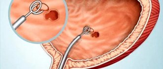
The mechanism of visualization of existing stenoses in the coronary arteries.
Vessel contrasting is performed with iodine-containing contrast, which is injected directly into the sinuses of Valsalva, located near the aortic valve leaflets, to visualize the coronary arteries. For convenience, special catheters are used with which you can enter the mouth of the right and left coronary arteries.
Access for this is usually used through the femoral or radial artery. The choice is based on the anatomical features of the arterial bed of each patient, as well as the preferences of the doctor.
Local anesthesia is used at the site of the artery puncture. A mild anxiolytic (eg, Diazepam) can be used to generally calm the patient in anxiety states, but its use is not widespread.
Important! The procedure is painless, with the exception of pain in the area of the selected access.
If we talk about planned angiography, it is usually customary for the patient to be hospitalized for one night after the procedure. In the absence of complications and only after assessment by a doctor, the patient can be discharged one day after coronary angiography. In some medical institutions, it is customary to leave it overnight before the procedure for closer attention to the patient and assessment of his condition.
As with most invasive medical interventions, this case requires patient preparation. The instructions that the patient must adhere to usually include not eating for at least 6-8 hours before the procedure, limiting oral fluid intake (medicines and small amounts of colorless liquid are an exception to this list), and also preparing as much as possible psychologically for the intervention.
Even minimally invasive interventions carry possible risks and complications.
The need for coronary artery revascularization is assessed on site, immediately after determining the blood flow in the right and left coronary arteries. If the condition of the vessels allows angioplasty, it is performed immediately during the procedure.
This allows you to save time in a hospital stay and at the same time improve your quality of life.
Stenosis in a branch of the left coronary artery.
If the patient is not suitable for stenting, but revascularization is needed, the doctor will decide when bypass surgery can be performed. The delay of surgical intervention depends on the degree of damage to the coronary bed - from 24-48 hours after angiography to several weeks.
Attention! If the question of prescribing bypass surgery to a patient is being decided, the patency of the internal mammary arteries, which can be used as a bypass during surgery, is usually assessed.
Research technique
At the very beginning, the patient signs consent to conduct this type of study. The patient is placed with an intravenous peripheral catheter to have immediate access to the circulatory system. Then premedication is carried out (approximately 20-30 minutes before the procedure): antihistamines, tranquilizers, painkillers are administered to minimize discomfort during the procedure and the risk of complications.
The patient is placed on the table and connected to devices (cardiomonitor, pulse oximeter). After treating the skin with local anesthetic and anesthesia, the corresponding vessel (carotid or vertebral artery) is punctured. Since it is not always possible to accurately get into these arteries, most often a small incision is made in the skin and a puncture of the femoral artery, followed by immersion of the catheter and passing it through the vessels to the study site. Advancement of the catheter along the arterial bed is not accompanied by pain, since the inner wall of the vessels is devoid of pain receptors. The catheter's progress is monitored using x-rays. When the catheter is brought to the mouth of the required vessel, a contrast agent in a volume of 8-10 ml, preheated to body temperature, is injected through it. The administration of contrast may be accompanied by the appearance of a metallic taste in the mouth, a feeling of heat, and a rush of blood to the face. These sensations go away on their own within a few minutes. After the administration of contrast, x-rays are taken in frontal and lateral projections almost every second several times (which allows you to see the arteries, the capillary phase, and veins). The photographs are developed and immediately assessed. If something remains unclear to the doctor, an additional portion of contrast agent is injected and the images are repeated. Then the catheter is removed, and a sterile pressure bandage is applied to the puncture site of the vessel. The patient should be monitored by medical personnel for at least 6-10 hours.
Decoding of received images
The results of the examination are presented in the form of an X-ray image, which is examined by the surgeon and the attending physician. The image shows the blood vessels and arteries into which the contrast agent was injected. It is strongly not recommended to independently decipher the obtained indicators and establish a diagnosis.
The conclusion of the disease is determined by the surgeon and the doctor who performed the radiation diagnostics. The image shows the presence or absence of vein blockage and thrombophlebitis. The condition of the veins and the integrity of the arteries are also determined. The narrowing of the lumen is noticeable, and the exact location of the vessel damage is diagnosed.
Constructing a picture of the vessels of the legs based on angiography results
Elimination of blood clots
If a blood clot is found in a vessel or artery, it is examined in detail to see if it can be opened. This procedure is called in medicine – angioplasty. A balloon is attached to the end of the catheter, which penetrates the blocked vessel. The balloon is then inflated to open the clot. Pressure is applied to the arteries and blood vessels, they dilate, normalizing healthy blood flow.
After the microsurgery, the doctor may decide to install a stent in the area of the blockage to prevent further formation of blood clots. The device is also attached to the end of the catheter and advanced through the vessels to the appropriate area. Another way to open a blood clot is with medication, without the use of surgical maneuvers. A drug that dissolves vein blockages is injected into the blood during examination and diagnosis.
In addition to medication, the doctor may decide to install a stent in the blood artery. In addition to this, medications that thin the blood are prescribed. After the procedure, the catheter is removed from the vessel, and a bandage is applied to the wound site to prevent bleeding. In some cases, the doctor may decide to use stitches. After inserting the catheter, physical pressure should not be applied to the area being used, nor should it be bent to avoid complications and bleeding.
After the procedure, the patient remains in the hospital under the supervision of medical personnel to eliminate side effects and complications. In case of bleeding or anaphylactic shock, doctors will be able to provide timely assistance. For two days after angiography, you should avoid physical activity and adhere to bed rest. If all recommendations are followed, the examination process will proceed without complications.
Complications
According to statistics, complications during this diagnostic method occur in 0.4-3% of cases, that is, not so often. Their occurrence may be associated both with the procedure itself (for example, bleeding from the puncture site of a vessel) and with the use of a contrast agent. It should be borne in mind that compliance with all conditions when preparing and performing angiography is the prevention of possible complications. The use of iodine-containing drugs of the latest generation (Omnipaque and Ultravist) is characterized by lower statistics of complications.
So, possible complications of cerebral angiography are:
- vomit;
- an allergic reaction to an iodine-containing drug: itching, swelling and redness at the injection site, followed by the appearance of shortness of breath (reflexive breathing disorder), a drop in blood pressure, and heart rhythm disturbances. In severe cases, anaphylactic shock may develop, which is a life-threatening condition;
- spasm of cerebral vessels and, as a consequence, acute cerebrovascular accident (up to stroke);
- seizures;
- penetration of the contrast agent into the soft tissue in the area of the puncture of the vessel (outside the vascular bed). If the volume of the drug spilled into the tissue is up to 10 ml, then the consequences are minimal, but if more, then inflammation of the skin and subcutaneous fat develops;
- leakage of blood from the puncture site of the vessel.
CT and MR angiography: what are the features?
CT and MR angiography of cerebral vessels are inherently a similar study as angiography. But there are a number of features of these procedures that distinguish them from angiography of cerebral vessels. That's what we'll talk about.
CT angiography
- it is performed using a tomograph rather than a conventional X-ray machine. The study also relies on X-rays. However, its dose is significantly less than with conventional angiography of cerebral vessels, which is safer for the patient;
- computer processing of information allows you to obtain a three-dimensional image of blood vessels at absolutely any point of the study (this applies to the so-called spiral CT angiography, carried out on a special spiral tomograph);
- the contrast agent is injected into the vein of the elbow, and not into the arterial network (which significantly reduces the risk of complications, since the administration of the drug becomes a routine intravenous injection through a peripheral catheter).
- To perform CT angiography, there is a restriction on the person’s weight. Most tomographs can withstand body weights of up to 200 kg;
- the procedure is carried out on an outpatient basis and does not require observation of the patient after its completion.
CT angiography on a multislice tomograph (MSCT angiography)
In multislice (multi-layer, multi-slice) computed tomographs (MSCT), unlike tomographs of previous generations, not one, but two or more rows of detectors are located around the circumference. This technology significantly increases the speed and information content of studies, and also reduces the radiation dose to the patient. MSCT has a number of advantages over conventional computed tomography:
- improved time resolution;
- improved spatial resolution along the longitudinal z-axis;
- increasing scanning speed;
- improved contrast resolution;
- increase in signal-to-noise ratio;
- effective use of the X-ray tube;
- large anatomical coverage area;
- reducing radiation exposure to the patient;