On traditional x-rays, the specialist is not able to visualize the arteries, venous network, lymphatic vascular system and capillaries, since these elements are not able to absorb x-rays, similar to the soft tissues in their surroundings.
Therefore, to diagnose these anatomical structures, vascular angiography is performed, accompanied by the injection of a contrast agent.
In this way, the coronary vessels of the heart are diagnosed or coronary angiography is carried out; the vascular network of the brain, neck and other vessels of the human body is examined.
Angiography of cerebral vessels: the essence of the procedure
Traditional angiography of cerebral vessels means an X-ray of the head after contrasting the vascular network of the brain using contrast agents.
This diagnostic method helps to examine all phases of the blood supply to the brain step by step, establish defects in the blood vessels of the head and their location, and is applicable in identifying neoplasms.
The procedure is carried out by puncture or catheterization of extracranial and intracranial vessels, injection of medication and subsequent output of photographs.
Among the X-ray contrast agents used are substances containing iodine (Verografin, Triombrast, Gipak, etc.). These agents are water-soluble and are administered in parallel.
Blood enters the brain from the following pools - carotid and vertebrobasilar (these are the carotid and vertebral arteries).
Consequently, one of the arteries is filled with contrast. Most likely it is the carotid artery.
The essence of the method
Angiography is a diagnostic method that helps identify diseases of the veins and arteries in tissues and internal organs. Previously, it could be used as part of radiography or computed tomography. But both methods involve irradiation with X-ray frequency waves. With the discovery of magnetic resonance imaging, it became possible to obtain images of the vascular network of the human body without harm to the patient. Let's find out how it works.
The essence of MRI in angiography mode is nuclear magnetic resonance. The fact is that the nuclei of hydrogen atoms vibrate at a certain frequency, creating a magnetic field around themselves. If they are placed in another magnetic field created by a tomograph, then these fields will overlap each other and intensify. If you ensure that the magnetic field inside the tomograph oscillates at the same frequency as the nuclei of hydrogen atoms, a phenomenon called resonance occurs. At this time, the strength of the magnetic field increases sharply, which is recorded by the device’s sensors. Collecting signals, the computer analyzes them, converting them into a graphical form of information. The result is a picture that accurately depicts the structure of veins and arteries, as well as their location.
Diagnosis using MRI is safe for the patient because it uses only a magnetic field and radio waves. Unlike X-rays, they do not irradiate the body.
Types of research
The methodology for performing the procedure determines two types of examinations:
- puncture (the contrast is poured exactly into the vessel using a puncture needle);
- catheterization (the agent is injected using a catheter connected to the local vascular bed).
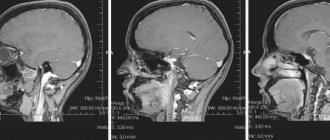
MRI
Based on the area examined, brain scans with contrast can be presented as:
- general angiography (visualization of vessels of different calibers is performed);
- selective angiography (means scanning of the vertebrobasilar, carotid region);
- superselective technique (a vessel with a caliber that does not correspond to any blood pool is examined).
The vascular system imaging pathway determines the following scan types:
- classical angiography - taking x-rays with preliminary introduction of contrast into the vessels of the head;
- CT angiography - scanning of the vascular system in the brain using an x-ray method with preliminary contrast and further 3D modeling of the display of the blood supply system;
- MR angiography involves examination using magnetic resonance imaging, often without prior contrast.
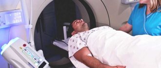
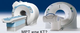
Computed tomography (MSCT) MRI
Preparing for the study
Examination of the vessels of the head using CT angiography does not require special preparation - you can eat food and take medications the day before. However, some diagnostic centers require the patient to refuse food 4-6 hours before the diagnosis, so this issue must be clarified with the clinic specialists when registering for the procedure. It may also be recommended for several days before the procedure to refrain from drinking alcohol and foods that change the composition of the blood (spicy, fatty, fried, etc.).
If the patient has renal impairment, it is necessary to undergo an analysis of blood biochemistry indicators (urea and creatinine), since with a contrast study there is a risk of additional damage to renal function.
Features of CT and MR angiography
Both research methods are characterized by a high level of accuracy and efficiency, however, each of them has a number of features that are taken into account when choosing a specific procedure.
MR angiography
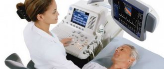
Vascular MRI has a small list of limitations and is safe for health, since it is often performed without contrast. This diagnostic method is applicable both for examining the vascular network and soft tissue elements.
However, in case of traumatic brain injury, MRI with angiography cannot be considered an effective measure. Under such circumstances, it is necessary to look for cracks in the cranial bone, vascular ruptures and malfunctions of the digestive system. Vascular MRI is not suitable for diagnosing bone and fluid structure.
CT angiography
Diagnosis is accompanied by the introduction of a contrast agent under the skin into the venous system of the forearm. The procedure will be a suitable diagnostic method in cases of detection of bone tissue pathologies or aortic aneurysm.
During the procedure, the doctor determines the extent of the pathological area, identifies the presence of blood clots and clots (provided they are visualized), and assesses the possibility of performing the operation.
MRA
CT
What is CT angiography of the brain?
One of the most popular studies in modern medicine is CT angiography, which is an X-ray method for visualizing the functional and morphological state of cerebral vessels. During the scan, a series of images of arteries, veins and cranial sinuses are obtained; these images can later be reconstructed into a three-dimensional model of the cerebral circulatory system.
This study allows us to make accurate conclusions about the places of narrowing and dilation of the vessel, blockage of the lumen of the vessel with a thrombus, compression of an artery or vein by a growing tumor, the presence of an inflammatory process and other disorders.
Note that CT angiography is always performed with intravenous administration of a radiopaque contrast agent containing iodine compounds - otherwise the examination will be uninformative.
Indications for use
Among the key purposes for performing cerebral angiography:
- assumption of the development of an arterial (arteriovenous) aneurysm of blood vessels in the local area;
- manifestation of symptoms of arteriovenous malformation;
- development of stenosis or blockage of cerebral vessels (the presence of atherosclerotic vascular deformations and the need for further surgery are revealed);
- determining the degree of contact of the brain’s blood network with the tumor for the purpose of planning surgical intervention;
- control of the location of clips on the vessels of the thinking organ.
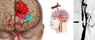
Cerebral aneurysms
Patient complaints of dizziness, migraines, and noise in the head are not considered direct indications for scanning.
A person with similar symptoms is examined by a neurologist, who determines whether angiography is justified in a particular clinical case.
Decoding images
Examination of blood vessels reveals the following pathologies:
- Aneurysms (pathological expansion of the walls of blood vessels) and their dissection;
- Congenital heart defects;
- Arterial atherosclerosis;
- Inflammation of the vascular walls (vasculitis);
- Stenosis (pathological narrowing) of blood vessels.
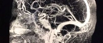
Unlike conventional tomography of the brain, angiography diagnostics can detect hemorrhagic types of strokes. This method, combined with contrast, is also good for detecting tumors. Inside them there is always a densely woven vascular network. When a contrast agent passes through it, the neoplasm stands out in the image as a bright spot with clearly defined edges.
MRI vascular angiography is completely safe for the body, but this type of diagnosis is very informative. It detects the presence of tumors and visualizes pathological structures. All this is necessary to make an accurate diagnosis and prescribe effective treatment for vascular diseases.
List of contraindications
In the table below we present a list of restrictions on conducting various types of research:
| Event | Limitation |
| X-ray CT angiography | — acute infection — allergy to contrast — diabetes mellitus (type 2) — pregnancy period — myeloma — thyroid disease — serious kidney, liver and cardiovascular diseases |
| MRI with angioprogram | — implanted pacemaker, orthopedic and orthodontic metal implants, vascular clips — heart failure — intolerance to confined spaces |
Additional contraindications for both diagnostic methods include: the weight of the person being scanned is more than 120 kg, mental illness (possible use of anesthesia), and poor health of the patient.
The essence of the examination
The use of X-rays in medicine has revolutionized everything. Small doses of radioactive radiation made it possible to visualize tissues and organs, which influenced the quality of treatment and helped to make a more accurate diagnosis.
Organs and tissues absorb ionizing radiation. The high density of the organ is reflected by a lighter area in the image. The network of brain vessels does not accumulate photons, since the fluid in them is in continuous movement. Therefore, the visualization in the image turns out to be quite weak and not informative during a standard study.
Cerebral angiography refers to a contrast diagnostic method using non-invasive radiography. An iodine-based contrast agent is injected into the bloodstream. The blood flow carries the drug throughout all vessels, including the capillary network.
Iodine actively absorbs X-rays, which helps to examine the vascular network in the brain and detect pathological changes. What angiography shows is that a series of images allows you to evaluate blood circulation in the brain and detect the cause of obstruction in the vascular network.
When is angiography prescribed?
The need for angiography will be determined by a medical council or one of the specialists (surgeon, neurologist, oncologist). Indications are the patient’s subjective complaints or an established diagnosis.
Objective reasons for diagnostics:
- Migraine;
- Fainting;
- Seizures;
- Vomiting that does not bring relief due to headache;
- Loss of coordination (wobbly gait);
- Stenosis of cerebral vessels;
- Atherosclerosis;
- Suspicion of the presence of a tumor formation or diagnostic control after surgery;
- History of vascular pathologies;
- Suspicion of acute cerebrovascular accident;
- Dynamics of the therapy performed.
Angiography of cerebral vessels using the classical method is possible only after undergoing a medical examination. If there are contraindications, the procedure is not prescribed. One of the fluoroscopic diagnostic methods is selected - computed angiography or MRA.
Advantages of the method
Angiography of the brain allows you to accurately determine the cause of pathological changes in blood vessels or identify congenital anomalies. The contrast agent penetrates even the smallest intracranial vessels and does not require exposure to high doses of radiation.
Visualization of cerebral vessels in the image is possible in several projections, which makes it possible to examine the dynamics of blood flow and its phases. This method has no age restrictions, complications after the procedure are rare.
Angiography is a highly accurate diagnostic method that allows you to track the extent of the pathological process, as well as identify rare chronic diseases.
The procedure takes a short period of time and is the most cost-effective in comparison with other methods.
Preparatory stage
Before cerebral vascular angiography (X-ray, CT or MRI diagnostics), laboratory tests are performed: general blood test, urine, biochemical analysis of biological fluid, determination of blood group, Rh factor.
Before the procedure (two days), the person being scanned is required to stop using medications that affect blood clotting.
Before the examination, the doctor makes a visual examination of the patient and obtains approval for the diagnosis.
A few hours (6-8) before performing angiography of the vessels of the brain and neck, the person being scanned should refuse to eat.
If there is hair at the needle insertion site, it needs to be shaved off. On the eve of the study, the person is recommended to use sedatives.
If it is planned to determine vascular diseases of the main organ of the central nervous system using contrast, the patient must be tested for the presence of an allergic reaction.
During the procedure, a small amount of medication is injected subcutaneously into the person, and then the patient’s well-being is monitored.
If adverse events occur (in the form of rash, nausea, vomiting, etc.), angiography is canceled. In such a case, it is advisable to use MRI of the arteries without contrast.
If you are planning an MRI in angio mode without contrast injection, there is no need to follow a diet on the eve of the procedure or stop using medications.
The only thing the patient should do before the examination is to get rid of metal objects and jewelry.
How is the examination carried out?
Before MSCT angiography of cerebral vessels is performed, you will need to sign an agreement regarding the study. Next, an intravenous catheter will be inserted into the patient's body to gain access to the bloodstream. After this, remedication is carried out, this is done about half an hour before the examination.
The person will be given antiallergic drugs and analgesics to reduce the likelihood of complications and reduce discomfort from the procedure. The person will be placed on a special table and then connected to devices: a heart monitor and a pulse oximeter. The doctor will treat the skin and then puncture the desired vessel.
Given the fact that it is not possible to reach the artery in all cases, it may be necessary to cut the skin. A catheter will be inserted through the femoral artery and then moved to the test site. This action does not cause pain, because the vascular wall is not capable of experiencing pain.
The resulting results are immediately displayed and analyzed. If any points remain unclear, you will need to use an additional portion of contrast and take an x-ray again. After this, the catheter is removed, and a sterile bandage is applied to the damaged area, exerting pressure. Now the patient must be observed by a doctor for 8 hours.
There is cerebral angiography of cerebral vessels, magnetic and resonance, computed tomography, and multispiral. The doctor will independently decide which type is needed for a particular person. If cerebral is prescribed, then the contrast will be administered through the femoral or carotid artery.
There are several different types of cerebral angiography.
Angiography using computed tomography (CTA) provides a detailed image of the vessels and shows patterns of blood flow. In this case, intravenous contrast enhancement is used.
After CTA, image reconstruction is performed.
A definite positive side of this method is the reduced radiation exposure to the patient’s body.
CT angiography is often performed for stenosis, thrombosis, aneurysms, and vascular developmental defects.
Contraindications are allergies to the contrast agent, diabetes, pregnancy, obesity, thyroid problems, myeloma, heart disease, uncontrollable arrhythmia and tachycardia.
The study is carried out on an outpatient basis. Approximately 100 ml of contrast agent is injected into a venous catheter, which is inserted into the cubital vein. The patient lies on the CT scanner table.
X-rays scan the area under study in parallel with the introduction of a contrast agent.
Magnetic resonance angiography (MRA) allows you to study the functions of blood flow and its anatomical features.
The basis of magnetic resonance imaging is to track energy changes in tissues, their structure and chemical composition. Contrast agents are practically not used in MRA (occasionally gadolinium-based to obtain high-precision images).
MRI angiography of cerebral vessels is used to diagnose aneurysm dissection, congenital heart defects, and vasculitis.
Contraindications include installed implants, pacemakers, nerve stimulators, blood-restoring clips, insulin pumps, prosthetic heart valves, heart failure, pregnancy, claustrophobia.
Cerebral angiography is a kind of “gold standard” for studying cerebral vessels.
The author of this method is Egas Monitz, who performed angiography for the first time in 1927.
The method is of the highest value because it allows you to accurately detect aneurysms, narrowing of blood vessels or the site of their blockage, and brain tumors.
The catheter is inserted into the vessel through the femoral artery and directed to the carotid artery. A contrast agent is injected into the blood vessels and X-rays are taken to determine the state of the inflow and outflow of venous blood.
During cerebral angiography, surgical intervention is possible. The information content of the method is much superior to CTA and MRA.
Arteriography
Arteriography involves the introduction of a contrast agent into the lumen of the vessel, which makes it possible to determine the presence of tumors located close to blood vessels, arterial pathologies and other circulatory disorders.
Most often, this method is used to study the limbs.
Arteriography is relatively simple, performed on an outpatient basis, but is painful because the contrast moves quite quickly through the arteries.
X-ray contrast agents (about 30-40 ml) are injected through a catheter or directly into the artery under strong pressure in the direction of the blood flow (less often against the blood flow).
This method allows you to diagnose changes even in the deepest arteries, which are monitored using the screen of an X-ray machine.
Venography
Another name for venography is phlebography. The essence of the method corresponds to its name.
Venography allows you to see the distribution of veins; it is actively used for varicose veins and thrombosis, as well as arrhythmia. The patient is advised to breathe calmly and relax during the procedure.
This is a simple and painless method, but in rare cases, your health may deteriorate after the procedure, and phlebitis may appear - inflammation at the site of contrast injection.
Venography involves the use of small amounts of a contrast agent that is injected directly into a vein (direct venography). After the procedure, an injection is made using 60 ml of saline to cleanse the vessels.
It is most justified to use venography before surgery on the veins.
Indirect venography can be performed in three ways:
- contrast is injected into the artery and then enters the veins through the capillaries;
- contrast is introduced into the tissue of the affected organ that needs to be examined, and the pictures show veins draining blood from the organ;
- contrast is injected directly into the medullary space.
What is the danger of post-traumatic encephalopathy of the brain - treatment and prevention of the disease. What is the most common cause of a retrocerebellar arachnoid cyst of the brain and what to do if there is a suspicion of this formation.
Lymphography
Lymphography is a method of studying the lymphatic system also using a radiopaque agent.
The study is carried out in three projections and the data is studied immediately after the administration of contrast (early lymphogram) and after 1-2 days (late lymphogram).
Early lymphograms make it possible to examine the condition of the lymphatic vessels, late lymphograms - the lymph nodes.
This method allows you to identify changes in the external and common iliac, inguinal, supra- and subclavian, lumbar, axillary lymph nodes; identify the presence of tumor processes and optimize cancer treatment.
During angiography, the patient is placed on a table, his position is fixed, and he is connected to a cardiac monitor.
Before administering contrast, premedication is performed, that is, injections of analgesics, tranquilizers, and antihistamines are given.
A special catheter is inserted into the vessel through which the study needs to be carried out (most often the femoral artery) using a puncture (puncture). Catheterization allows you to introduce a contrast agent, usually iodine. The puncture site is numbed.
Angiography is performed within 40 minutes. The doctor controls the actions using X-ray television. Medical conclusions are made after developing and viewing the images.
Possible complications may be:
- introduction of a contrast agent into tissues outside the vessel (extravasation), which leads to damage to the skin and subcutaneous tissues;
- allergic reaction to contrast agent;
- disorders of the kidneys.
Modern angiography uses digital technologies, which makes the study less traumatic for the patient and highly informative for the doctor.
Important Terms
- Aseptic conditions for the procedure,
- Availability of a team of doctors: radiologist, anesthesiologist, cardiac resuscitator.
The patient examination process itself takes approximately half an hour to an hour. The procedure is considered invasive because a puncture is made to access the artery, where a special catheter is inserted. Therefore, cerebral angiography is often combined with other interventions in the body that occur through access through large blood vessels, for example, with the removal of an aneurysm.
To avoid complications associated with infection through the catheterization site, the skin is treated with an antiseptic solution. Next, local anesthesia is performed. Puncture (puncture) of the vessel is carried out with a special needle. A flexible catheter is inserted through this site to supply contrast. As a rule, the puncture is done in places through which it is easy to “get” to the necessary vessels.
A contrast agent is injected into the blood through a special catheter. After contrast, a series of X-ray images of the cerebral vessels is taken.
When the filming is completed, the patient's catheter is removed and the bleeding stops. Next, the received information is decrypted. A vascular surgeon and a radiologist are involved in deciphering and establishing or clarifying the diagnosis.
After the angiography procedure, the patient must remain under the supervision of medical professionals for some time.
Progress of the examination
The event begins with the administration of contrast medication (if necessary). The drug enters a vein in the elbow or forearm. No more than 100 ml of the drug is used.
Medical actions do not cause pain to the person; some people being scanned experience a slight sensation of heat.
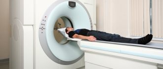
At the next stage, the person being scanned changes into disposable clothes and lies down on an equipped table, which will move into the ring part of the device during the examination. During the procedure, the person must lie still.
Injection of contrast agent
The diagnostic process does not cause discomfort to the patient. Oxygen enters the tomograph, the machine makes noise, and a cracking sound may appear; if necessary, the patient is asked to use earplugs or headphones.
If the patient needs help, he can always contact a specialist using the built-in microphone or a button inside the equipment.
The examination takes on average about half an hour. After the table is outside the cylinder, the subject can get up, get dressed and leave the room. The results of the study are given to the patient.
The examination being carried out is not a traditional surgical intervention; it is a rather difficult procedure that puts a strain on the patient’s organs. This statement is especially relevant in relation to contrast research.
Therefore, after diagnosis, the patient is under the supervision of specialists to prevent undesirable consequences.
Mandatory rehabilitation appointments include regular monitoring of the patient’s body temperature and inspection of the puncture area.
Contraindications and restrictions
MRI with vascular angiography is not always possible. The procedure has the following contraindications:
- The presence of implants in the middle ear (if they are made of metal from the ferromagnetic group);
- Installed pacemaker (the magnetic field will simulate the heart rhythm);
- Foreign metal bodies in the examined area of the body;
- Clips installed in the brain to stop bleeding in order to exclude subarachnoid or intracerebral bleeding;
- Body weight is greater than the maximum permissible load for which the tomograph is designed (usually the limit for MRI is 120-140 kg);
- Individual intolerance to gadolinium (when performing a procedure with contrast).
The above are absolute contraindications, but there are also relative ones:
- Claustrophobia;
- Pregnancy in the first trimester;
- The presence of dental implants, which can distort the real picture;
- Colds and runny nose, as well as other inflammatory diseases with very pronounced symptoms (diagnosis loses some information content);
- Kidney failure and bronchial asthma (if a substance is administered to obtain clearer images).
Tomography is safe for women during pregnancy at any stage (and for the fetus too). However, in the first trimester, when all the organs are developing, it is better to play it safe. Especially if we are talking about a study with the introduction of a contrast agent.
MR angiography allows you to look inside the vessels, but unlike other diagnostic methods that involve x-rays, it does not visualize calcium deposits inside them. Therefore, the results of CT and MRI may differ from each other. In addition, the image of small vessels and capillaries will be blurry and not clear enough.
Possible complications
Despite the relative safety of angiography, scanning can lead to a number of undesirable reactions, including:
- the appearance of an allergy to the administered contrast agent, leading to anaphylactic shock;
- the formation of an inflammatory process (neurosis) of tissues surrounding the vessel, which develops due to the penetration of contrast into it;
- development of acute renal failure.
An allergic reaction is the most common of the rare unwanted deviations. The reaction to iodine-containing drugs appears unexpectedly and progresses sharply. Possible manifestations of this adverse reaction include:
- swelling;
- redness of the skin;
- itching;
- low blood pressure;
- lethargy;
- fainting.
The use of non-ionic contrast agents will help prevent the occurrence of anaphylactic shock.
Extravasation is the result of poor technique for puncturing the arterial wall. Under such circumstances, the artery is punctured through - the contrast drug enters the soft tissue near the artery, provoking an inflammatory process or, less commonly, necrosis syndrome.
Acute renal failure manifests itself with previously diagnosed malfunctions in the functioning of the renal apparatus. This adverse reaction occurs when contrast is used.
Since the kidneys are responsible for removing the auxiliary agent, the organs undergo significant stress. The result of this is parenchymal ischemia and renal dysfunction.
To reduce the intensity of the load on the excretory organ and speed up the process of removing contrast from the body, after diagnosis the patient is advised to drink a lot.
Assessing the condition of the urinary system is a mandatory technique that precedes angiography of the cerebral vascular system.
Possible contraindications
Despite the relative safety and information content of the manipulation, it is not always permitted . The main contraindications will be the following:
- period of pregnancy and breastfeeding;
- allergic reaction to iodine and drugs that contain it;
- coma, when the patient is unable to respond to the doctor’s instructions;
- mental illnesses accompanied by emotional agitation, when the patient’s behavior cannot be controlled;
- thyroid diseases, such as hyperthyroidism;
- severe form of renal failure;
- blood pathologies with impaired blood clotting and a tendency to bleed;
- the presence in the patient’s body of titanium implants, piercings or tattoos made with paint containing metal particles.
Diagnostics are not carried out without the consent of the patient. A relative contraindication would be any pathology in the acute stage. In this case, the specialist postpones the study until the patient’s condition improves.
Complications usually do not occur, but sometimes the patient may experience nausea caused by the contrast agent, headache, weakness, and drowsiness. As a rule, symptoms disappear within a few hours. If the condition does not improve, symptomatic therapy is used.
A question of cost
The cost of an MRI or CT scan of the brain veins varies depending on the specifics of the scan. The average price of a CT scan in Russia is 2,500 rubles, and an MRI diagnostic is 3,000 rubles.
Angiography of the vessels of the head and neck is a reliable and fairly safe diagnostic method and reveals many local undesirable processes. Based on the method of tissue visualization, classical, CT and MR diagnostics are distinguished.
In the first two cases, the direct examination is necessarily preceded by the administration of a contrast drug; with MR angiography, contrast is often excluded. There are a number of contraindications to each type of diagnostic procedure.
The examination takes about 30 minutes. Possible complications include: renal failure, tissue inflammation (necrosis), allergic reaction to contrast. The cost of the study is determined by the specifics of the event.
What organs are examined and in what cases are they used?
Magnetic resonance angiography can provide images of blood vessels in any organ. But most often there is a need:
- In the study of cerebral vessels;
- In the study of veins and arteries of the head and neck;
- In the diagnosis of cardiac vessels.
Indications for angiography using MRI are:
- Traumatic brain injury;
- Vasculitis (inflammation of the vascular walls);
- Vascular atherosclerosis;
- Phlebeurysm;
- Aortic dissection;
- Congenital heart defect;
- Visual and hearing impairments;
- External vascular compression syndrome;
- Frequent headaches;
- Narrowing of arteries in diameter;
- Suspicion of cancer.
Most brain pathologies have a cause-and-effect relationship with the condition of the vessels of the neck and the brain itself. Therefore, the possibilities of angiography are extensive: it can reveal not only the disease, but also the causes that caused it.
Are there any contraindications
Angiography has contraindications depending on the method used. There are restrictions that are the same for all methods:
- pregnancy;
- mental disorders;
- lactation;
- thyroid pathology;
- kidney failure;
- allergy to iodine;
- heart failure;
- diabetes;
- poor blood clotting;
- obesity (the patient does not fit into the device).
A contraindication for the classical method and computed tomography is the ban on x-ray exposure. Magnetic resonance angiography may have limitations due to the use of a magnetic field. These include:
- cardiac pacemaker implant;
- claustrophobia;
- electronic ear implants;
- metal parts in the body - plates, joints.
- Cupcake recipe with photos step by step. How to bake beautiful cupcakes at home
- Blood Rh factor
- APTT - what is it in a blood test. Normal indicators, reasons for increased or decreased APTT
Types of angiography
It is not only the vasculature that can be visualized using angiography. Depending on the indications, a study of the intercellular fluid (lymphatic system) may be performed.
Angiography allows you to study the intracranial vessels in the brain, which are responsible for the vital functions of the organ. Intracranial vessels are examined using one of the radiological techniques:
- Classic radiograph;
- MRI angiography of cerebral vessels;
- CT angiogram.
Iodine spreads throughout the circulatory system in phases:
- Arterial;
- Venous;
- Capillary.
The method of introducing radiocontrast has several types:
- Artery puncture - a puncture is carried out in one of the arteries (carotid or vertebral);
- Installation of a guidewire - a disposable catheter is inserted into the vessel through a puncture. Used to diagnose peripheral vascular diseases, the conductor reaches the mouth of the artery. When examining the arteries of the neck and brain, the guide is installed in the largest vessel (aortic arch in the sternum).
Angiogram is widely used to diagnose various vascular disorders throughout the body.
Types of angiogram:
- Coronary angiography is an accurate diagnostic method for heart diseases. A radiopaque contrast agent is injected alternately into the coronary arteries (left and right) through a guidewire (catheter). The water-soluble substance rapidly fills the lumen of the artery. Ionizing radiation displays on the image the relief of the vessel and the degree of damage to the coronary arteries;
- Phlebography – the venous network of the brain, lower extremities or pelvic organs is examined. In the image, it is possible to examine changes in the vascular wall and determine the location of the blood clot. With the ascending venography method, the patency of the vascular network is determined. Venography performed opposite the blood flow (retrograde) tests the functioning of the valvular vascular system;
- Angiogram of internal organs - the image shows the hepatic artery. Used for the diagnosis of cirrhosis and neoplasms. Splenic venography is used to examine the portal vein;
- Arteriography is the study of the entire circulatory network in a certain area.
With the introduction of a radiopaque substance, it is possible to examine the brain vessels in a certain area or study the entire structure of the bloodstream.
Type of angiogram depending on the area being examined:
- General angiography – the image shows all the vessels of the brain. It is considered the most difficult diagnostic method;
- Selective angiography - selective examination of the vessel branches is carried out. The method combines both diagnosis and treatment.
Selective angiogram is divided into:
- Lymphogram – visualization of the lymphatic system.
- Cerebral angiography – visualization of the venous network of the brain. A water-soluble substance is injected through an artery, followed by exposure to ionizing radiation (images in several projections).
- Superselective angiography is a study of a specific vessel. In the image, it is possible to examine in detail the deformation of the vascular wall or other pathological changes. The study combines the possibility of microsurgical treatment.
MSCT angiography of cerebral vessels is a non-invasive diagnostic method. It is performed for various brain pathologies. The procedure involves the administration of an iodine-containing substance and diagnostics using tomography.
Multislice CT is a type of radiological examination. The method allows you to obtain highly accurate data, since images can be obtained in any projection and layer by layer.
If there are contraindications for the classical research method or it is impossible to gain access to the vessel, the diagnosis is carried out without the use of contrast. The procedure is carried out using magnetic resonance imaging.
Resonance angiography allows one to evaluate not only the anatomical structure of blood vessels, but also the functional characteristics of blood flow in the brain.
Angiography techniques
Angiography of the brain is prescribed only by a doctor after a thorough examination and preparation of the patient. A brain angiogram using a contrast agent is performed after a preliminary test for an allergic reaction.
The choice of technique depends on the indications and contraindications:
- Standard method;
- MRI diagnostics;
- CT diagnostics.
Before the procedure, the patient is warned about possible complications. To monitor the condition, the patient may be offered medical supervision on the day of the study; this requires hospitalization for 1 to 3 days. After the examination, you may feel general weakness and chills.
CT angiography
CT angiography of the vessels of the brain and neck is performed using a radiocontrast agent. The method is similar to the classical one, but is considered more informative. Visualization of brain vessels occurs using a tomograph, which allows you to view the image on a computer screen or simulate a three-dimensional image on film.
CT scan of cerebral vessels has less radiation exposure to the body, unlike an X-ray machine. The study has minimal risks of developing serious complications.
A subtype of computed tomography is multispiral (MSCT). The general diagnostic principles of the methods are absolutely identical. The difference lies in the equipment.
Most often, MSCT is chosen for diagnosing cerebral vessels. Angiography with this method takes only a few minutes, unlike CT, which allows the study to be carried out on seriously ill patients who cannot maintain a motionless body position for a long period of time.
The sensors in a multislice tomograph are located around the entire circumference of the device. During scanning, the machine turns slowly, producing a smooth spiral-like movement - which is where the procedure gets its name.
Only one revolution is needed to scan the brain's vasculature. In a few minutes, the doctor receives a clear, informative image. Computed tomography is inferior to MSCT in terms of information content, but is considered more accessible (budget).
Advantages of multislice CT:
- A wider range of settings in the device, which allows you to change parameters for each patient;
- Lower ionizing radiation (30% difference);
- Simultaneous examination of soft tissues and bone fragments;
- The minimum study time allows the procedure to be performed on children and seriously ill patients (including those connected to life support equipment);
- Detects neoplasms from the moment of inception (up to 1 mm);
- Visualizes hematomas in the brain.
MSCT can be performed on patients with somatic diseases (mental) or people suffering from claustrophobia. The speed of the procedure does not provoke panic attacks when the patient is placed in a tomograph.
Multislice computed tomography allows you to obtain a larger number of images in a short period of time. This is achieved through thinner slices, unlike CT scans.
The average duration of a CT examination is at least 15 minutes, provided that the diagnostician’s instructions are followed during the examination (hold your breath, do not move). The study is stopped after the information obtained is reconstructed into a clear three-dimensional image.
MR angiography
MR angiography of cerebral vessels is considered a more modern diagnostic method. Visualization of the circulatory network in the image is obtained using the action of electromagnetic fields.
What is it and what is the essence of the technique? A strong magnetic field is created in the tomograph, as the positions of the hydrogen nuclei change. The magnetic field and radio frequency radiation act with a certain force, which leads to rotation of the nucleus around the created axes.
Energy release and absorption creates its own magnetic field. The change in pulses is recorded by the tomograph, thus creating an image. Registration of energy pulses is most informative in cavities filled with liquid.
Angiography of the arteries and veins of the brain is performed without radiocontrast. Obtaining a clearer image is achieved by introducing gadolinium-based contrast.
The advantage of MR angiography is that it provides ultra-clear images in any plane. The method does not require invasive intervention.
Diagnosis is carried out using one of the following options:
- Time-of-flight angiography – examines the arteries of the brain and neck. The pulses pass sequentially (perpendicular sections);
- Phase-contrast angiography - MR venography of the brain is performed. The speed of blood flow is assessed. Takes a longer period of time to study. The signal conveys amplitude and phase information;
- Four-dimensional angiography – examines the arteries and veins in the brain. The dynamics of blood flow are visualized.
The tomograph power and parameters may vary. There are only two types of tomograph:
- Open – allows diagnostics for children and seriously ill patients. The open tomograph does not have sealed walls, which allows examination of patients with phobias. An open device is used to diagnose people who do not fit the parameters of a standard tomograph (weight, height);
- Tunnel apparatus - a movable couch slides into a kind of tunnel (wide pipe). The device has a built-in ventilation system. Data is output to the computer through wires located parallel to the neck. Communication with medical personnel is carried out via a microphone. During the examination, the patient is immobilized (limbs are fastened with belts to a movable platform).
The duration of the procedure depends on the quality of the transmitted image (from 20 minutes to 1 hour). The use of a contrast agent increases the duration of the procedure. This diagnostic method is considered absolutely harmless and has no complications. There is no need for an inpatient rehabilitation period.
Absolute contraindications for MR angiography:
- Metal-based structures in the body (screws, knitting needles, plates), pacemakers;
- Clipped vessels in the brain (risk of bleeding).
Pregnancy is a relative contraindication, since there is not enough research on the effect of the magnetic field on the fetus. The study is not recommended for people suffering from heart failure (even in the stage of decompensation).
Pathological panic attacks are also a relative contraindication. When stopping attacks with sedatives, it is possible to conduct a study.
Angiography sequence
Detailed knowledge about the upcoming steps of the procedure will help the patient avoid unnecessary stress. The algorithm of actions used when examining the brain is described below:
- The patient signs a document confirming his consent to the procedure.
- The person takes a lying position, lying down on the couch.
- Local anesthesia is given and the area of the body is treated. Then a puncture of the brain artery is taken. To insert a catheter, you first need to make a puncture with a needle, then a straight tube is inserted - a flexible thin plastic probe that gradually reaches the aorta. The movement of the catheter is controlled by a program on a computer monitor.
- A contrast agent pre-warmed to body temperature is injected through the catheter. This action may be accompanied by a rush of blood to the face, a metallic taste, and fever.
- After introducing the dye, pictures are taken with an X-ray machine, at right and side angles.
- The resulting images are immediately checked and studied. If questions or ambiguities arise, an additional portion of contrast liquid is injected, after which the procedure is repeated again.
- At the end of the procedure, the catheter is removed and a sterile pressure bandage is applied in its place.
- After the procedure, the patient is under the supervision of medical personnel for six to ten hours. This is necessary to prevent possible complications.
It is recommended to drink plenty of fluids after the test to remove the contrast agent from the body. Also, you should not bend or strain your leg after inserting the catheter to avoid bleeding.
Methodology
Multispiral scanning (MSCT) is performed in the position:
- lying on your back;
- hands pressed to hips;
- the head is placed in the headrest and, if necessary, fixed in it.
A catheter is inserted into a vein of the upper limb.
- First, marking scans are performed.
- The operator selects a monitoring area.
- For this purpose, a tomogram is performed just below the angle of the lower jaw.
- The trigger is placed on the internal carotid artery or vertebral artery in the middle or upper third of the neck.
- Then the contrast injection begins.
After contrast appears in the artery and the density value exceeds a preset parameter (150-200 Hounsfield units), scanning is started.
- The scanning direction is along the blood flow in the arteries - towards the head.
- 50-100 ml of contrast is injected at a rate of 4-5 ml/sec.
- After receiving the data, post-processing is carried out.
For this purpose the following are used:
- maximum intensity projection (MIP);
- three-dimensional reconstructions reformatted in different image planes.
Progress of the procedure
A standard angiographic study of cerebral vessels begins with premedication.
Painkillers, sedatives and antihistamines are administered intravenously. To reduce risks and facilitate diagnostics. Then, after 20-30 minutes, the person is placed on the table. Connected to a heart monitor. This allows you to monitor cardiac activity during the process.
Survey options are available. Depending on the chosen technique.
- Puncture of the femoral artery under the control of an X-ray machine.
- Puncture of the vertebral or carotid arteries.
Regardless of the tactics, the specialist moves towards the cerebral structures under the constant control of the apparatus. A contrast agent in an amount of 8-10 ml is injected into the bed.
Upon completion, scanning begins and visual indicators are assessed. Everything takes about 20-40 minutes, give or take.
MR modification does not require punctures, so it is much easier.
Classic angiography, how the procedure is performed
The study using the classical method is carried out using x-rays. The introduction of a radiopaque substance into the vascular bed helps the rays to be reflected. Thus, the required section of the riverbed appears in the image. If there is a pathological area in the brain, then an iodine-based substance stains it.
The reflection of rays in the picture has light and dark areas. This depends on the passage of the X-ray beam to tissues of different densities. Inhomogeneous attenuated radiation hits the X-ray film and a peculiar reflection of the vessels is obtained.
How is angiography of cerebral vessels performed? Diagnosis is carried out only in a hospital setting. The patient is examined in the X-ray room.
The patient takes a comfortable position on the couch (lying down) or angiographic table, followed by fixation. Sensors are placed on the chest to track heart rate (cardiac monitoring). The area for installing the conductor is treated with a disinfectant liquid, and the hair is shaved off with a machine.
An injection catheter is installed in the cubital vein to administer the necessary medications (before the study). Next, a puncture of the femoral artery (carotid, vertebral) is performed, after which a conductor is installed in it.
All actions are performed under visual control of special equipment (X-ray television). Next, a contrast agent is injected and an X-ray scan is performed.
After the procedure is completed, the guidewire is removed. A sterile pressure bandage is applied to the puncture area (for a day). The patient remains in the hospital under medical supervision.
Preparatory measures
Classical cerebral angiography requires a thorough examination of the patient. Both laboratory and non-invasive research methods are used:
- General blood analysis;
- Determination of blood group and Rh factor;
- Coagulogram (clotting);
- Ultrasound diagnostics of kidneys and liver;
- ECG;
- Examination of the lungs (radiography).
Particular attention is paid to the results of the kidney examination. The introduction of contrast creates additional stress on the organ. After the examination results, the doctor prescribes classical angiography or MRI.
Two weeks before the procedure, it is prohibited to drink alcoholic beverages. Blood thinners should be stopped 3 to 4 days before the test.
A few days before the angiography, a test for an allergic reaction is performed. The iodine preparation is administered subcutaneously or intravenously in small quantities. Even with a weak positive reaction (redness, rash), the procedure is canceled or the radiocontrast agent is replaced.
Preparation on the day of the test
The patient is given intravenous hydration. Saturating the body with fluid will help dilute the concentration of the X-ray contrast agent and remove it from the body faster.
Preliminary drug preparation is carried out. The contrast is administered under the cover of antihistamines, which eliminates allergic manifestations. To reduce anxiety, “minor” tranquilizers are administered. Analgesics are administered to relieve pain.
Before the study, it is forbidden to take food and liquids (exclude 8-10 hours before, water 4 hours before).
MR angiography without contrast does not require preliminary preparation. To reduce anxiety, you can take sedatives.
Contraindications to the procedure
Angiography of the vessels of the neck and brain using a radiocontrast agent has many contraindications in contrast to MRI.
Contraindications:
- Diseases of the urinary system and liver;
- Pregnancy and lactation;
- Mental illness (related to self-control);
- Infectious and viral diseases;
- Allergic reaction to iodine;
- Endocrine pathologies;
- Heart attack;
- Impaired blood clotting rate;
- Heart failure.
The patient's refusal to study is also a contraindication. In case of emergency, the decision on the advisability of diagnosis is made by the doctor.
Possible complications
Serious complications after the procedure are extremely rare. Before the procedure, a possible risk assessment is carried out (based on contraindications).
Possible consequences:
- Vessel rupture;
- Allergic reaction of immediate type (AS);
- Hyperthermia;
- Nausea and vomiting after the procedure;
- Skin hyperemia and itching (mild allergic manifestation);
- Increased heart rate;
- Inflammatory process at the puncture site;
- Necrosis of tissue surrounding the vessel or inflammatory process;
- Loss of consciousness due to decreased blood pressure;
- Vascular spasm;
- Impaired blood flow and the development of stroke;
- Convulsive syndrome.
According to statistics, complications after the procedure have a very low percentage (3-5%). The most common is considered to be extravasation (with insufficient qualifications of the diagnostician). The walls of the vessel are pierced with a needle on both sides, which leads to the drug getting into the surrounding tissues and the development of inflammation.
Too rapid administration of contrast leads to leakage of the drug from the puncture site. A large amount of contrast when it comes into contact with tissue leads to necrosis (rupture of a vessel with rapid administration).
After research
After completing the procedure, you can wait for the results in the clinic or go home. It is necessary to drink as much fluid as possible, since the contrast is excreted by the kidneys. No other action is required.
Results and conclusion
The waiting time for results can vary from several hours to a day. The answers are given in the form:
- printed report;
- disk;
- films (optional).
Patients in the hospital do not always need to receive a report in hand. The radiologist can communicate the results to the treating physician without the patient's involvement.
