Ultrasound dopplerography of the cerebral vessels (transcranial Dopplerography of the vessels of the head, ultrasound Dopplerography of the vessels of the head) is a method of studying the vascular system of the head, carried out using the capabilities of ultrasound based on the Doppler effect. The blood flow through the veins and arteries supplying the brain is assessed.
The examination procedure is prescribed for any disorders of a neurological nature, for changes in vision, hearing, consciousness, for hallucinations, strokes, and panic attacks.
The accessibility and simplicity of the technique allows you to conduct an examination even at home and helps to assess the condition of the arteries and veins, predict the further development of the disease, as well as prescribe and monitor treatment.
What does ultrasound examination of the vessels of the neck and head show?
The Doppler ultrasound effect is based on the ability of ultrasound waves to reflect from blood platelets, which is projected on the device’s monitor as an image of arteries and veins with blood flowing in them. Visualization of the blood flow of the vessels of the neck and head or duplex scanning allows you to see and evaluate the condition of the large arteries that feed the brain, vertebral and carotid arteries, the speed of blood movement within the vascular bed and pathological deviations from the norm.
Doppler ultrasound of the head and neck vessels is correlated with the localization of arteries or veins that need to be considered:
- if it is necessary to directly visualize the vessels of the brain, then the sensor is installed directly on the bones of the skull, where they have a minimum thickness - this is called transcranial Dopplerography;
- if large vessels of the neck are to be assessed: the jugular veins, carotid, vertebral and subclavian arteries, then they are scanned (the sensor is placed on each object separately) - this study is called extracranial Dopplerography.
As a result of the research it becomes possible:
- assess the overall picture of blood outflow;
- see the structure of blood outflow through the veins;
- analyze the potential of collaterals;
- identify pathological tortuosity of the vascular network, arteriovenous anomalies, intravascular tumors;
- obtain data on the degree of obstruction of the patency of veins and arteries.
All this is necessary for the correct diagnosis of vascular pathology and the selection of adequate therapy.
Uzdg btsa: what is it, indications, implementation, interpretation of results
Ultrasound Dopplerography of brachiocephalic arteries and vessels, or Doppler ultrasound of the BCA and BCS - what is it?
This is an ultrasound diagnostic method that allows you to assess the condition of the vessels of the neck and the nature of blood flow in them. With its help, you can assess existing disorders of the blood supply to the brain and determine the likelihood of developing such disorders in the future.
Doppler ultrasound is still used today, but it can be called outdated compared to other research methods - duplex and triplex scanning of the brachiocephalic arteries.
The essence of the study
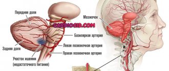
The brachiocephalic arteries are the vessels that are responsible for supplying blood to the brain, tissues of the head, shoulder girdle and arms. They are separated from the aorta at shoulder level. The research method is based on the use of the Doppler effect, which is named after the Austrian scientist Christian Doppler who discovered it.
Its essence lies in the fact that ultrasonic waves from moving objects are reflected with a changed frequency: if the object moves towards the sensor, the frequency increases, when moving away from the sensor it decreases.
The main advantages of Dopplerography of neck vessels:
- Dopplerography is a non-invasive and therefore painless method;
- it does not require much time - the examination takes only a few minutes;
- it is an inexpensive study;
- At the same time, Doppler ultrasound is informative and will help the doctor make the correct diagnosis.
Ultrasound of the BCA allows you to determine:
- condition of the vascular walls;
- vascular damage even in the early stages;
- presence and severity of atherosclerosis;
- presence and severity of stenosis;
- speed and nature of blood flow.
Doppler ultrasound also shows the anatomical features of the vessels of the neck - for example, how strongly they twist around the spine.
With all these advantages, keep in mind that Doppler ultrasound is a more primitive method compared to duplex and triplex vascular scanning. They are more informative, but require more sophisticated equipment and are more expensive.
Doppler ultrasound cannot show the cause of blood flow disturbance, which is located outside the vessels, that is, if blood circulation is not obstructed due to the formation of atherosclerotic plaques or blood clots.
A duplex or triplex scan will provide more information that will help your doctor choose the right treatment. The information content is further enhanced by the use of color mapping method during ultrasound diagnostics.
Indications and contraindications
The risk group for the development of cerebrovascular accidents includes people with diseases such as:
- diabetes;
- hypertension;
- cervical osteochondrosis;
- autoimmune vascular lesions (vasculitis);
- obesity;
- angina pectoris;
- cardiac ischemia;
- history of heart attack and stroke;
- heart attacks, strokes, hypertension and atherosclerosis in the anamnesis of relatives;
- age over 40 years.
In the presence of these diseases , periodic (once a year or once every two years) scheduled ultrasound examination of the vessels of the neck is indicated . It is also recommended that people with a smoking history of 15 years or more undergo this examination regularly.
Direct indications for undergoing ultrasound scanning of the brachiocephalic trunk are also:
- headache;
- dizziness;
- fainting;
- sleep disorders;
- permanent or transient visual disturbances;
- flickering of flies, a veil before the eyes;
- tinnitus, ringing in the head;
- coordination problems;
- disturbance of tactile sensations, weakness in the arms and legs;
- impaired concentration;
- memory impairment.
The only contraindication to the study is a wound in the neck. A referral for Doppler ultrasound is usually given by a neurologist.
For more information about the indications and examination, watch the video:
Rules for preparing for the procedure and procedure
To prepare for the study, you need to adjust your diet. The day before the study, it is necessary to exclude foods that affect vascular tone:
- drinks containing caffeine (tea, coffee, energy drinks);
- alcohol;
- salty foods.
Two hours before the procedure, refrain from taking hot baths and smoking, as well as staying in smoky rooms: in the first case, arteries and veins expand, in the second, they contract.
If you do not adhere to these recommendations before the study, the reliability of the results will significantly decrease and you will not be able to find out the real condition of the vascular walls.
In addition, the results of BCA ultrasound are distorted by drugs that change vascular tone and affect the nature of blood flow . You should consult with your doctor about which medications need to be stopped before testing.
As noted above, Doppler ultrasound takes very little time . An experienced ultrasound specialist will only need a few minutes to perform all the necessary manipulations.
To carry out the procedure, you must undress to the waist. During the examination, the patient lies on the couch, periodically changing position - on his back, on his side, on his stomach - so that the diagnostic specialist can examine all the vessels that need to be checked.
Also, during ultrasound diagnostics of neck vessels, additional functional tests can be performed:
- holding your breath;
- changing the horizontal position of the body to a vertical one;
- taking certain medications (most often nitroglycerin).
Typically, such additional tests are prescribed in the presence of genetic abnormalities in the structure of the vascular system.
Principles for deciphering results and normal indicators
A diagnostic specialist can prepare a conclusion immediately after the procedure is completed . When interpreting the results, the data obtained during the study is compared with normal indicators of the condition of the arterial walls and the nature of blood flow in each of them.
The conclusion states:
- condition of the walls of blood vessels;
- description of pathological changes, if any;
- nature of blood flow.
Normally, there are no narrowings or atherosclerotic plaques in the vessels that would impede the movement of blood; the blood flow has no turbulence. In this case, the blood moves freely through the arteries and veins , providing the brain, tissues of the head and arms with sufficient oxygen and nutrients.
At the same time, in a young body, the vessels are elastic, which protects against the development of a stroke, but with age they lose their ability to stretch.
The ultrasound specialist’s conclusion must be shown to the attending physician , who will compare the data obtained with the clinical picture, advise you on all issues and, if necessary, prescribe treatment.
Deviations from the norm and possible diagnoses
With changes in blood vessels and the nature of blood flow, the following concepts may appear in the conclusion:
- stenosis is a pathological narrowing of the lumen of a vessel, as a result of which blood cannot pass freely through them;
- aneurysm - a protrusion of a vessel wall as a result of its thinning, in which the wall of an artery or vein expands more than twice (learn about carotid artery aneurysm);
- atherosclerosis - the formation of cholesterol plaques on the walls of blood vessels, which impede blood flow, resulting in an increased risk;
- occlusion - disruption of the patency of a vessel due to damage to its wall;
- turbulent flow - when blood moves through a vessel, turbulence is formed.
Average prices in Russia and abroad
The price of Doppler ultrasound varies in Russia from 500 to 5000 rubles. The specific cost depends on the region and varies from one hospital or clinic to another, but the average price is 1000-1500 rubles. In neighboring countries the price is lower - for example, in Ukraine it is about 300 hryvnia.
At the same time, as noted above, ultrasound examination of the BCA is rarely used . In our country, it is mainly used in public clinics for treatment under the compulsory medical insurance policy. Paid clinics both in Russia and in other countries today use more advanced methods - duplex and triplex scanning of the brachiocephalic arteries with color mapping.
Doppler ultrasound is a non-invasive method for studying the vessels that supply blood to the brain.
It does not require special preparation, with the exception of minor adjustments to the diet and the withdrawal of certain medications the day before the procedure, and at the same time it allows you to accurately assess not only the condition of the arteries, but also the nature of the blood flow in them. The procedure will allow the doctor to choose the right treatment to restore normal blood supply to the head and arms if it is disrupted, and to avoid the development of serious health problems - for example, to prevent a stroke.
However, this research method can already be considered outdated , and if possible, it is worth paying attention to more modern duplex scanning of the brachiocephalic arteries.
Source: //oserdce.com/diagnostika/sosudov/uzdg-bca.html
Indications for Dopplerography of head and neck vessels
A Doppler examination of the vessels of the brain and neck is prescribed to differentiate neurological symptoms of unknown etiology:
- vertigo, ataxia, sleep disturbance, ringing in the ears;
- for arrhythmias of various origins; to identify provoking factors for the formation of cholesterol plaques, at the first signs of cerebral atherosclerosis;
- hypertension;
- VSD;
- transient ischemic attacks;
- previous AMI or stroke;
- diabetes mellitus or osteochondrosis of the cervical spine.
Duplex scanning of the vessels of the head and neck is indicated if cardiac surgery is planned; there is a suspicion of thrombosis, vascular disorders; A pathologically pulsating neoplasm in the neck area and rapid fatigue of unknown cause were detected.
The reason for performing Dopplerography of blood vessels may be intoxication, trauma, a sharp decrease in visual acuity, congenital pathologies, poor posture; compression of the vertebral artery, lack of blood pressure or pulse in the arteries of the arms; use the procedure to monitor patients and evaluate treatment outcome.
In children, ultrasound examination of the arteries of the neck and head is recommended for scoliosis or delayed mental development of the baby.
Recommendations for carrying out
In principle, this examination method does not have special conditions for preparation.
This applies to scanning the neck or head areas. If we are talking about studying the veins of the lower extremities or the pelvic area, then there are several important points.
Three days before the scan, you need to adhere to a strict diet: exclude milk, meat, black bread and cabbage.
This also applies to all other foods containing large amounts of fiber.
If you adhere to these conditions, the doctor will be able to conduct a high-quality duplex ultrasound scan and get a clear picture of the disease.
It is prohibited to take medications that stop the formation of gases in the body . The result obtained directly depends on quality preparation. Therefore, every patient should take all the doctor’s recommendations seriously.
Contraindications for use
We emphasize that for ultrasound examination of the vessels of the head there are practically no such examinations, since the procedure is absolutely safe for the patient. The only limitation may be a violation of the integrity of the skin. In this case, the examination is postponed until the wound heals, so that there is no feeling of discomfort during it.
However, there is an exception to every rule. In this case, these are the options that make Doppler ultrasound difficult:
- the object under study is hidden under the bone or hypodermis;
- arrhythmia, which changes blood flow in healthy veins and arteries;
- naturally slow blood flow.
Such cases require the professionalism of the doctor, changes in the usual installation of sensors, and recalculation of the results taking into account the initial blood flow speed.
Advantages of ultrasonic testing
Ultrasound scanning (ultrasound), what is it? Today, this is a modern type of diagnostics of blood vessels of the whole body. Ultrasound duplex scanning has a number of advantages over other procedures:
- speed of examination;
- lack of any preparation before the procedure;
- identification of natural pathologies of veins and blood vessels (excessive tortuosity or deformation);
- identification of possible cholesterol plaques or blood clots;
- the speed of blood flow is established even in the smallest vessels of the body;
- has no contraindications;
- the ability to repeat several times a day;
- can be used for women during pregnancy and lactation.
The only disadvantage is the relatively high cost compared to simpler diagnostic methods (ultrasound or Doppler ultrasound).
Progress of the study
The procedure is painless. The doctor lubricates the patient’s skin with a special gel over the projection of the arteries being examined and passes an ultrasonic sensor along them. Moreover, the doctor places the sensors so that the directed ultrasound is reflected from the blood cells inside the vessel. Doppler ultrasound is essentially an ultrasonic reflection wave that is redirected to a computer, where its trajectory is processed, which leads to the formation of the Doppler effect. In the absence of blood flow, the effect does not occur.
Ultrasound scanning of extracranial vessels begins with examination of the right lower segment of the common carotid artery, then the sensor is moved up the neck to the angle of the mandible. This makes it possible to determine the course of the artery, its depth, the place of division into internal and external branches, and to see atherosclerotic layers. Then they turn on the color mode, which shows areas of impaired blood flow and deformation of the vascular walls. The vertebral arteries are sequentially examined and the blood flow velocity is measured.
Transcranial Dopplerography of the vessels of the brain and neck is carried out through the temporal windows near the auricle. This is how all the main arteries of the brain are visualized, with the exception of the ophthalmic artery, which is viewed with the eyelid closed.
Variations of ultrasound examination
Sometimes the duplex of the vessels of the head and neck requires additional tests: using vascular drugs, hyperventilation, pressure on the capillaries with fingers for accurate data on the mechanism of regulation of intravascular blood flow.
In emergency situations, continuous Doppler ultrasound is used, when reflected ultrasound signals are transformed into standard sound. This examination is practiced in severe, bedridden patients using portable devices. The maximum duration of the examination does not take more than 45 minutes, on average it is about a quarter of an hour.
How is it different from ultrasound?
Doppler ultrasound is considered a rather outdated method for studying blood vessels . The doctor cannot fully examine the condition of their walls, blood flow speed, etc.
This method is based on the so-called Doppler effect, thanks to which it is possible to record the patency of blood vessels or identify a malfunction of the venous valve .
How does ultrasound examination differ from ultrasound examination? One might say, in almost everything. Let's start with the fact that the ultrasound method can only establish the problem itself, and not the cause of its occurrence .
The USDG method can only be used for the primary diagnosis of vessels; for a more detailed study of them, it is worth using an ultrasound scan.
Using a special ultrasonic sensor, a general picture of the movement of blood through the vessels, the condition of their walls, etc. is displayed on the monitor screen.
Data decryption
When performing Dopplerography of the vessels of the head and neck, the following are assessed: the thickness and diameter of the vascular wall, phases and symmetry of blood flow, minimum (occurring in diastole) and maximum (initiated by systole) blood flow velocity, the ratio between them, vascular indices (pulsation and resistive). The results of the study are then compared with norms and conclusions are drawn.
Normally, when scanning, the average values are:
- the thickness of the vascular wall is no more than 0.9 mm (in exceptional cases, individual thickening up to 1.1 mm is possible);
- the examined arteries and veins are free throughout their entire diameter, there is no external compression;
- blood flow turbulence, malformation – not determined;
- the lumen of the vertebral arteries is the same, no more than a couple of millimeters;
- blood flow through the vertebral veins up to the 6th vertebra inclusive - no higher than 0.3 m/s.
Ultrasound scanning of vessels is not the only criterion for a correct diagnosis: small veins are beyond its control, angiography is needed. In addition, the accuracy and reliability of the study directly depend on the quality of the equipment and the qualifications of the doctor.
Doppler ultrasound decoding is carried out in several directions: atherosclerosis, thrombosis, deformation.
Atherosclerotic disorders
Most often, ultrasound of the head and neck diagnoses atherosclerosis of the cerebral arteries, which is characterized by thickening of the vascular walls, plaques on the endothelium, and loss of homogeneous structure (echogenicity). If the artery loses more than 20% of its lumen, it is referred to as stenosis. Symptoms are turbulence of linear blood flow and increased pressure. Diagnostics is carried out by duplex or triplex scanning.
When identifying the first symptoms of a lesion of this nature, a complete examination of the vascular bed is necessary: internal organs, lower extremities, since atherosclerosis is total in nature.
Signs of thrombosis
Disorders of hemostasis in the body are manifested by arterial thrombosis, when the lumen of the artery is blocked by a blood clot. On Doppler ultrasound, inflammation of the vascular wall with a homogeneous (hypoechoic) formation inside is clearly visible. The contours of the blood clot are clear; if it stays in place for a long time, arterial atrophy forms.
Deformation
A common finding during ultrasound examination of the vessels of the head and neck is deformation of the vascular bed, which shows hidden congenital or acquired malformations: excessive tortuosity (S-shape), bends (the angle of inflection can reach 90 degrees or be sharp, which disrupts hemodynamics), loops - circular configurations of arteries and veins (a consequence of the pathology of fetal angiogenesis), which do not in any way affect the quality of human life.
Sometimes hypoplasia of the vertebral arteries occurs with a decrease in diameter to 2 mm or less.
Other pathologies
When examining the vessels of the head and neck with ultrasound, the doctor may find:
- autoimmune aortoarteritis (Takayasu disease);
- temporal arteritis (vasculitis);
- metabolic angiopathy;
- post-traumatic dissection of the arterial wall;
- anomalies of communication between veins and arteries.
What is the price
It is impossible to accurately determine the cost of MAG ultrasound, because prices in the Russian Federation differ significantly depending on the chosen institution: public or private clinic. On average, you will have to pay about 1,500 rubles for the study. A similar procedure in Ukraine will be half the price. When choosing a clinic for diagnostics, it is important to pay attention not to the cost of the procedure, but to the quality of the equipment and methods - only duplex and triplex scanning with color mapping can be called modern diagnostics.
The role of ultrasound in the prevention of vascular diseases of the neck and head
The advantages of the ultrasound technique include carrying out preventive Doppler ultrasound in vascular risk groups, during clinical examination of different groups of the population. The indication is:
- elderly age;
- alcohol and nicotine abuse;
- obesity, especially against the background of atherosclerosis;
- occupational hazards: capillary intoxication with varnishes, paints, chemicals;
- diabetes;
- professions with excessive mental or physical stress;
- genetic predisposition to vascular diseases;
- hormonal fluctuations of unknown origin;
- pregnancy, childbirth, puberty, taking hormonal contraceptives.
How is ultrasound examination performed?
Dopplerography of neck vessels is done both on an outpatient and inpatient basis. The duration of the procedure does not exceed 20 minutes. The patient takes the required position (sitting or lying), after which the doctor begins the examination. A gel is applied to the entire neck area to improve contact between the sensor and tissues.
The procedure begins with an ultrasound of the carotid arteries. The examination method makes it possible to determine the depth at which the arterial trunk is located, its course, and the presence of deformation. After the carotid artery, the doctor proceeds to examine other extracranial vessels. Other important parameters: the course of the artery, the nature, speed of blood flow, the presence of pathological elements in the wall.