Before the advent of MRI, the arteries and veins of the cerebral vessels had to be studied using radiographic methods, which were not always informative. Magnetic resonance imaging is characterized by higher accuracy, due to which many pathologies of the circulatory system can be detected in the early stages.
Medical indications for MRI of the head vessels:
- symptoms of increased intracranial pressure (feeling of nausea, vomiting, headaches in the morning);
- dizziness;
- suffered traumatic brain injuries;
- convulsions;
- noise or ringing in the ears;
- periodic nosebleeds;
- blurred vision;
- sleep problems;
- difficulty concentrating;
- memory disorder;
- change in consciousness, behavior;
- surgery planning;
- control after surgery;
- dynamic monitoring of the identified disease.
MRI of cerebral vessels has certain contraindications. Absolute prohibitions are associated with the presence on the body of electronic devices or metal elements with pronounced magnetic properties: artificial pacemaker, neurostimulator, middle ear implant, prosthetic heart valves, insulin pump, etc.
Conditional contraindications to performing MRI include:
- claustrophobia;
- pregnancy up to 12 weeks;
- patient conditions in which it is difficult to maintain a stationary position;
- early childhood;
- body weight more than 120 kg.
What is a cerebral aneurysm?
A cerebral aneurysm is a potentially life-threatening condition associated with bulging and weakening of the wall of an arterial vessel, most often localized at sites of bifurcation (bifurcation). Rupture of the defect without immediate treatment can lead to massive intracranial hemorrhage. This outcome occurs in 20-50% of patients, of which 40% of people die from paralysis of the respiratory center due to cerebral edema or blood compression of important centers.
Aneurysms are clearly visualized on MRI of the head, so it is wiser to undergo examination at the first complaints, without waiting for serious consequences. Not all cerebral vessel wall protrusions require therapy, but follow-up with magnetic resonance imaging of the head with contrast is vital to assess the progression of the disease. Management tactics can be conservative (small aneurysms with a low risk of rupture) and surgical, and the prognosis is more favorable with planned intervention.
Types of aneurysms: on the left - saccular (saccular), on the right - fusiform (fusiform)
Currently, aneurysms are classified according to location, size and shape, which is important for the choice of treatment. The location of dilatation is determined relative to the large vessels that form the circle of Willis. There may be several aneurysmal formations, their localization varies. The size of vascular dilatation: small - up to 10 mm, medium - 25 mm, large - over 25 mm. Giant aneurysms are known, reaching a diameter of 50 mm. According to their shape, saccular (saccular), fusiform and dissecting vascular protrusions are distinguished; by origin - post-traumatic, infectious, mycotic, resulting from persistent hypertension, vasculopathy, atherosclerosis, etc.
Aneurysms with a maximum diameter of less than 7 mm are statistically unlikely to rupture, but this cannot be completely ruled out. The risk of a vascular accident within 5 years from the moment of detection of the pathology increases in proportion to the size of the protrusion of the vessel wall.
Classification of the disease
>The disease can be classified according to its location. Aneurysms occur:
- Internal carotid artery;
- Anterior cerebral artery;
- Vertebro-basilar system;
- Middle cerebral artery;
- Multiple aneurysms affecting several vessels at the same time.
The latter type of disorder occurs in approximately 13% of patients.
In addition, aneurysms are distinguished by shape:
- Fusiform;
- Saccular, occurs fifty times more often. It can be multi-chamber or single-chamber.
By size they are distinguished:
- Giant - more than 25 millimeters;
- Large – 16-25 millimeters;
- Medium – 11-15 millimeters;
- Small – up to 10 millimeters;
- Miliary - up to 3 millimeters.
Causes of aneurysm
A sharp jump in blood pressure leads to aneurysm rupture
Cerebral aneurysms can be congenital or acquired (most often resulting from traumatic brain injury). Autosomal dominant polycystic kidney disease is an inherited disorder that disrupts the renin-angiotensin-aldosterone system and often leads to persistently high blood pressure. Typically, cysts form not only in the kidneys, but also in the vessels of the brain, which aggravates the condition and weakens the arteries and veins.
Inherited Marfan syndrome is characterized by a pathology of connective tissue in the body, which contributes to modification of the vascular wall. Serious autoimmune diseases, for example, systemic lupus erythematosus, in some cases cause cerebrovascular disorders.
The problem of rupture of cerebral vessels is more often encountered by patients aged 55-60 years; the first symptoms appear in the fifth decade; in children, the pathology is rare and accounts for less than 2% of cases. Factors that provoke rupture of an existing aneurysm include:
- sudden lifting of weights;
- straining during bowel movements;
- intense sexual contact;
- severe stress with the release of adrenaline and increased blood pressure;
- hacking cough;
- physical stress.
The immediate cause of rupture is often a sharp increase in intracranial pressure, leading to a violation of the integrity of the weak vascular wall. Scientists have identified predisposing conditions:
- persistent hypertension, its inadequate therapy or refusal of treatment;
- chronic alcohol and nicotine intoxication, drug use;
- primary or metastatic neoplasms;
- congenital connective tissue diseases, for example, with Ehlers-Danlos syndrome, fibromuscular dysplasia;
- arteriovenous malformation of cerebral vessels;
- congenital narrowing of the aorta (coarctation);
- hereditary hemorrhagic telangiectasia;
- alpha-1 antitrypsin deficiency;
- generalized atherosclerosis;
- hypoestrogenism, obesity and female gender;
- generalized mycotic infection.
Symptoms of a cerebral aneurysm
The most common location of pain from an aneurysm is in the center of the head
Small protrusions of the vascular wall are asymptomatic. Full-blown clinical manifestations develop mainly after the rupture of the aneurysmal sac or when the formation reaches significant volumes, which is associated with compression of nearby structures. With a medium and large aneurysm, in the absence of a cerebrovascular accident, the following signs are typical:
- ophthalmological disorders: diplopia (double picture), deterioration of visualization, loss of visual fields;
- pain in the eyeball area;
- numbness or weakness on one half of the face or body;
- periodic headaches of varying degrees of intensity, dizziness;
- imbalance, unsteady gait;
- difficulty concentrating, problems with short-term memory.
The following symptoms indicate a possible hemorrhagic stroke due to a ruptured aneurysm:
- sudden severe (dagger) headache;
- stiff neck;
- sudden deterioration of vision;
- sensitivity to light;
- ptosis (drooping eyelid);
- numbness, weakness on one side of the face;
- nausea, vomiting;
- problems with speech or changes in consciousness (up to its complete loss) and mental state;
- convulsive syndrome.
Which method of diagnosing an aortic aneurysm to choose: MRI, CT, ultrasound, angiography
Selection method
- MSCT.
What can be seen in the images of an abdominal aortic aneurysm
- Limited or generalized dilatation of the aorta
- With local dilatation, the possibility of a mycotic or traumatic etiology of the aneurysm should be taken into account.
- In the dilatation zone, signs of perfusion are determined, as well as the presence of a thrombotic component.
Why is Doppler ultrasound of the aorta performed for an aneurysm?
- An informative diagnostic technique that allows you to determine the size and extent of the aneurysm for the tactics of further surgery for abdominal aortic aneurysm
- Screening technique.
Is CT angiography and MR angiography of the aorta informative?
- Visualization in MPR mode allows you to determine the size of the aortic aneurysm
- The features of the syntopy of the aneurysm to the branches of the aorta are visualized
- CT scan identifies crescentic calcification
- Inflammatory changes in surrounding fatty tissue
- CTA and MRA are very informative in terms of determining the nature and extent of subsequent treatment
- Blurred contours of the aortic wall
- Signs of threat of perforation of an aortic aneurysm: increased density according to imaging data, the presence of a contrast agent outside the lumen of the vessel (in a thrombus or in the perivascular space).
Diagnostic indicators
| Abdominal aorta | Outside diameter |
| Norm | 1.8-2 cm |
| Aneurysm | >3cm |
Why is angiography prescribed for abdominal aortic aneurysm?
- The use of DSA is indicated only when it is impossible to perform a sufficiently high-quality CTA or MRA, as well as when installing a stent
- Only the perfusion zone in the lumen of the vessel is visualized
- DSA does not allow visualization of the lumbar arteries and thereby indirectly assess the extent of the thrombotic process.
Is a cerebral aneurysm visible on an MRI?
MRI: cerebral aneurysm
Cerebral aneurysm is usually diagnosed using MRI, CT, magnetic resonance angiography, or computed tomography angiography. Each method has its own indications and contraindications. Magnetic resonance scanning does not involve the use of X-rays, so it is completely safe for most people, including pregnant women and children. Another type of diagnosis is justified for patients with metal in the body; these can be orthopedic structures, insulin pumps, cochlear implants, removable dentures, etc. A powerful magnetic field will cause equipment failure, and artifacts (blurriness) will appear on the pictures.
For better visibility of details, the doctor will prescribe contrast. Modern drugs based on gadolinium chelates rarely cause significant side effects and are well tolerated. World practice has not recorded any long-term consequences from MRI and contrast administration.
Magnetic resonance scanning is preferable for routine examinations, since the procedure itself takes 40-60 minutes and decoding requires the same amount. In emergency cases, the doctor does not have this time: the sooner the nature of the pathology can be determined, the greater the chance of correct treatment, and the higher the likelihood of a favorable outcome. Non-contrast CT, the study of choice for urgent situations associated with subarachnoid hemorrhage, allows assessment of the location and volume of the hematoma in a ruptured aneurysm.
Is a cerebral aneurysm visible on an MRI? Research opportunities are limited more by the financial component. The thickness of the sections during examination is from 1.0 mm - theoretically, everything that is larger can be identified with the careful approach and experience of a specialist. Practice shows that small aneurysms (less than 3-4 mm) may remain unrecognized. Magnetic resonance angiography, a more sensitive method that complements MRI of the brain, is being considered as a way to detect minor protrusions. MR scanning may also show small amounts of parenchymal blood surrounding the aneurysm; this allows you to determine which of the numerous vascular dilations (occur in 10-30% of cases) has ruptured.
General overview
Speaking about the operating principles of MRI and CT used to examine the brain, it is worth noting that the devices are very similar in appearance. They are a narrow couch embedded in a huge tube-shaped structure. But the phenomena they study are different.
The first research apparatus operates on the basis of magnetic fields. The electromagnetic poles are directed perpendicular to the acting field. Resonance occurs due to the vibration of atoms. In this way, the device is controlled. As a result of the procedure, data is transferred to a three-dimensional quality image.
The second one works on the basis of X-ray radiation. During the examination of the brain, the scanner begins to make rotational movements around the head, while scanning the organ from different angles. The images are transferred to a computer and processed. As a result, the doctor can observe the image in three-dimensional quality.
Features of CT
Using the device, aneurysms or other pathologies can be detected. The presence of abnormal formations is reflected on the image in the form of light spots. The result is obtained due to the fact that the density of brain tissue increases, which is what the device recognizes.
All deviations can only be correctly deciphered by a radiologist. The patient is sent for tests by a therapist, who, after examination, can assume that the patient has inflammation.
Features of MRI
This device also detects abnormal tumors. But its difference from another device is that the supply of signals depends on how intense the signals are received. This signal comes from hydrogen contained in the brain.
Bone and fat tissues cannot give such strong signals, so the presence of atoms in the head fluid is very easy to determine. Pathological areas are shown on the image as dark zones.
MRI machine
Indications
In order for a patient to be sent for an MRI or CT scan, he must have abnormalities that raise suspicion of brain pathology. These health problems include:
- neoplasms;
- disruption of blood flow;
- presence of lesions;
- suspected deviations in the condition of the organ.
The device is most often used to detect aneurysms. It is this method that guarantees their accurate determination.
Contraindications
Since these devices are examination methods that affect the body through irradiation, it is natural that there are certain contraindications for the procedures.
It is radiation that is the reason why women who are carrying and breastfeeding cannot undergo brain examinations.
The study is also excluded if the patient is suspected of having the following pathologies:
- if the patient has metal objects in his body (knitting needles, bullets, disks, etc.);
- claustrophobia;
- when the body has an electronic device;
- some types of mental illness;
- body weight exceeding two hundred kilograms.
To avoid risk, the patient must undergo a thorough examination before either procedure begins.
Computed tomography machine
Interpretation of MRI of arterial aneurysm
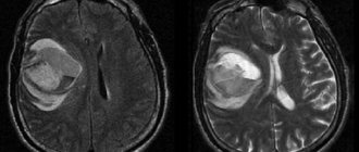
MRI: arrow points to a significant aneurysm
An aneurysm can be suspected by visualizing a formation associated with an artery and filled with blood. Additional signs include: swelling of adjacent tissues, suspected accumulation of blood in the ventricles of the brain, areas of hemorrhage, necrosis. Rupture of the aneurysm confirms the presence of fluid in the subarachnoid space. By its localization, the location of the burst formation is judged. In emergency situations, the diagnosis in 90% of cases is beyond doubt; during a routine examination, another pathology with similar symptoms is excluded: a benign or malignant tumor, trauma and its consequences, vascular malformation.
In large diagnostic centers, interpretation of an MRI of a cerebral aneurysm is included in the total cost, so if you have any questions, ask them to the radiologist who performed the study. Sometimes the uncertainty of the situation in suspicious patients causes a headache after undergoing an MRI procedure, which has nothing to do with either the effect of the magnetic field or the administration of a contrast agent.
T1-weighted magnetic resonance image (MRI) of the brain of a patient with progressive headaches, speech impairment, and right-sided hemiparesis: a large intracerebral mass with significant edema is a giant aneurysm of the internal carotid artery.
CT without contrast injection: giant aneurysm of the left internal carotid artery in the intracavernous segment with dense calcification.
additional information
Exactly how the diagnosis will be carried out depends not only on the doctor’s testimony, but also on the patient’s wishes. Naturally, the doctor may recommend undergoing a certain study, but the choice still remains with the patient.
The differences between the devices also include the price costs of the procedures. CT has a lower monetary value than MRI. This is due to the fact that the operation of the second device requires more expensive devices. There are also different time periods.
X-ray scanning is three times faster than magnetic scanning. Sometimes, in order to get an accurate result from the device, neither the patient’s financial condition nor his free time will play a role. Contraindications and the individuality of each case may have an impact.
Possible consequences of an aneurysm
The white arrow shows the location of the aneurysm rupture in the Circle of Willis.
The consequences of aneurysm rupture depend on many factors:
- amount of bleeding;
- location of education;
- patient's age;
- general health and the presence of severe concomitant pathologies;
- the quality of subsequent medical care and the speed of its provision.
Unfortunately, about 15% of patients die before reaching hospital, and 40% will not survive the first two weeks, even with the best treatment. Of the remaining patients, more than half will become disabled. Only less than 20% of people will recover to their original level. Most of the damage caused by a ruptured aneurysm appears within the first minutes of bleeding. Therapeutic measures are aimed at preventing repeated hemorrhages and restoring brain function.
The body tries to compensate for the rupture of a vessel in a thin place with bleeding by a sharp spasm of the arteries and veins; the spilled blood prevents the adequate outflow of cerebrospinal fluid and leads to occlusive hydrocephalus, some areas of the brain become necrotic, and their functions are lost. Long-term consequences include:
- Pain syndrome. After a rupture of an aneurysm, a person suffers from attacks of headache, which vary in intensity and duration and are resistant to the effects of drugs.
- Cognitive impairment. In severe cases, the patient loses the ability to process incoming information, logical thinking, criticality, etc. Complete collapse of the personality implies constant care from loved ones and medical staff. The consequences of aneurysm rupture can manifest as depressive disorders, irritability, aggressiveness, and insomnia. Speech function suffers.
- Disruption of the pelvic organs. Patients are unable to empty the bladder, which requires continuous or intermittent catheterization, when urine is released through an installed tube. Another “delicate” problem is the tendency to constipation: bowel movements occur only after an enema. In some patients, dysfunction of the pelvic organs manifests itself in urinary and fecal incontinence.
- Motor disorders. Muscle weakness, paralysis and paresis, impaired coordination are typical consequences of aneurysm rupture. After massive hemorrhage, in some cases the patient is doomed to complete immobility.
Diagnostics
Photo: pillsman.org
This diagnosis is made by a neurologist during the initial examination. Also, the diagnosis of cerebral aneurysm occurs through an X-ray examination of the skull, examination of the spinal cord fluid, and a tomographic examination. An MRI examination allows identifying signs of a cerebral aneurysm much more quickly.
Symptomatic signs of cerebral aneurysm.
Very often, the signs of a cerebral aneurysm are not expressed in any way until it becomes particularly large or ruptures.
If symptoms of the disease occur, then, as a rule, they are expressed in the following manifestations:
- aching eyes;
- paralysis syndrome;
- weakening of the facial muscles;
- blurred vision;
- enlarged pupils.
Symptoms of a ruptured cerebral aneurysm are expressed in intense and piercing pain in the head, gagging, nausea reflex, nuchal rigidity (increased tone of the neck muscles), and in some episodes, fainting. Sometimes the symptoms of the disease in a patient are expressed in migraines, which can be long-lasting. Less commonly, signs of a cerebral aneurysm may include:
- drooping eyelid;
- increased sensitivity to bright light;
- violation of mental stability;
- increased restlessness;
- convulsions.
All these symptoms are an “alarm bell”; in this case, you should urgently seek medical help. It must be remembered that only a specialist can make a diagnostic conclusion; All of these signs of cerebral aneurysm do not completely determine the presence of this disease. Any conclusions can only be drawn by a neurologist, based on the examination and the results of the examination.
Diagnosis of signs of cerebral aneurysm
Signs of a cerebral aneurysm require a medical examination; only a doctor can confirm or deny the presence of the disease in a patient.
The examination is very important, since the risk of hemorrhage from the detected pathology is very high. The likelihood of this negative prognosis is influenced by many factors: the magnitude of the pathology, its location, the condition of the blood vessels, as well as general anamnesis. Recurrence of hemorrhage occurs in a more complex form and increases the risk of death. That is why signs of a cerebral aneurysm are a serious reason to seek medical help. If the symptoms become more and more pronounced, then when the patient contacts specialists, the following types of examinations are possible:
- When examining a patient, a neurologist makes appropriate conclusions. A doctor's examination helps identify meningeal (symptoms of irritation of the meninges) and focal (deficiencies that begin due to local damage to the brain) symptomatic signs. Using them, a specialist can confirm that the observed problems are signs of a cerebral aneurysm.
- Signs of a cerebral aneurysm are confirmed or refuted by an X-ray of the skull. The procedure “shows” clots in the vessels, as well as a violation of the integrity of the bones at the base of the skull, which helps to identify the disease.
- CT scans make it possible to quickly scan the brain structure and its structure. Diagnosis of signs of cerebral aneurysm using this method allows us to record the slightest abnormal changes in the brain and determine the disease. A CT scan will immediately “see” signs of a cerebral aneurysm; MRI also helps to cope with this task.
- MRI also helps to identify signs of cerebral aneurysm in the early stages. The procedure makes it possible to “examine” the structure of an organ (brain) and “see” abnormal formations. MRI usually detects signs of cerebral aneurysm from the first procedure, except in cases where the pathology is negligible. Then the diagnosis of signs of cerebral aneurysm is carried out using CT. However, with primary signs of a cerebral aneurysm, specialists most often prescribe an MRI.
- Signs of a cerebral aneurysm are grounds for a doctor to prescribe a cerebrospinal fluid examination. Diagnosis of signs of cerebral aneurysm using the presented method is carried out using laboratory tests. Experts check how clear the liquid is.
- If there are signs of a cerebral aneurysm, angiographic examination of the vessels is also prescribed. It determines where the pathology develops, determines its shape and dimensions, and scans the veins of the brain.
So:
- signs of cerebral aneurysm do not appear for a long time;
- The diagnosis can be made by studying the signs of a cerebral aneurysm only when examining the patient with special equipment;
- if signs of a cerebral aneurysm appear, it means that the disease has acquired a serious form;
- the signs of cerebral aneurysm indicated on the website do not determine the presence of the disease; only a specialist can determine the diagnosis. Only a doctor can diagnose a cerebral aneurysm.
In addition to all these methods, the collection of information based on anamnesis is of great importance in diagnosing the disease. Before prescribing any examinations, the neurologist asks the patient or his relatives and the following important factors:
- symptoms that worry you most at the moment;
- first manifestations of the disease;
- concomitant chronic or acquired diseases;
- treatment performed previously, whether it was performed at all;
- injuries;
- allergies;
- hereditary diseases.
Sometimes this disease can be discovered completely by accident when the patient undergoes an examination due to complaints about other circumstances. Similar diagnostic examinations are also carried out if tumor formations in the brain are suspected. Even more often, this disease, unfortunately, is discovered only after the aneurysm ruptures, in which case the patient is urgently hospitalized.
Brain aneurysm: how to treat?
Endovascular surgery - insertion of a coil into a cerebral aneurysm. A - a microcatheter was inserted via transfemoral access through the femoral artery, occlusion of vascular dilatation was carried out using a microspiral (B)
Treatment tactics for wide-necked aneurysms include balloon (above) and stenting techniques (below)
Aneurysm clipping during open surgery
Today, neurosurgeons are able to help many patients with cerebrovascular aneurysms of the brain. Several techniques have been developed and are used, the most effective of which is surgery. Perform clipping and strengthen the vascular defect. During minimally invasive endovascular manipulation, special microscopic coils are inserted into the aneurysmal sac and a blood clot is formed, preventing rupture of the weakened wall. Each intervention has its own indications. Clipping is possible for a saccular defect, and wrapping the vessel with surgical gauze, which promotes the development of connective tissue, is preferable for a fusiform aneurysm. This procedure is less traumatic because it does not require craniotomy and is performed under angiography control. The prognosis for life is much better with planned operations, but in emergency cases, successful surgical treatment is possible: the dilated sac is switched off from the general blood flow or the weak wall is strengthened, while large vessels are left patent.
If the doctor assesses the likelihood of a cerebrovascular accident as low, the patient is monitored over time. Maintaining blood pressure at normal levels plays a huge role; antihypertensive therapy is often prescribed for life.
Surgery as the only treatment option
Once an aneurysm is found, no doctor can accurately predict whether or when it will rupture. But in medical practice it is believed that with sizes less than 5 mm it is unlikely, and the risk of complications exceeds the risks of rupture. In any case, the decision about surgery is made individually for each patient.
If access is possible, surgery will help restore or cut off blood flow to the aneurysm.
This prevents its further growth or rupture. Operations include:
- Surgical “switching off” of a vessel from the bloodstream (microvascular circumcision), in which the aneurysm is closed using a metal clamp (clipping). The operation requires craniotomy and direct access to the vessel.
- Endovascular embolization, in which blood flow is blocked using a coil or balloon through a catheter passed through the artery to the aneurysm. The procedure is performed under CT control and is less traumatic than clipping.
Before, during and after surgery, the focus is on protecting the brain and blood vessels from potential further damage.
Prevention of cerebral aneurysm
Having an MRI if you suspect a neurological or neurosurgical pathology can save your life
Prevention involves periodic examination of patients at risk with a hereditary predisposition to cerebrovascular disorders. Sometimes it is not possible to eliminate all aneurysms during surgery; the intervention consists of treating the most dangerous defect. For these patients and those diagnosed with low-risk pathology, MRI for follow-up evaluation is recommended. Equally important is commitment to a healthy lifestyle: giving up alcohol, smoking, proper nutrition and feasible physical activity, without overexertion. Products that increase cholesterol levels and change glucose tolerance are excluded from the diet, which is the prevention of diabetes mellitus and atherosclerosis. For patients with high anxiety, consultation with a psychiatrist (psychologist) is indicated to stabilize the emotional background. If an attempt to change behavior is unsuccessful, antidepressants and sedatives are prescribed.
Performing dynamic MRI makes it possible to timely diagnose newly emerging or growing aneurysms and carry out adequate therapy aimed at preventing cerebrovascular accidents and preserving a full life.