According to medical indicators in the field of urology, one of the most common urological diseases is urolithiasis - urolithiasis.
One of the forms of this disease is the development of compactions (calculi) in the urinary system - in the kidneys. This pathology is called nephrolithiasis. The formation and growth of stones can be caused by various factors, ranging from congenital predisposition to alcohol abuse and physical inactivity.
The phenomenon of kidney stones has been known since ancient times. According to data obtained as a result of excavations, this pathology was discovered in Egyptian mummies.
- Nephrolithiasis at risk
- 01congenital predisposition
- 02Alcohol abuse
- 03Sedentary
In this article we will reveal what causes stones, and also consider the first manifestations of the disease, methods of determination, and what to do if a sad diagnosis is made.
Types of kidney stones
There are four main types of kidney stones.
Calcium stones
Calcium stones are the most common type of kidney stones and contain calcium oxalate, calcium phosphate, or a combination of both.
Calcium oxalate stones consist of calcium oxalate dihydrate and calcium oxalate monohydrate.
A calcium oxalate dihydrate stone will have jagged edges. However, a calcium oxalate monohydrate kidney stone, which is more common, will have a smooth surface.
Swelled again
Swelling of the eyes and legs immediately suggests kidney problems. However, renal edema is different from edema that occurs with heart failure, impaired venous or lymphatic drainage, or allergies.
Renal edema is softer, dough-like, and symmetrical. And often “floating” (moving quickly when the body position changes). In the afternoon, swelling usually decreases or disappears. In this case, there is no other choice but to check the condition of the kidneys.
Uric acid stones
Uric acid stones account for 3-10% of kidney stones. Uric acid stones usually look like pebbles. Some of the stones may be hard on the outside but soft on the inside because they are composed of different types of uric acid and calcium oxalate monohydrate.
Struvite stones
Struvite stones are the next most common type of kidney stones (7-8% of cases) and are larger than the others.
Cystine stones
Cystine stones are the result of cystinuria
, which can be inherited. The kidneys produce cystine, a type of amino acid.
Cystine stones are compact, partially opaque, and amber in color.
Urinary disorders
Painful urination is hard to miss and is the most obvious symptom of kidney problems. But you need to pay attention to changes in the type of urination.
For example, you started going to the toilet often and little by little. Or she is forced to get up at night, although she did not drink anything before going to bed. Pay attention if the urine suddenly changes color: it becomes darker, foamy, or there is blood in it.
How do kidney stones pass?
Kidney stones can pass without medical intervention. Smaller stones are more likely to pass, but larger ones may block the urinary tract. The stones can take up to 6 weeks to pass; the patient should drink plenty of water during this time.
People often experience pain when a kidney stone passes through the ureter. If the pain is unbearable, you should consult a doctor. The doctor may prescribe strong pain medications, anti-nausea medications, and a drug called tamsulosin, which relaxes the ureter, making it easier for the kidney stone to pass.
Passage of stone depending on size
The average size of the ureter is 3-4 millimeters wide. The chances of passing stones depend on their size:
| Stone size(mm) | Probable percentage of passage through the ureter (%) |
| Less than 4 | 80 |
| 4–6 | 60 |
| More than 6 | 20 |
Shortness of breath appeared
Nocturnal shortness of breath and suffocation are observed mainly in people with heart failure. But lack of air may also indicate kidney problems.
We usually need time to regain our breath after climbing stairs, running or walking at a fast pace. This is completely normal and is caused by the active consumption of oxygen and the accumulation of carbon dioxide in the lungs. Shortness of breath in renal failure is associated with anemia and insufficient transport of oxygen to the organs.
- They asked a urologist: how to remove sand from the kidneys
Treatment of urolithiasis
If a patient has large kidney stones or stones that are blocking the urinary tract, the doctor may recommend surgical removal.
There are three types of surgical interventions:
- Shock wave lithotripsy
: Kidney stones are broken into smaller pieces, allowing them to pass through the urinary tract. - Cystoscopy and urethroscopy
: these procedures use a camera to detect stones in the urethra or bladder. Once the urologist finds kidney stones, he can remove them completely or break them into small pieces, which will then pass on their own. - Percutaneous nephrolithotomy
: The urologist inserts a camera directly into the kidney through the back wall to remove stones. If the stones are very large, a laser is used to break them into pieces.
Instrumental methods
To find out absolutely everything about the condition of the kidneys, it is important to supplement the diagnosis with instrumental research methods.
Ultrasound
A complete examination of the kidneys cannot be done without an ultrasound.
Thanks to ultrasound, you can determine the size of the kidneys, evaluate their structure, understand how correctly they are located, how mobile they are.
In addition, the specialist can see the expansion of the renal pelvis, as well as the calyces, and, if necessary, find out the stage of the pathology. It is ultrasound that allows you to detect stones at the earliest stages.
Also, using an ultrasound examination, it is possible to examine the blood flow in the organ and, based on this, draw conclusions regarding the functional state of the kidneys.
Diagnosis of polycystic kidney disease cannot be done without ultrasound.
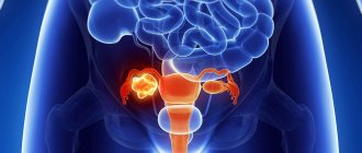
Separately, it should be said about the possibility of diagnosing tumors, including small ones, in which the patient does not complain.
Thus, ultrasound examination is indicated for absolutely all people, even those who do not have unpleasant symptoms from the urinary system. The procedure should be performed for preventive purposes, as, for example, fluorography is performed.
If a patient has kidney pain and tests are normal, then he undergoes an ultrasound of the pelvic organs. In women, this problem may be due to pathologies of the reproductive sphere. A nephrologist probably knows everything about kidney diseases in women, symptoms, diagnosis. You should not engage in amateur activities. This will help avoid a number of unpleasant complications. It is the doctor who should be entrusted with kidney diagnosis and treatment.
Biopsy
Instrumental methods for examining the kidneys include puncture biopsy. With the help of a biopsy, the doctor can find out what exactly the patient is suffering from and what exactly caused the development of the pathological process, as well as make a prognosis of the disease.
A biopsy complements the sclerotic analysis of the kidneys and makes it possible to see the full picture of the disease.
The essence of the procedure itself is that the doctor inserts a needle into the abdominal cavity and then removes a piece of tissue from the kidney. The specialist controls all actions using ultrasound.
There is no need to be afraid of such a kidney examination, since before it begins, the patient is given local anesthesia, so he does not feel pain or discomfort.
After the biopsy, the patient should monitor blood pressure for 3 days, avoid heavy physical work and drink plenty of fluids (preferably plain water).
In the first days after the procedure, there may be some blood in the urine. This is the absolute norm. However, if after the biopsy the patient has an increased temperature, difficulty urinating, or pain in the kidney area, he needs to seek medical help.
Endoscopy
This method of examining the kidneys involves the use of a special device - an endoscope. It is introduced into the body through the ureters.
During the procedure, the doctor evaluates the condition of the kidneys and calyces. In some cases, the study is combined with a biopsy.
Using this method, you can detect a variety of diseases and the causes of their occurrence. However, it is important to say that patients often experience complications after endoscopy. For this reason, the procedure is prescribed only in exceptional cases.
CT
A CT scan is usually performed to confirm the preliminary diagnosis.
Kidney examination is based on the use of x-ray waves, which tend to scan the human body. After the procedure is completed, the doctor receives a 3D image of the kidneys.
To get reliable results, doctors recommend that patients a few days before the test avoid foods that contribute to increased gas formation, and cleanse the intestines immediately before the procedure.
More accurate diagnostic results can be obtained if you do a CT scan with contrast agents that are injected directly into a vein. Therefore, CT with contrast is prescribed more often.
MRI
If we talk about methods for studying kidney function, we cannot forget about MRI.
MRI is one of the most modern, informative and safe methods for examining the kidneys.
The principle of the procedure is similar to CT, but unlike it, during MRI the patient’s body is not exposed to harmful radiation. The essence of the study is the use of magnetic fields, so MRI is safer than CT. The procedure has virtually no contraindications.
The tomograph scans the kidneys layer by layer, and then a special program processes the results and displays them in the form of a 3D image.
Using visual diagnostics of the kidney, you can not only assess the condition of the organs, but also identify pathology, as well as determine its exact location.
Chromocystoscopy
A complete kidney diagnosis may include chromocytoscopy. This is a method of functional diagnosis of kidney diseases. It can be used to diagnose surgical pathologies of the kidneys, as well as the upper urinary tract.
If your kidneys hurt, diagnosis by tapping will not always help. Thus, it is with the help of chromocystoscopy that renal colic is diagnosed.
Contraindications include acute diseases of the urethra, prostate, scrotum and testicles.
This method of examining the kidney goes like this: the patient is injected with an indigo carmine solution, after which the doctor inserts a cystoscope into the bladder and examines it.
If the patient's kidneys and upper urinary tract are healthy, blue-colored urine will begin to flow into the bladder. If the drug was injected into a vein, it will take about 4 minutes, and if into a muscle - about 12.
As a result, the kidneys should excrete 2-5 ml of the drug.
During the study, the doctor pays attention to the intensity of the color of the urine, the frequency of contractions of the ureter, as well as the peculiarity of the ejection of colored urine.
X-ray
An examination of the kidneys in this case will help to assess their general condition, detect inflammation and pathological inclusions, and identify disturbances in their functioning. If a patient is suspected of having renal hypertension, X-ray diagnosis will also help.
In order for the diagnostic results to be reliable, the patient is recommended to abstain from food that contributes to increased gas formation a couple of days after the examination, and to empty the intestines immediately before taking the pictures.
Risk factor
Some factors that may increase your chance of developing kidney stones include:
- the person does not drink enough fluids
- doing too little or too much exercise
- is obese
- eats foods containing too much salt or sugar
- consumes foods containing fructose
People are more likely to develop kidney stones if they have:
- urinary tract abnormality
- obesity
- cystinuria
- gout
- recurrent urinary tract infections
- cystic kidney disease
- digestive problems
- hypercalciuria - an inherited condition in which the urine contains too much calcium
- hyperoxaluria - a condition in which the urine contains too much oxalate
- hyperparathyroidism, a condition in which the body produces too much parathyroid hormone, with extra calcium in the blood
- hyperuricosuria - a condition in which the urine contains too much uric acid
- renal tubular acidosis, a condition in which the kidneys do not excrete acid into the urine, leaving acid in the blood
Use of drugs
People taking the following medications are more likely to develop kidney stones:
- diuretics, which help the body get rid of excess water
- calcium-based antacids
- indinavir, a drug that treats HIV
- topiramate, antiepileptic drug
The temperature has risen
An increase in body temperature is characteristic of the acute form of pyelonephritis. But, as a rule, pyelonephritis is accompanied by some other symptoms - chills, problems with urination and others.
- Low hemoglobin and diseased kidneys: how can you tell about diseases by the appearance of your hair?
By the way, pyelonephritis today is second only to ARVI in prevalence. And, alas, it quite often becomes chronic. If you have been diagnosed with pyelonephritis at least once, you need to undergo tests every three months for the first year to keep the disease under strict control.
Isotope diagnostics
Information about the performance of the kidneys can be obtained using radioactive isotopes. On this basis, the principle of two studies is built - scintigraphy and renography. In the first case, a special pharmacological drug is injected into a vein. The kidney has the ability to capture a radioactive isotope from the bloodstream and store it. The resulting picture provides valuable information about the state of various parts of the organ against the background of the disease. This method is effective in diagnosing inflammatory diseases, hydronephrosis, and malignant tumors.
Scintigraphy examines the functioning of the kidneys
If scintigraphy produces a picture of the accumulation of the drug in the tissues of the kidney, then renography reflects the ability of the organ to excrete the isotope in the urine. The latter is first introduced into the bloodstream by intravenous injection. The resulting graph will help the specialist find out at what stage of blood purification the failure occurred - at the level of the vascular glomeruli or convoluted tubules. Renography is used to diagnose chronic pyelonephritis, hydronephrosis and other common kidney diseases.
Renography presents the results of the study in the form of a graph
Classification of kidney stones by number and shape
Experts distinguish between multiple or single stones. In addition, two- or three-concrete conglomerates are sometimes found.
They differ in volume and weight. The patient encounters both sand and stones ranging in size from one millimeter to fifteen centimeters, as well as up to two kilograms and above. They affect one or both kidneys.
Stones come in a wide variety of shapes, smooth or crystalline. Sometimes their outlines correspond to the volume of the calyx or pelvis when it fills it entirely.
The location of the stones also differs very noticeably. They can be located in the bladder, ureters or kidneys.
- coral-shaped;
- round;
- multifaceted;
- flat;
- with spikes.
What does a doctor do first during an appointment?
A kidney examination is carried out by a nephrologist - a doctor who treats pathologies of this organ. You can come to see him either independently or with a referral from a therapist/urologist. After collecting anamnesis, the doctor examines the person using palpation and percussion. During this test, the area where the kidneys are located is felt and tapped. With the help of palpation, you can determine the prolapse of the kidney, and pain when pressing or tapping will be evidence of problems with the organ.
After the examination, the nephrologist will tell you where to start the diagnosis. Its stages and methods depend on the diagnosis that the doctor suspects during examination.
What symptoms can be observed with kidney pathologies?
Every doctor should know how to check the kidneys, besides palpation. If an inflammatory process is suspected, special functional tests are performed to assess its presence or absence. The most widely used method is the "tapping symptom". It is carried out by a general practitioner who wants to rule out kidney disease. In addition, this method is used in any hospital during the doctor’s daily rounds. The test is carried out with the patient standing or lying on his stomach. The doctor places one palm on the kidney area, and with the other makes light tapping movements on it. After this you need to change sides. The test allows you to assess the presence of pain in the right or left kidney. Pain indicates an inflammatory process. Most often, a positive reaction to the “tapping symptom” is observed with pyelonephritis, a pathological condition in the tubules.
Basic methods of therapy for urolithiasis
How to treat kidney stones is decided by the urologist after carrying out all diagnostic procedures. Only after drawing up a complete clinical picture will he be able to decide on the choice of treatment tactics, which may include both surgical and conservative therapy.
Until recently, the treatment of kidney stones was most often carried out using open surgery, but now this method of treatment is applicable only in rare cases. For kidney stones, treatment involves the use of minimally invasive technologies, such as remote, contact and percutaneous lithotripsy.
Kidney stone disease also requires treatment with the use of special medications for the treatment of urolithiasis. Most of them are aimed at dissolving stones, as well as at their independent passage. In particular, the following may apply:
In addition, such methods of therapy as physiotherapy, balneotherapy and herbal medicine are widely used. They correct metabolic disorders and are excellent preventive measures for recurrent stone formation.
Review from our reader Natalia Barkovskaya
I recently read an article that talks about the drug “Renon Duo” for COMPLEX treatment of KIDNEYS and URILOSTICAL DISEASE. Using this remedy, you can PERMANENTLY get rid of kidney stones at home.
I’m not used to trusting any information, but I decided to check and ordered a package. I felt relief already on the 3rd day: the pain and pain when urinating subsided, the swelling began to subside, and after 3 weeks of taking it, I already felt great. My mood improved, my lower back pain went away, my desire to live and enjoy life appeared again! Try it too, and if anyone is interested, below is the link to the article.
If renal colic occurs as a result of the presence of stones in the ureter, analgesic and antispasmodic drugs should be taken (Nimesulide, Diclofenac, No-shpa, Papaverine). Sometimes in such cases the use of lytic mixtures with Promedol is indicated. If renal colic occurs as a result of DLT, then in such cases it is necessary to use NSAIDs (Voltaren) and antioxidant therapy (Enterosgel).
Pyelonephritis can be a concomitant disease with urolithiasis. In this case, you should use medications that improve microcirculation processes (Curantil), calcium antagonists (Nifedipine, Verapamil) together with uroseptics (Canephron).
Therapy using antibacterial drugs should be carried out only on the basis of the results of urine culture for microflora, establishing the degree of bacteriuria and sensitivity to antibiotics.
If the results of a urine test reveal slight bacteriuria, then therapy is carried out using drugs of the nitrofuran series (5-NOK), as well as sulfonamides (Biseptol).
If the inflammatory process is more pronounced, then it is necessary to use antibiotics that have a broad spectrum of action (Augmentin, Ospamox). The advantage of modern antibacterial drugs is that they are often available in the form of tablets or capsules, so therapy can be carried out on an outpatient basis.
The exception is severe complications of ICD, for example, pyelonephritis. In this case, therapy is indicated only in a hospital setting with the use of aminoglycoside antibiotics (Neomycin), beta-lactam antibiotics (Amoxicillin), and fluoroquinolones (Ofloxacin).
Important! It is not possible to completely get rid of the infection when urolithiasis develops, so antibiotic therapy is prescribed both before and after surgery.
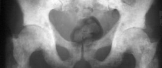
Survey X-ray
This is an x-ray of the abdominal cavity, which allows you to detect the presence of stones without the use of a special contrast agent. The method is successfully used to identify radiopaque stones. Kidney stones are a pathological condition that develops due to disturbances in the properties of urine. Sometimes they are not visible even on a plain X-ray.
Urolithiasis disease
Superposition of the shadow of the stone on the shadow of the vertebrae prevents diagnosis. Also, the formation of gases in the intestinal cavity can obstruct vision, which will not allow the medical professional to see the presence of pathology. There are a number of urological phenomena that mimic the presence of a calculus. These include calcified lymph nodes or the presence of gallstones. Among the positive aspects of this diagnostic method are:
- the ability to establish the presence of stones consisting of calcium in the patient’s body;
- low cost of research;
- accessibility, x-rays can be taken at any medical institution.
Changes in the qualitative composition of urine
If kidney disease is suspected, many tests are prescribed to detect changes not only in the quantity, but also in the quality of urine. Such laboratory tests include the Nechiporenko, Amburge, and Kakovsky-Addis tests. All these tests involve taking an average portion of urine. The material is then examined for the presence of leukocytes, red blood cells and casts. In all cases, an accurate count of the formed elements is carried out, and then a conclusion is given.
The samples differ from each other in that each of them has different normal values. The Nechiporenko analysis is considered good if there are less than 2000 leukocytes and less than 1000 erythrocytes in the field of view. Cylinders with normal indicators are rarely determined, the norm is up to 500. In the analyzes according to Amburge and Kakovsky-Addis, the shaped elements are the same. The difference is that in the first the norms are 200 and 100 units, and in the second - 2 million and 1 million.
X-ray and CT
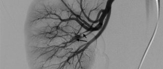
X-ray examination is still one of the main methods for diagnosing kidney diseases. Since the kidneys do not have significant density (like bones) or airiness (like lungs), a regular X-ray will not provide much information about the structure of the kidneys, their vessels and ureters. Therefore, a modification of x-ray examination is more often used - excretory urography. A contrast agent helps make the kidneys visible. Before the procedure, the drug is injected into the blood, after which, according to the scheme, pictures are taken at certain time intervals. In each of them, the picture of the kidneys and urinary tract is slightly different. With its help, you can evaluate not only anatomy, but also the performance of organs.
Urography - examination of the kidneys with a contrast agent
Computed tomography is a method that is significantly more informative than conventional radiography. A CT scan is essentially a series of x-rays. They are given stunning clarity by a computer program that processes the numerous images received. This technology is not inferior in accuracy to magnetic resonance imaging. However, the need to use x-rays in some situations limits the use of the method (for example, in pregnant women). To study the vessels and urinary tract, contrast technology is used using Omnipaque or Optirey.
Optireum is a contrast agent used for CT scanning.
CT - video
Contrast examination of the kidneys: excretory urography
The method of examining the kidneys, excretory urography, is based on the ability of the kidneys to secrete certain substances (radiopaque, iodine-containing) introduced into the body, resulting in an image of the kidneys and urinary tract on radiographs.
The indication for excretory urography is the need to determine, first of all, the anatomical and functional (to a lesser extent) state of the kidneys, pelvis, ureters, and bladder. Contrast agents for kidney examinations are administered mainly intravenously.
The main contraindication to excretory urography is intolerance to iodine drugs.
Before examining the kidneys using this method, the patient’s preparation is the same as for a plain radiograph.
But still, these types of examinations have a number of disadvantages, in particular, they require preparation of the patient, and the patient himself receives, albeit a small, dose of X-ray radiation. Therefore, more modern types of examination are being replaced.
What tests are the most informative?
After conducting an external examination of the patient and listening to his complaints, the urologist will tell you where to start the examination and what tests are necessary. As a rule, the results of laboratory and biochemical tests of urine and blood are needed to make an initial diagnosis. The content of white and red blood cells, as well as epithelial tissues, is determined in urine. The clarity, color and specific gravity of urine must be assessed. The concentration of casts, proteins and glucose is a determining factor in the presence of an infectious focus in the kidneys.
An informative method for studying the functioning of paired organs is the daily volume of urine. It is necessary to submit to the laboratory urine collected during the day, excluding the first emptying of the bladder. In this way, it is possible not only to establish the presence of an inflammatory focus, but also the location of its localization in one of the organs of the urinary system.
In order to determine the type of pathogenic pathogen, laboratory technicians inoculate a biological sample in a nutrient medium. This method reveals the sensitivity of microorganisms to antibacterial drugs that will be used in treatment.
Before donating blood for analysis, the doctor recommends that the patient refrain from physical activity, smoking and drinking alcoholic beverages for 2-3 days. To check the kidneys, blood is taken from a person in the following ways:
Normal kidney ultrasound findings
- from a finger to establish or refute the inflammatory process and the extent of its spread;
- from a vein to determine the concentration of proteins and urea.
For reliable test results, you must not eat food 12 hours before the procedure. If you suspect an endocrine etiology of decreased functional activity of the kidneys, you should not drink any liquid or even brush your teeth. People with systemic diseases need laboratory tests every 6 months.
Kidney structure
The kidneys are paired organs located in the lumbar region. Their main function is the formation of urine. The kidneys maintain the oncotic pressure of the blood and produce erythropoietin. The main structural unit - the nephron - consists of the vascular part (glomeruli) and tubules. The former are responsible for filtering the blood and forming primary urine. The second ones are involved in the reabsorption of substances necessary for the body. Ultimately, what remains is a processed waste product – secondary urine. If at any stage an obstruction occurs, then kidney function weakens. This is expressed in a change in the qualitative or quantitative composition of urine. To understand how to check the kidneys yourself, you need to know about diuresis disorders, which almost all patients have. These may include a decrease or increase in the urge to urinate, a change in the color of urine, or visiting the toilet more often or less often.
Chronic kidney disease on ultrasound
Ultrasound is used to diagnose and monitor patients with chronic kidney disease. With glomerulosclerosis, tubular atrophy, interstitial inflammation or fibrosis on ultrasound, the renal cortex is hyperechoic, corticomedullary differentiation is smoothed. As the disease progresses, the parenchyma becomes thinner and the size of the kidneys decreases.
Drawing. Ultrasound shows chronic pyelonephritis (1): the kidney is reduced to 74 mm, the contour is uneven due to a local decrease in the thickness of the cortical layer. Ultrasound shows chronic glomerulonephritis (2): kidney size is 90 mm, corticomedullary differentiation of the parenchyma is smoothed, a thin cortical layer of increased echogenicity. On ultrasound, nephrotic syndrome (2): hyperechoic kidney without clear differentiation into cortex and medulla.
Drawing. Ultrasound of a patient with chronic renal failure (1, 2, 3): the kidneys are reduced in size to 70x40 mm, the thickness of the parenchyma is 7 mm, the corticomedullary differentiation is smoothed. On ultrasound, end-stage chronic renal failure: the kidney is very small - 36 mm, echogenicity is significantly increased, it is not possible to distinguish between parenchyma and sinus.