The X-ray technique for examining the renal arteries (translumbar or transfemoral aortography) began to be used in the first half of the last century. In 1942-1945 and beyond, the method was actively introduced into the medical practice of urologists around the world.
From the beginning of this century to the present day, renal angiography has been widely used for various diseases, which makes it possible to identify the anatomical and physiological features of the functioning of the blood vessels of the organ. Its significant diagnostic value attracts more and more supporters of this type of research.
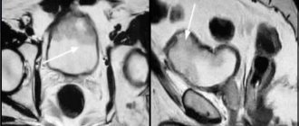
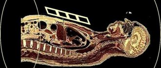
Angiography image of renal vessels
Angiography of renal vessels
Renal angiography is a highly informative research procedure aimed at studying the condition of blood vessels using contrast agents and radiography. This technique has been used in clinical medicine for many years. Moreover, with the help of angiography of the renal vessels, specialists make the most accurate diagnosis, while other methods do not give an unambiguous and clear answer.
The kidney structures are distinguished by a fairly developed system of blood vessels, so the normal functioning of the organ depends on the functionality of the vascular network. Any disturbance of vascular circulation negatively affects renal functionality. Angiographic examination allows us to assess the functional and anatomical state of the renal vessels.
Benefits of renal angiography
Aortography gives a good examination result where other research methods have proven ineffective. It makes it possible to determine the presence and position of additional vessels in the kidneys, compression of the aorta, the local area of blood supply to organs, and determine the type of stenosis. This type of study makes it possible to judge not only specific changes in the functioning of the system, but also the condition of the kidneys as a whole. Based on the data obtained during the examination, a preliminary diagnosis can not only be confirmed, but also excluded altogether. Data obtained during the procedure helps determine and predict the course of kidney surgery. Thanks to modern technologies, errors in processing aortography results are the lowest among all other studies.
Nuances of the procedure
A correct conclusion based on angiography can only be made if the contrast agent was injected evenly and distributed throughout all vessels, the aorta and renal arteries. An isolated examination of certain vessels and canals is carried out only if the condition of the other kidney is reliably known based on a set of other studies. If a certain section of the artery was not filled evenly, then a repeat study may be prescribed to obtain the final result. Recently, aortography is often used to diagnose problems with the renal system in children and newborns. The procedure is often performed under general anesthesia, but otherwise is no different from adult aortography.
Indications and diagnostic value
Renal angiography is prescribed to patients in the following clinical situations:
- Neoplasms of a cystic nature;
- In order to clarify the nature of the disorders that occurred in the renal arteries;
- Complex renal disorders, various functional abnormalities of renal structures;
- If you suspect cancer of the renal cortex;
- Hydronephrosis, in which the kidneys become filled with fluid accumulations;
- In order to identify the type of tumor process that has arisen in the abdominal cavity;
- In severe cases of renal tuberculosis, when removal of the affected kidney is indicated;
- Hematuria of unknown etiology, in which other diagnostic methods have not helped determine the reasons for the appearance of blood in urine;
- In clinical cases in which the diagnosis remains unclear after other types of examination;
- To assess blood flow in the transplanted kidney;
- With renal arterial embolism or stenosis.
The procedure of vascular renal angiography has enormous diagnostic significance. It helps determine the diagnosis when other diagnostic methods remain uninformative.
Angiographic examination helps determine the extent of vascular and tissue damage in the kidneys, allows one to accurately determine their etiology and assess the condition of vascular structures. This diagnosis is also effective in oncology, when it is necessary to examine the location of blood channels.
Indications
Renal angiography is prescribed in the following cases:
- kidney development abnormalities;
- renal hematuria (presence of blood in the urine) of unknown nature;
- kidney tuberculosis, especially if there are indications for removal;
- hydronephosis;
- suspicion of the presence of neoplasms in the cortical part;
- suspicion of cysts;
- unknown cause of hypertension, development of hypertension against the background of disorders in the vessels of the kidneys;
- the presence of tumors in the adrenal glands, as well as organs of the retroperitoneal space.
Angiographic examination of the kidneys allows you to determine the degree of damage to tissues and blood vessels, helps to identify their cause, the structure of the vascular network. The method is considered effective in thoroughly examining the location of blood vessels in cases of oncology.
Kinds
Renal angiography is divided into several types, depending on the method of introducing the contrast component: transfemoral and translumbar angiography. In retrograde (transfemoral) angiography, contrast is injected through an artery in the thigh. And during a translumbar angiographic study, a contrast agent is injected into the lower back through an aortic puncture.
In addition, there is also multislice computed tomography angiography of the renal arteries (MSCT). This is a non-invasive study of blood flow in the arteries using high-speed multi-slice vascular scanning. Multispiral angiography after CT is needed to detect congenital vascular anomalies, as well as to determine tumor pressure on the vessels, renal infarction or intraluminal arterial stenosis. CT angiography of the renal vessels uses x-rays for diagnosis.
There is also ultrasound angiography, which involves several methods for obtaining ultrasound images of vascular tracts, such as energy mapping or Doppler color mapping, artificial contrast with intravenous contrast, three-dimensional vascular reconstruction, etc. Vascular angiography is often performed after CT, especially when planning angiosurgery on the renal vessels. Video of CT angiography for renal artery aneurysm:
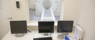
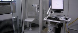
General information about the procedure
Renal angiography has been around for a long time, but has become popular recently as medicine has developed and X-ray equipment has improved, making the procedure safe and effective.
Angiography of renal vessels is a method of diagnosing blood vessels, used to study changes in the vascular system of the kidneys in various diseases. The kidneys have a well-developed vascular system, which transports blood throughout the body. Any changes in the structure of the kidneys provoke disruptions in the functioning of an entire part of the body.
And all these changes occurring in the kidneys can be studied using angiography in combination with other diagnostic methods. The data obtained as a result of these examinations are very significant not only from the diagnostic side, but also for drawing up a further prognosis and the necessary treatment. Renal angiography is used for the following purposes:
- detection of tumors in the kidneys;
- determining the cause of the development of renovascular hypertension;
- studying the structural features of the renal vascular system before surgery;
- monitoring the condition of the kidney vessels in case of renal failure or chronic diseases;
- assessment of the condition of the organ after injury;
- identification of complications after transplantation;
- in order to distinguish between a cyst and a tumor.
Preparation for the procedure
The angiographic diagnostic procedure is not carried out without specialized training, which involves:
- Alcohol should be avoided 14 days before the test;
- For 7 days, avoid taking blood thinning drugs like aspirin, etc.;
- 5 days before the planned procedure, you need to do an ultrasound diagnosis of the heart and fluorography, an electrocardiogram and a coagulogram;
- 2 days before the procedure, you need to test for contrast tolerance;
- One day before, if necessary, you need to remove hair from the area indicated by the doctor;
- Before the procedure, you need complete rest and a good night's sleep, so you can take a sedative;
- In the morning you need to do an enema to cleanse the intestines, you can’t eat or drink anything, and before the test you need to go to the toilet.
Stages of angiography of renal tissue
In medical practice, there are two ways to study a problem area. These include:
- translumbar aortography;
- transfemoral aortography.
In the first case, a puncture is taken from the lumbar aorta. After this, they move on to the procedure itself. It includes several important steps:
- 25 minutes before the administration of the reagent, the patient is given anesthetic anesthesia.
- Next, the patient is placed on his stomach. The left hand is placed along the body of the body. The right one is pushed to the side. It allows you to control blood pressure levels and monitor the patient’s pulse.
- After this, local anesthesia is administered.
- When the drug anesthetic takes effect, the reagent composition begins to be administered. During the administration of liquid, the patient is recommended to hold his breath for 6 seconds.
- In the first 2 minutes, a nephrogram and an arteriogram are performed.
- At the end of the procedure, a venogram of the venous network is made.
- After 6 minutes, a repeat radiograph and excretory ultrasound are performed.
- Next, they begin to administer a drug that prevents blood clots.
Transfer aortography has some differences in the procedure. The puncture is made in the area of the femoral artery. Manipulation includes the following actions:
- The patient is placed on his back.
- Next, an anesthetic substance is injected, which prevents spasm of the venous network.
- The periods of radiographs are carried out similarly with the same interval.
The advantage of this technique is the absence of subsequent medical sutures at the puncture sites.
At the end of the examination, the patient should be in a lying position for 2 days. Any movement negatively affects the patient’s well-being. In addition, there are several criteria that require careful adherence. These include:
- Ultrasound of renal vessels: Indications, Indicators, Preparation, Methodology, Explanation
- complete refusal of water procedures within 15 hours from the moment of aortography;
- the patient should drink up to 4 liters of clean water. It promotes rapid removal of the reagent outside the body;
- do not touch the bandage at the puncture site;
- exclude any movement for 3 days;
- It is prohibited to drink alcohol;
- eliminate smoking tobacco products;
- It is recommended to adhere to bed rest.
How they do it
The process of angiography of the renal vessels varies somewhat depending on the method of administration of the contrast agent. During translumbar angiographic examination, Omnopon and Morphine are administered before the procedure. Anesthesia is used locally, contrast is injected into the lumbar region while breathing is held. In the first minutes, an arteriogram and nephrogram are performed, and then a venogram. An excretory urogram is performed 5 minutes after the first radiograph. Together with the contrast agent, Heparin is administered to prevent thrombus formation, as well as Novocaine for aortic spasm.
In transfemoral aortography, contrast is injected into an artery in the thigh. The patient is placed on his back and novocaine is administered. Otherwise, the radiograph periods are similar to translumbar angiography. No stitches are required at the puncture site; a sterile bandage is sufficient. The difference between these methods is the recovery period.
During a retrograde study, standing up and walking is allowed only after three days, and after translumbar angiography - on the next day.
Indications for renal angiography
In some cases, doctors have difficulty making the correct diagnosis. And when all other diagnostic methods do not help in determining the location of the renal vessels, and also do not provide the necessary information about their functional activity, renal angiography is recommended. But it should be understood that angiography is used only as a supplement to other studies or to clarify the diagnosis when there is a lack of information, and this procedure is not used as an independent and only diagnostic method.
The need for research is determined only by the attending physician. Typically, angiography of the renal arteries is prescribed if the following indications exist:
- presence of traces of blood in the urine for an unknown reason;
- suspicions of oncology in the kidneys;
- abnormal kidney development;
- inability to make an accurate diagnosis based on research results;
- renal tuberculosis in severe form, which involves removal of the organ;
- arterial hypertension;
- establishing the type and nature of neoplasms;
- the presence of a large amount of excess fluid in the kidneys;
- nephrogenic hypertension.
Contraindications
Angiographic diagnostics has a number of specific contraindications:
- Presence of pregnancy;
- Severe form of kidney failure;
- Intolerance or negative reaction to iodine-containing drugs;
- Active pulmonary tuberculosis;
- Tendency to form blood clots;
- Liver or myocardial failure;
- Thyrotoxicosis, etc.
Within 2-4 hours after the study, adverse reactions may occur, which are considered normal. They manifest themselves as headaches and dizziness, a iron taste in the mouth and mild fever. But soon these reactions go away on their own.
Contraindications for carrying out
Dynamic indirect angiography of the kidneys is a rather complex procedure that cannot be prescribed if the following contraindications are present:
- pregnancy (all trimesters);
- pulmonary tuberculosis;
- poor blood clotting;
- vascular atherosclerosis;
- renal, pulmonary, liver failure;
- allergy to iodine;
- serious condition of the patient (caused, in particular, by cancer);
- arrhythmia, angina pectoris and other pathologies of the cardiovascular system in severe form.
How to prepare for angiography of the renal vessels
Angiography of the renal vessels provides for appropriate preparation of the patient for manipulation. 2 weeks before the procedure, you should avoid alcoholic drinks, including low-alcohol ones. 7 days before angiography, you should completely stop taking medications that thin the blood, such as Aspirin. Therefore, the patient should consult a doctor if there is a need for its use.
5 days before the diagnosis, the patient undergoes a number of studies such as: ultrasound of the heart, cardiogram, fluorography. Laboratory blood tests are prescribed for coagulation, syphilis, hepatitis, and HIV. General blood tests are performed for sugar, Rh factor and biochemical test.
2-3 days before the examination of the blood vessels of the kidneys, a contrast agent tolerance test is done. If during the procedure an allergic reaction or negative changes in the functioning of the heart are detected, the main operation may be cancelled.
If all indicators meet the standards, the day before the procedure, hair should be removed from a certain area of the body where the puncture will be made and the catheter will be inserted. You should get a good night's sleep the night before the test. In case of anxiety and fear, it is recommended to take a sedative only as prescribed by a doctor.
In the morning you should completely cleanse your intestines. For this, an enema and special suppositories are administered. Immediately before the procedure, you should empty your bladder. On the day of diagnosis, you must completely abstain from water and food. Only if all recommendations are followed, the procedure will be successful.
Features of the procedure
Angiographic examination of the kidneys can be performed in two ways:
- translumbar aortography;
- retrograde aortography.
Both of these methods of angiographic diagnosis involve the insertion of a catheter into the aorta, that is, a kind of small operation occurs. And each case has its own peculiarities of the procedure. Which type is suitable in a particular case can only be determined by the attending physician.
Angiographic examination of the kidneys can be carried out in the following ways:
- 20 minutes before translumbar aortography, the patient is injected with a dose of morphine, and before the study itself, an omnopon solution is administered. The patient should take the following position: lying on his stomach, the right arm is moved to the side, and the left one lies along the body. The pulse and pressure are recorded on the right hand, after which local anesthesia is given. The first 2 x-rays are taken during the process of introducing contrast into the body, the third x-ray is taken while the contrast is fully in the body, and the fourth – 5 minutes after recording the first x-ray. Along with contrast, a heparin solution is injected dropwise into the aorta throughout the procedure, which prevents the formation of blood clots in the vessels. In order to prevent the response of the aorta in the form of a spasm, novocaine is administered. Immediately before contrast is introduced into the body, the patient is asked to hold his breath.
- Retrograde aortography is characterized by a puncture on the thigh. The patient lies on his back, after which local anesthesia is administered, and a novocaine solution is injected into the vessel. Then specialists place a special probe into the aorta, with the help of which the contrast agent is released at high speed. Only 3 pictures are taken, two of which are recorded at the beginning of the contrast agent injection, and 1 at the very end of the study. After the probe is removed, small hematomas and swelling may appear on the patient’s body, although the wound is very small. But if tissue was dissected to expose the femoral vein, a small suture may be necessary.