Pneumothorax is a disease that occurs as a result of injury or against the background of a complicated course of another disease. Numerous cases of diagnosing the disease have been recorded around the world.
Patients often include newborn children and young men under 30 years of age, especially if they are thin and short in stature. The risk group also includes cigarette lovers and people suffering from lung pathologies, for example, pneumonia (pneumonia), tuberculosis, etc.
The danger of the pathological phenomenon is a reduction in the mobility of the lung, as a result of which the human body lacks oxygen. If the disease is not detected in a timely manner, there is a risk of cardiac arrest. The corresponding diagnosis necessarily includes an x-ray examination.
Classification of pneumothorax
During X-ray diagnostics, three types of disease are detected. Each of them has a number of features:
| Type of illness | general characteristics | Signs in the picture |
| Open | Accompanied by the formation of a canal connecting the pleural cavity and the external environment - the pressure in the chest is compared with atmospheric pressure. The lung tissue collapses, the organ loses functionality. The disease has a poor prognosis. | -lung tissue is compressed; - flattened diaphragm dome; -mediastinum is displaced. |
| Closed | The main characteristic is the presence of a “barrier” along the path of air to the organs in the chest. The pleural cavity either has no damage or is covered with tissue particles, blood clots, and pus. This type of pneumothorax responds well to treatment and is eliminated without serious consequences. | -there is no pulmonary pattern; - the edges of the lung are visualized. |
| Valve | A valve is formed that does not interfere with the passage of air from the atmosphere into the lungs, but does not allow it to escape outside. Internal pressure rises, increases above atmospheric pressure. The blood vessels, trachea, and heart are compressed—the patient develops arrhythmia, and the person goes into shock. | - mediastinal organs are significantly displaced to the side opposite to the developing pathological process |
Valve pneumothorax is different in that the degree of danger to the patient’s life depends on the rate of penetration of atmospheric air.
How the disease develops
The rate of development of the disease and the degree of involvement of lung tissue in the pathology depends on the severity of the primary pathology, the time that has passed since the penetration of air into the cavity, and the dynamics of the inflammatory reaction.
The spontaneous form of the disease is characterized by the presence of a hole in the pleura. The penetration of air into the pleural cavity causes an increase in pressure in it, a collapse of the lung tissue, which provokes the collapse of the organ. As a result, the mediastinal organs shift and move synchronously with the act of breathing.
5 hours after the gas is released, the inflammatory process begins. Inflammation is characterized by blood filling of small vessels and effusion of serous exudate. After 48 hours, pleural edema begins, which increases, it is accompanied by the loss of fibrin on the surface of the serous membrane.
If the inflammatory process is not stopped, the area of the lung loses its ability to straighten. The addition of infection threatens suppuration of the affected area and the formation of fistulas.
X-ray examination techniques
If pneumothorax is suspected, visual examination and x-rays are the first choice.
When examining the patient, the diagnosis is confirmed in the following cases:
- the person tries to be in a forced half-sitting position (in this position the pain is not so pronounced);
- the spaces between the ribs are widened;
- cold sweat is visualized on the skin, a blue tint to the skin;
- there is severe shortness of breath.
Among other symptoms identified during the initial diagnosis: low blood pressure, displacement of the borders of the heart to an intact organ, tympanic sound.
The final diagnosis is made after the conclusion of a radiologist.
Diagnosis and diagnostic criteria for pathology
Diagnosis of the disease consists of examining the patient and instrumental research methods.
During the interview, the doctor pays attention to the presence of concomitant pathologies, manifestations of the clinical picture, and the position occupied by the patient (half-sitting, leaning on the painful side). During the examination, the doctor notes the lag of the diseased half of the chest during the respiratory act, difficulty breathing, and during percussion - a boxy sound. After the examination, instrumental research methods are used.
We recommend reading! Follow the link: Allergy to injected tuberculin
Alternative diagnostics
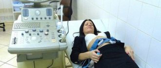
If pneumothorax on x-ray does not provide specialists with the necessary information, additional ultrasound examination and blood gasometry are performed.
An alternative to radiography is computed tomography, and in rare cases MRI. This technique is most justified:
- if necessary, detect small pneumothorax;
- to identify emphysematous bullae - disease provocateurs;
- when identifying the cause of a secondary disease (cyst, etc.).
Pneumothorax on CT
After both studies (X-ray and CT), specialists receive information about the degree of collapse of the affected organ.
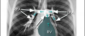
X-ray
To determine the apical localized air collection, fluoroscopy of the lungs (real-time x-ray) is prescribed. During the rotation of the patient, the specialist can recognize the movement of the air mass.
The described situation is typical when there is an insignificant change in pressure in the patient’s chest. In this case, a certain area of the organ is already collapsed. The displacement of the mediastinum has not yet been identified; the diaphragmatic dome is slightly flattened.
If the disease is not examined in a timely manner on an x-ray, the pressure will increase, and the tissue surface of the lung will completely collapse - an acute deficiency of oxygen will appear in the body.
Radiation research methods
It is better to take x-rays in the anteroposterior projection, with the patient in a vertical position. With this technique, fluid will collect under the influence of gravity in the lower parts of the pleural cavity, and its level will be detected. Air, on the contrary, will collect in the upper sections. With hydropneumothorax, a horizontal fluid level is observed.
Signs of pneumothorax are clearing of the pulmonary fields and the absence of a pulmonary pattern.
Left-sided pneumothorax
The visceral edge of the pleura may be visible, as a very thin white line, which is separated from the parietal layer by a gas space. The mediastinum shifts towards the lesion (unless we are talking about tension pneumothorax). Subcutaneous emphysema may also be seen on x-rays. With a tense form, a downward displacement of the diaphragm is also observed.
In doubtful cases, a lateral view may be taken. Then the lung will “fall away” from the chest wall. When taking pictures during inhalation and exhalation during the expiratory phase, the lung becomes smaller and even denser, the size of the pneumothorax does not increase.
In cases where the patient is in a serious condition that does not allow an X-ray to be taken in a direct vertical projection, a picture can be taken with the person in a horizontal position. Then the X-ray signs are poorly visible. Deep costophrenic angles and the presence of changes in lung size are all that can be seen on the image in this case.
Bilateral pneumothorax
What is the importance of diagnosis
It is preferable to diagnose pathology using X-rays in the initial stages of the disease. The disease develops slowly and occurs against the background of the following symptoms:
- inflammatory process in the pleura;
- respiratory failure;
- concentrated serous exudate in the deepest sinus;
- accumulated fibrin on the pleura;
- thickening of lung tissue;
- purulent cavities;
- signs of hemorrhage.
If organ tissue is weak, physical stress, such as coughing or sudden body movements, can result in disruption of the integrity of the organ and the release of gas.
The disease becomes a consequence of repeated atelectasis (collapse of the damaged lung) with gas concentration in the local area.
An X-ray is necessary to save the patient. In the event of a rapid lung rupture, specialists have very little time left to provide the patient with appropriate medical care.
The gradual accumulation of pleural air (development of pneumothorax) is accompanied by a number of characteristic symptoms, including:
- sharp chest pain;
- severe shortness of breath, dry cough;
- pain in the heart area;
- fainting (in the case of a pronounced pathological process);
- increasing the distance between the ribs.
The manifestation of such signs helps a specialist assess a person’s condition based on an x-ray and reliably formulate a conclusion taking into account the specifics of the disease.
If a pneumothorax is suspected, an x-ray is an integral part of diagnosing the disease. The basic manifestation of the disease is the concentration of air in the pleural tissue.
On the image, this sign is visualized as an undetected pulmonary pattern or air space. The specificity of the manifestations in the resulting image is dictated by the type of developing pathological process. X-ray helps diagnose closed, open and valve pneumothorax.
As part of diagnostic procedures, in addition to radiography, a visual examination is performed. Sometimes an ultrasound or blood gasometry is required. Computed tomography is used as an alternative method for making a diagnosis.
How can a pneumothorax be seen on an x-ray?
The description of a radiograph for pneumothorax, as for any other pathology, includes a number of points, including the pulmonary pattern. Of all the characteristics, the pulmonary pattern is the most important in this case. It is formed by shadows from blood vessels, and sometimes bronchi. The main radiological signs of pneumothorax:
- If the lung or part of it has collapsed, and there is air in this place, then there is no pulmonary pattern in the projection of such an area.
- In the same place, the transparency of the pulmonary field is locally increased.
- Almost always, this area is clearly visually delimited by a strip of lung tissue, on which the pattern is condensed due to the fact that the lung is “tucked in”.
- Sometimes a linear shadow from the thickened pleura is visible at the border.
These signs apply to any form of pneumothorax. Another symptom that may indicate tension pneumothorax is displacement of the mediastinal shadow and widened intercostal spaces on the affected side.
The role of the procedure in its treatment
Using an x-ray, the doctor not only diagnoses pneumothorax, but also determines the location of the air “bubble” in order to make a puncture in this exact place to remove it, and also estimates the approximate volume of gas to be evacuated.
If it is impossible to immediately remove air, the patient may need to install drains. X-rays are used to monitor the correctness of their position and the dynamics of the disease.
Symptomatic manifestations
Signs of pneumothorax depend on the severity of the underlying disease, in most cases it is tuberculosis. If the disease proceeds in a long latent stage, then signs of pneumothorax may not be detected, since in most cases they are classified as tuberculosis. However, if proper therapy is not carried out for tuberculosis or the disease progresses, then specific symptoms may appear.
Pain is the main symptom of the development of pneumothorax. The pain may be squeezing or stabbing in nature and develops in the affected part of the chest. In most cases, the pain is localized in the upper chest, but sometimes it radiates to the lower back, arm, shoulder, abdomen or neck. Along with pain, the following unpleasant symptoms of pathology occur:
- Arrhythmia, breathing becomes deeper and faster.
- Severe dry cough.
- Shortness of breath, which increases as the pathology develops.
- Due to an excess of carbon dioxide in the circulatory system, the skin takes on a bluish tint.
- With pneumothorax, the patient experiences compression in the chest.
- Due to the development of pneumothorax, the patient may experience panic and anxiety.
Basic principles of therapy
Therapy for pneumothorax that develops as a result of tuberculosis is complex. During treatment, the doctor performs chemotherapy and puncture of the pleural cavity. During puncture, all accumulated air is removed from the lungs.
If tuberculosis occurs at an early stage, the patient may have an artificial pneumothorax, in which 300 ml of gas is injected into the organ. Thanks to this procedure, the lung is quickly restored and expanded.
If pneumothorax is detected, treatment must be started immediately, otherwise this can lead to rapid progression of the pathology and the development of irreparable complications.