This disease is dangerous not only due to disruption of the functioning of the organ, but also due to numerous complications (oncological processes, bleeding, ascites, problems with the kidneys and lungs). Below we will look at whether cirrhosis of the liver is visible on ultrasound, what its echographic signs are, and how the picture differs from the norm.
Ultrasound of the liver will show cirrhosis of the liver or not
With 100% probability, ultrasound of the abdominal organs allows you to determine cirrhosis at stages 3-4. On compensatory and subcompensatory - an experienced specialist can detect only indirect manifestations of pathology, and this is the reason for prescribing other more accurate methods: biochemical blood test, elastometry, Dopplerography.
If these methods do not allow an accurate diagnosis to be made, and doctors have doubts about cirrhosis, liver tissue is collected for examination (liver biopsy).
But, with the help of ultrasound, it is possible to detect cirrhosis at stage 1 with a 70% probability if destructive processes begin to occur on the surface of the organ (nodular form). In other cases - 20-50% depending on the experience of the doctor and the correct preparation of the patient and the sensitivity of the equipment.
What is liver cirrhosis
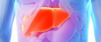
Liver cirrhosis is a chronic disease that is accompanied by significant death of hepatocytes and their replacement by connective tissue. Usually this process is a consequence of other diseases:
- viral hepatitis;
- excessive consumption of alcoholic beverages (chronic alcoholism);
- heart failure;
- obstruction of the biliary tract (cholelithiasis, chronic cholangitis);
- taking hepatotoxic drugs;
- hereditary metabolic disorders in the liver;
- occlusion or thrombosis of the portal vein;
- autoimmune disorders;
- parasitic infestation that was not cured in time.
About 10-20% of all cases of cirrhosis remain without an identified cause. A genetic predisposition to the development of this disease has been identified.
The essence of ultrasound diagnostics
Ultrasound examination (ultrasound) allows you to determine the size, and modern equipment and density of the organ being diagnosed. The color image is supplied to the display in high resolution - if in doubt, the image can be enlarged without loss of quality.
Unlike radiographic methods, ultrasound does not harm soft and bone tissue. The study can be carried out daily, tracking the dynamics of changes in key indicators.
Since ultrasound is a dynamic method, there is no blurred image effect. Unlike MSCT, a color image, and on modern equipment - an echo function - the density of the surface layer.
Reasons for examination
Cirrhosis is a gradual transformation of living hepatocytes (liver cells), which ensure the functioning of the liver, into connective tissue that does not carry a functional load. Since the process of degeneration occurs slowly, the signs of the disease at the initial stage are weakly expressed. The main symptoms are:
- loss of appetite and weight loss;
- chronic drowsiness and weakness;
- heaviness in the epigastric region and right hypochondrium;
- indigestion and intense gas formation;
- bitter taste in the mouth.
Many potential patients do not pay attention to the listed signs and seek medical help only at the stage of yellowing of the eyeballs and skin. Early diagnosis of cirrhosis can increase the patient's life expectancy to 10–12 years. Detection of liver pathology at the second stage reduces this period by half. Before performing an ultrasound examination of the organs of the hepatobiliary system, the patient is prescribed blood microscopy, including general clinical and biochemical tests.
In the presence of cirrhosis of the exocrine gland (liver), the following changes in blood composition are determined:
- elevated levels of liver enzymes AST (aspartate aminotransferase), ALT (alanine aminotransferase), Alpha-Amylase, alkaline phosphatase (ALP), bilirubin (the main component of bile);
- high concentration of gamma globulins and IgM immunoglobulins (moderate IgG and IgA immunoglobulins);
- decreased level of platelets and protein fractions;
- high ESR (erythrocyte sedimentation rate) and leukocytosis;
- significantly increased levels of lipids and cholesterol (hyperlipidemia and hypercholesterolemia).
Depending on the stage of the disease, pathological changes in the blood progress.
Preparing for the study
Meticulous preparation is 75% of the effectiveness of the procedure.
| During the week |
|
| Per day |
|
Preparation
A responsible approach to research on the part of the patient will allow you to see even minor changes in the condition of the organ. The subject is required to follow simple recommendations, namely:
- Stick to a diet for several days before the procedure. You should exclude foods that promote gas formation or increase the load on the liver (fatty, smoked, fried, fermented milk products, foods containing large amounts of carbohydrates). Preference should be given to easily digestible foods, steamed or baked if possible. The doctor who gave you the referral for ultrasound diagnostics of the liver will tell you more about the diet.
- If necessary, take medications prescribed by your doctor.
How is diagnostics carried out?
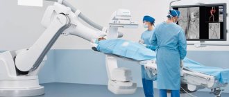
Optimal positions: on your back, left side, with your right hand behind your head. The contact point of the sensor is lubricated with a special gel. While passing the sensor, hold your breath. The doctor moves the sensor slowly, evenly until he gets a general picture of the condition of the area being examined.
Next - jerky movements with a sensor delay of 1-5 seconds in doubtful areas. After the examination, the gel is wiped off with a swab containing a special solution. Afterwards, use a wet napkin (it is advisable to have them with you).
Watch a video about preparing for a liver ultrasound and normal indicators:
Recommendations for preparation
To increase the accuracy with which an ultrasound examination detects cirrhosis of the liver, you should properly prepare for the procedure. Full preparation takes at least 3 days. It includes following a diet and taking medications as needed.
A diet is needed to eliminate gas formation and bloating. Removed from the diet:
- cabbage;
- peas and beans;
- black bread;
- baking;
- sweets;
- carbonated drinks;
- alcohol.
During the entire preparation period, the diet should consist of low-fat soups, buckwheat porridge, and boiled meat. On the eve of the procedure, a light dinner is allowed, after which you can only drink water.
If flatulence is severe, there are difficulties with bowel movements, medications are prescribed:
- enzymes - “Mezim”, “Creon”;
- laxatives - “Nutrifiber”, “Slabilen”;
- carminatives - “Espumizan”, “Bobotik”.
People come for the study on an empty stomach, with empty bowels.
To prepare for a liver test, watch the video:
Survey results
The results are printed on a special form or on 2 or 3 sheets of A4 format. Depending on the clinic, the doctor enters his explanations in a specially designated field or next to each parameter; less often - with a ballpoint pen on a printout.
In 90% of cases, the doctor announces the preliminary results and sends the patient with a printout to a therapist. The results are added to the epicrisis. The patient is sent for additional examination. Treatment is prescribed by a hepatologist and or gastroenterologist. As a rule, they are also nutritionists.
Symptoms of cirrhosis
The clinical manifestations of cirrhosis are very extensive, since the disease affects the functioning of not only the digestive system. Most often they include:
- loss of appetite;
- a feeling of heaviness or periodic aching pain in the right hypochondrium;
- weight loss;
- an increase in the size of the abdomen;
- itchy skin;
- intolerance to many types of foods (meat, fried, high fat);
- increase in body temperature to low-grade levels;
- decreased performance;
- joint pain;
- tremor of fingers;
- decreased intellectual abilities;
- development of cough and shortness of breath with moderate physical exertion;
- peripheral edema (mainly legs);
- yellowness of the skin;
- white coating on the tongue;
- an increase in the size of the spleen;
- increased production of gases in the intestines;
- nausea, tendency to constipation or diarrhea;
- the appearance of “jellyfish head” - enlarged veins of the anterior abdominal wall.
It has been reliably established that as liver failure progresses, the frequency of cardiovascular events (myocardial infarction, stroke, vascular thrombosis of the lower extremities) increases. There is also a decrease in the filtration function of the kidneys (hepatorenal syndrome).
Ultrasound indicators for cirrhosis
First of all, the blood-purifying function of the liver is disrupted. With normal liver parameters, at the compensatory stage of cirrhosis, an enlargement of the spleen is observed. The reason for other diagnostic methods is length 120, width 60 mm. Dead red blood cells and macrophages accumulate in the spleen.
It is easier to diagnose the nodular form - destructive processes begin to occur on the surface - lumpy formations 1-3 mm of dark red, rich brown or dark brown.
At stage 1, a heterogeneous echo structure may be observed - with a normal image, a loose structure in density.
Another indicator is the amount of fluid in the abdominal cavity - ideally up to 0.3 l; if the spleen is enlarged and the echo is disturbed, 0.5 l is already an excess.
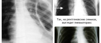
Important! Liver enlargement is often not observed, especially with necrotic cirrhosis. The necrotic form has a brownish surface, and if it is deep, it can only be diagnosed by x-ray - increased density of necrotic tissue.
If the pathology occurs deep in the organ - an increase in the diameter of the portal vein to 9 or more millimeters, fluid in the abdominal cavity and an enlargement of the spleen.
Methods for diagnosing cirrhosis
Is it possible to detect cirrhosis of the liver by ultrasound? Suspicion of a disease arises based on the patient’s complaints and symptoms. After laboratory tests and tests for viral hepatitis, visualization of the organ is required.
Ultrasound diagnostics is the simplest and most effective method. It allows you to accurately measure the size of the liver and determine the presence of structural abnormalities in the parenchyma of the organ. It is after this that cirrhosis is first discovered in most patients. It is also possible to measure the diameter of the veins and diagnose the condition of the portal system. The study has no contraindications. It can be performed without restrictions during pregnancy and childhood.
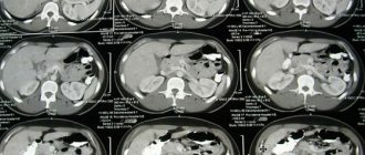
Computed tomography (CT) is also used to diagnose cirrhosis. To increase the information content of research, contrast is often used. Liver imaging allows for a definitive diagnosis. However, this technique is not suitable during pregnancy due to radiation exposure to the fetus.
In case of liver cirrhosis, it is recommended to carry out “Fibrotest”. This is a non-invasive technique that allows you to determine the degree of organ fibrosis, which is key for assessing the patient’s prognosis and choosing the optimal treatment tactics.
Liver biopsy, which is often performed under ultrasound guidance, is of great importance. Then a small puncture is made in the front wall of the abdomen and a small section of organ tissue is taken. The sample is sent for cytological examination. It allows you to definitively confirm the diagnosis of cirrhosis, as well as detect malignant processes at an early stage.
Types of pathology
From the point of view of ultrasound examination, the following types of liver cirrhosis are distinguished.
| Nodular | Micronodular up to 2 mm; large nodular – more than 2 mm. Dark tissue between the nodules; internal tissue under the dark areas is difficult to diagnose. It can be diagnosed at the compensatory stage if the pathology occurs on the surface. |
| By location | Superficial – easily diagnosed at stage 1; internal - using ultrasound only at stage 3; at stages 1-2 by indirect signs and other methods. |
| According to the structure of the degenerated tissue | Fibrous - manifests itself from stage 1 with nonspecific symptoms, difficult to diagnose using ultrasound; necrotic – the spleen enlarges from the moment the pathology occurs; symptoms from stage 3. |
| Unknown etiology | Nodules and fibrous tissue – easily diagnosed at stage 1, rapid development; hepatocytes do not function and do not degenerate - at stage 4, there is a high probability of cancer from stage 1. |
Differential diagnosis
Ultrasound can show similar results for different diseases, so it is very important to carry out differential diagnosis. Formed cirrhosis with a pronounced clinical picture, as a rule, does not cause difficulties; it is usually differentiated only from liver cancer. Asymptomatic cirrhosis, especially in the inactive phase, has similar symptoms to chronic hepatitis.
Cirrhosis should be distinguished from the following diseases:
- Hepatitis. Common signs are tissue tuberosity.
- Liver fibrosis. The cells of the organ are replaced by rough connective tissue.
- Budd-Chiari syndrome is an obstruction (obstruction) of the hepatic veins.
- Metastases. They imitate nodes and fibrous areas. Decreased liver volume.
- Sarcoidosis of the liver. Simulates cirrhosis. Presence of nodules and lymphadenopathy. A comparison with the clinical picture is required.
- Portal vein thrombosis. Atrophy of the peripheral part, hypertrophy of the central parts, varicose veins.
- Nodular hyperplasia. Leads to the development of portal hypertension and the appearance of nodes.
Related Issues
There are a number of objective reasons that make it difficult to make a correct diagnosis.
- Congenital anomalies of the shape and size of the liver, gall bladder, and bile ducts.
- Steatosis, untreated fatty hepatosis.
- Inflammatory processes in the hepatobiliary system.
- The course of an infectious disease or its complications - hepatocytes work in emergency mode; increased temperature - enlarged liver; additional load on the kidneys, temporary accumulation of fluid in the abdominal cavity.
- Working with harmful chemicals for more than 6 months and or playing professional sports – liver enlargement.
What can be seen on an ultrasound
Ultrasound for liver cirrhosis is prescribed for diagnostic purposes. For this purpose, an ultrasound examination of the abdominal organs is prescribed. During the procedure, you can evaluate the general characteristics of the liver:
Liver size is normal according to ultrasound
- the size of its parts;
- circuit;
- structure;
- diameter of the bile duct and portal vein.
In addition, ultrasound shows the condition of nearby organs.
During the examination you can find:
- changes in the location of the organ, its shape and size;
- presence of neoplasms;
- signs of inflammatory processes.
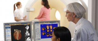
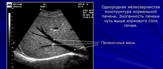
Ultrasound technology involves passing ultrasonic waves through organ tissue. Sensors record the speed and intensity of their back reflection. The results are converted by a special program and visualized on the monitor. The doctor takes measurements of the necessary parameters, evaluates ultrasound conductivity, vascular patterns, and the pattern of changes. The results are assessed by comparison with conventionally normal values characteristic of a healthy liver. Non-compliance with normal values indicates pathological changes in the liver.
The parenchyma (collection of cells, tissue) of a healthy liver has a fine-grained, uniform (homogeneous) structure. The boundaries of the liver contour are clear and even, there are no depressions or bulges. The main evaluation criterion is echogenicity, that is, the speed and degree of absorption of ultrasound waves by liver cells. Ultrasound will show the ability of the parenchyma to transmit or reflect ultrasound. Hyperechogenicity, or increased absorption, indicates an increase in the amount of fluid or thickening of the tissue. With increased echogenicity, the liver appears on the monitor as a white area. Hypoechogenicity, or reduced absorption, occurs when the amount of fluid decreases and healthy tissue is replaced by scar tissue (connective tissue). With reduced echogenicity, dark, sometimes almost black areas are visible on the monitor.
Tables of general evaluation criteria and normal values for the liver in mm:
| Length | Transverse size | Thickness |
| 140-180 | 190-230 | 100-120 |
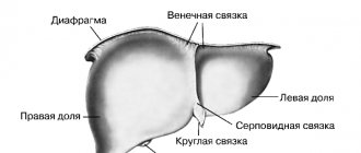
Normal values for the right and left lobes of the liver in mm:
| Thickness | Oblique vertical dimension | Length | Angle of the right lobe | |
| Right lobe | 110-130 | 150 | 110-150 | 75o |
| Height | Thickness | — | Angle of the left lobe | |
| Left lobe | 100 | 70 | 45o |
Normal values for hepatic veins, arteries, ducts in mm:
| Portal vein | vena cava | Hepatic artery | Splenic artery | Common bile duct |
| 13 | 15 | 10 | 7 | 7,5 |
Are ultrasound errors possible and why?
The main cause of errors is inadequate preparation or lack thereof. Sleeping on the stomach - dark spots, nodes not related to pathology, abnormal sharpening of the edges, oval bulge in the middle, muscle tension, enlargement of the organ due to lack of fluid.
The patient does not hold his breath during the procedure - a cloudy image, running blurry spots, heterogeneous contours of the organ.
Strong emotional stress 24 hours before the procedure and longer - enlargement of the organ due to excess adrenaline and accelerated hormonal metabolism.
Features of liver ultrasound
Liver ultrasound is prescribed to patients who contact an infectious disease specialist or hepatologist with certain complaints or are in the so-called risk group. People who have had viral hepatitis B or C are required to undergo an organ examination every six months.
It is advisable to do ultrasound in specialized clinics where doctors treat liver diseases. The likelihood that abnormalities in the structure, shape, and size of an organ will be detected in a regular clinic is extremely small if the disease has not yet progressed.
You can first contact your GP and describe your warning symptoms. He will prescribe an initial examination and give a referral to a specialist.
The problem with diagnosing most liver diseases is that they do not manifest themselves until a late stage of development. Such pathologies are often called “gentle killers” because patients do not experience pain or other unpleasant sensations, and when symptoms appear, it is no longer possible to help the patient.
The only manifestations of most liver diseases are dark urine and discolored feces. If you experience these symptoms, you should contact your doctor immediately. They may indicate minor renal failure, which is easily treatable. But there is a possibility that these are the first signs of hepatitis C, cirrhosis or liver cancer.
Self-diagnosis methods
Alcoholic cirrhosis is independently diagnosed at the compensatory stage - sunken, pointed contours of the liver upon palpation; swelling in the center; dry skin and dry mouth; the abdomen enlarges from the stomach.
The onset of internal venous bleeding; when you press on the swelling, the pain radiates to the stomach and or heart, compresses near the liver - a hematoma with the likelihood of developing into cancer.
Necrotic is diagnosed from the subcompensatory stage; loose liver, formations are clearly visible on palpation; as a rule, there are no nodes, an increase in temperature above 38 at the end of the wakefulness cycle, loss of strength from the compensatory stage; the spleen is palpable.
Fibrous - if there has been no alcohol abuse and prolonged contact with harmful chemicals - only by clinical methods.
Important! Palpation should only be performed by an experienced massage therapist or doctor. Possible rupture of internal organs, disruption of the liver structure; Strong pressure on fibrous tissue provokes cancer.
Technique
Ultrasound diagnostics is based on the ability of waves to pass through the skin and muscles and be reflected from internal organs. The returning waves are captured by the sensor and transmitted to the computer. There the sound is transformed into a picture.
The denser the fabric, the more it reflects sound. This determines how the organ looks on an ultrasound. Areas with high density are represented by light spots, and areas with low density are represented by dark spots. The liver in the ultrasound image is gray in color, as it has an average density.
Research technique:
- the patient lies down on the couch, exposing the upper abdomen;
- the doctor applies a gel to the skin to improve contact with the sensor;
- using a sensor, examines all parts of the liver.
The procedure lasts 15-20 minutes, then the doctor evaluates what he saw and makes a written conclusion. The examination protocol is given to the patient along with a printed image.
The doctor who conducts the examination does not diagnose cirrhosis, he only gives a description. The final diagnosis is made by the attending physician.
What are the echo signs of cirrhosis and how is it described?
The main manifestations on ultrasound are the following echographic signs of cirrhosis:
- Changes in liver size. First, an increase in size is observed, then, in the terminal stage, liver atrophy, mainly of the right lobe. There is a decrease in the size of the right lobe relative to the left, and an increase in the left lobe of the liver. Normally, the ratio of the width of the right lobe to the width of the left lobe in cross section is about 1.44. Liver cirrhosis is diagnosed if this ratio is less than 1.3. The sensitivity of diagnosing liver cirrhosis is 75%, specificity is 100%.
- Enlargement of the caudate lobe: its thickness is more than 3.5 - 4.0 cm.
- The lower edge of the liver becomes blunt - more than 75° in the right lobe of the liver and 45° in the left lobe with liver enlargement.
- Tuberous contour of the liver due to regeneration nodes. In micronodular cirrhosis, this sign is absent. In the case of small nodules located under the capsule, a “dotted line symptom” is possible, which manifests itself as an uneven and intermittent image of the liver capsule. When registering this symptom, the presence of regeneration nodes is convincingly verified by the uneven contour of the liver. In approximately half of patients with liver cirrhosis, regeneration nodes are not visualized by ultrasound, but if they are found, the likelihood of liver cirrhosis is practically beyond doubt when other pathologies are excluded. That is, if the problem of “hepatitis or cirrhosis” is being solved, the presence of a lumpy surface of the liver clearly indicates cirrhosis.
- Depletion of the vascular pattern of the liver. In healthy people, the vessels are normal: the pattern is small, but in sick patients the pattern is different, which allows us to draw appropriate conclusions.
- Irregular width of intrahepatic veins.
- Changes in the structure of the liver parenchyma. Changes in the echostructure of the liver parenchyma are a consequence of cicatricial degeneration of the liver tissue and the formation of regenerative nodes in it, the sizes of which vary from several millimeters (small- and medium-nodular cirrhosis) to several centimeters (large-nodular cirrhosis). Cirrhosis contributes to the heterogeneity of the parenchyma, the density of which can vary significantly. The echogenicity of the parenchyma is usually moderately increased, although it can be significantly increased with the formation of cirrhosis against the background of fatty hepatosis. More specific for liver cirrhosis are changes in the liver structure, which becomes granular and diffusely heterogeneous. The heterogeneity of the parenchyma can reach such a degree that in this “echostructural chaos” it is easy to view the tumor or, conversely, to misdiagnose it. It should be borne in mind that even with advanced liver cirrhosis, especially small-nodular cirrhosis, the echostructure may turn out to be completely normal. However, most often in ultrasound reports the liver parenchyma is described as mixed or increased.
- Enlargement of the spleen. The area of the spleen becomes more than 50 cm2. Sometimes 5-10% of patients do not have splenomegaly.
- Signs of portal hypertension. An increase in the diameter of the portal vein is more than 12–14 mm, the splenic vein is more than 9 mm. Reducing the speed of blood flow in the portal vein until it stops or reverse flow.
- Visualization of dilated veins that are not visible in healthy people. With portal hypertension, the umbilical vein is restored in the round ligament of the liver. In addition, there is an expansion of the gastric veins and the development of additional connections (anastomoses) between the vessels in the abdominal cavity.
- There is an increase in the diameter of the left branch of the portal vein in comparison with the right. Normally, the diameter of the right portal branch is greater than the diameter of the left.
- Doppler ultrasound records changes in hemodynamics in the liver vessels. An increase in the speed and volume of blood flow in the hepatic artery, a change in the shape of the portal flow, and slow, sometimes reverse portal blood flow are detected.
- Dilatation of the hepatic artery.
- Swelling of the wall of the gallbladder, stomach, intestines.
- Free fluid in the abdomen is ascites. It is detected at the final stage of liver cirrhosis. In this case, the small liver resembles a wooden block floating in water.
- With an autoimmune cause of liver cirrhosis, an increase in regional lymph nodes is observed, sometimes to large sizes, when their length reaches 50-60 mm. Their shape is oblong, the ratio of length to diameter exceeds 2, in contrast to cases of malignant tumor with metastasis, when the lymph nodes have a round shape.
Lumpy formations on the liver in this patient indicate cirrhosis
Decoding the results
Another significant advantage of ultrasound in diagnosing liver cirrhosis is that the patient receives the results of the examination immediately. He does not need to languish in waiting and prolonged ignorance.
You will receive a photograph and a detailed description. There will be no final diagnosis here, because the verdict is made by the attending physician after studying the results of ultrasound diagnostics and other research methods performed.
If you suspect cirrhosis of the liver, it is extremely important to undergo examination in the most concise form. Often the disease occurs in a reactive form, that is, the patient’s condition worsens very quickly. The sooner a final diagnosis is made and treatment is started, the greater the likelihood of the disease transitioning to a compensated form.
Despite the fact that the liver is the only organ that has the ability to regenerate, cirrhosis cannot be completely cured. The connective tissue that surrounds the gland cannot transform back into healthy cells. If the prognosis is favorable, surgery can be performed to remove a lobe of the liver and treatment can be continued.
Sometimes a patient can only benefit from a complete transplant. Transplant operations are carried out in Russia, but not all patients wait their turn. This once again proves that early diagnosis is extremely important. The earlier the diagnosis is made, the higher the likelihood that drug therapy will help bring the disease into a compensated form.
Why does cirrhosis develop?
Liver cirrhosis is a disease that is characterized by the proliferation of connective tissue in the liver, which leads to pathological processes that can ultimately lead to liver failure and portal hypertension. The normal physiological structure of the liver is also disrupted. Cirrhosis is chronic.
Dead liver cells are removed by cells of the immune system and replaced by fibrous tissue. The remaining living cells begin to actively increase in number.
https://youtube.com/watch?v=iis5e27zJAg
If individual cells die and fibrotic changes are minimal, then complete restoration of the liver occurs. However, if cell loss is significant and the structure of the hepatic lobules is disrupted, disordered cellular complexes (regenerative nodes) appear, which have an irregular structure and therefore cannot fully perform their functions like healthy liver tissue.
Liver cirrhosis is one of the main causes of mortality in patients with gastrointestinal diseases. Cirrhosis of liver tissue quite often leads to liver cancer. Moreover, cirrhosis of viral origin (provoked by hepatitis B and C viruses) often transforms into liver cancer.
There are many different reasons leading to this liver condition:
- viral hepatitis,
- alcohol abuse,
- accumulation of toxins,
- pathologies of an immune nature,
- biliary tract diseases,
- metabolic disorders,
- thrombosis of liver vessels,
- and others.
It is impossible to determine the cause of cirrhosis using ultrasound data, although individual symptoms may help in this regard.
The ultrasound image of liver cirrhosis depends on the type and stage of development of the pathology, however, ultrasound of the liver when diagnosing this disease shows an accuracy value of around 75%.
Quite often, ultrasound may show splenomegaly (enlarged spleen) and hepatomegaly (enlarged liver) along with moderate indicators of portal hypertension. It would seem a reliable diagnosis: liver cirrhosis.
But no, a similar picture is possible in other cases, for example, with lymphoproliferative diseases
The replacement of normal liver tissue with small fibrosis nodes initially does not cause pronounced changes in the structure of the liver on ultrasound. The appearance of fibrous tissue is accompanied by an increase in the echogenicity of the liver parenchyma, which also happens with other liver pathologies.
When performing an ultrasound scan of a patient with cirrhosis of the liver, you should be aware that not all ultrasound symptoms of the disease must be present. At the same time, key evidentiary symptoms should be recorded.
Despite the many ultrasound signs that are characteristic of liver cirrhosis, the data obtained from echography are insufficient to confidently establish a diagnosis of liver cirrhosis in the initial stages of the disease and when morphological changes in the organ are not clearly pronounced.
uziprosto.ru