Ultrasound scanning of internal organs is a reliable and safe method for identifying diseases. For accurate data, both the qualifications of the doctor and the availability of modern equipment, as well as proper preparation for ultrasound of the liver, are important. During diagnosis, neighboring organs can also be assessed. Depending on this, the preparation differs somewhat.
Ultrasound of the abdominal organs - interpretation of the results
Ultrasound of OBP - which organs are checked?
Standards for ultrasound
When performing an ultrasound, the doctor examines the structure and size of organs and assesses their condition. Ultrasound of the gallbladder determines its mobility, wall thickness, duct lumen and contractile function. This technique allows you to refute or confirm the presence of stones, polyps and tumors in the gallbladder.
During an ultrasound of the liver, both lobes are examined, the emphasis is on monitoring the condition of the veins, showing the condition of the bile ducts.
Liver norms on ultrasound:
- contours are clear and even;
- the structure of the liver is homogeneous;
- width – from 23 to 27 cm;
- length – from 14 to 20 cm;
- diameter within 22.5 cm;
- left lobe – from 8 to 10 cm;
- right lobe – 10 – 12.5 cm;
- diameter of the hepatic duct – 5 mm;
- The width of the lumen of the liver vein is 1.5 cm.
The standard sizes for ultrasound scanning of the gallbladder in adults are as follows:
- gallbladder length – 10 cm;
- width – 50 mm;
- transverse size – up to 35 mm;
- gallbladder wall thickness – 4 mm;
- duct diameter – up to 8 mm;
- the diameter of the lobar ducts is 3 mm.
The given sizes of the liver and gall bladder are optimal for adults. Deviation from the described norms may indicate the development of liver pathologies.
Why is the organ enlarged?
The liver is said to be enlarged when its dimensions at the intersection of the organ with the right midclavicular line start from 12 cm, and the left lobe is located in the epigastric region. Such formations can be provoked by both formations and:
- infectious liver lesions,
- alcohol damage to organ cells,
- hepatitis,
- cirrhosis,
- lipid metabolism disorder,
- heart failure,
- parasites,
- cholelithiasis.
An increase can be assumed by the appearance of heaviness in the right side, emotional instability, changes in color and stool. Dangerous consequences include oncological processes, cirrhosis and the development of liver failure.
Liver enlargement is not a disease. This is a symptom of the disease, indicating that the organ ceases to perform its functions.
What diseases can be detected
Deviations from the norms identified during liver ultrasound may indicate various diseases. With cirrhosis and hepatitis, the size of the affected liver increases, and echo signals weaken or increase. The appearance of abnormalities also indicates tumor damage to the liver vessels and the development of atherosclerosis, the presence of primary tumors and their foci from other organs. The method shows cystic neoplasms, abscess, fatty hepatosis and other existing changes.
With cholelithiasis (GSD), light spots appear in the cavity, confirming the presence of stones. However, small stones are not detected during gallbladder diagnostics. Enlarged ducts indicate a similar process. If there are polyps in the gallbladder, round formations appear on its walls. If they are more than 2 cm in diameter, the organ is deformed and a tumor process is possible. During ultrasound, congenital anomalies of the structure of the abdominal organs can be detected.
Developmental anomalies
An ultrasound machine can detect a large number of anomalies, including congenital liver hypoplasia in children.
The hardware diagnostic method allows you to determine:
- Agenesis of the right lobe of the liver and the left. The latter is more common. When the disease occurs, one lobe or part of it is missing. Additionally, other diagnostic methods are used to make a diagnosis.
- Riedel's share. It is characterized by a change in the shape of the organ. The doctor may detect the formation of a tongue shape.
- Additional shares. They are located above the diaphragm or in the hernial sac. They are connected to the main organ by a fibrous cord.
- Cystic and polycystic diseases. the latter appear on the walls of organs during intrauterine development. Diseases may not manifest themselves for many years.
Hepatocyte
Diffuse parenchymal changes
With advanced processes, they indicate the presence of a serious pathological process. Anomalies and changes in liver tissue can occur in the event of disorders and severe damage to the organ.
Usually, with diffuse changes in the parenchyma, deformation or thinning of the walls of the parenchyma and surrounding tissues is formed. This leads to disruption of the integrity and normal functioning of the liver.
Any type of hepatitis, cirrhosis, an increase in the fat layer in the tissues, a sharp increase or decrease in body weight, and a long course of antibiotics can lead to such changes.
Signs are aching pain on the right side of the abdomen, the appearance of a yellow color on the sclera, and a coating on the tongue.
Cysts
This is a focal cavitary change in the liver, manifested by pain, asymmetry of the abdomen, and nausea. Using ultrasound, you can find such benign formations in various segments, lobes and ligaments of the liver. The diameter usually ranges from a few millimeters to 25 cm.
Cysts in the liver are found in 0.8% of the population. They occur more often in women than in men. This disease is often combined with cholelithiasis, liver cirrhosis, and polycystic ovary syndrome.
Echinococcal
Echinococcosis is a liver disease caused by parasites and tapeworms. Sometimes the disease occurs without symptoms, so it is discovered by chance during ultrasound. An hydatid cyst is initially located in the liver.
There are two forms of the disease:
- The hydative form has the form of cysts.
- Alveolar – tumor-like formations.
Sometimes both types are combined. On an ultrasound, the doctor will see rounded limited areas of altered liver tissue that contain fluid.
Additionally, to clarify the diagnosis, an immunological test is prescribed.
Traumatic
They have a spherical or oval shape and are free from echoes. Traumatic ones develop after a central or subcapsular rupture of the liver; they can appear after treatment of a liver abscess.
Such a benign formation appears with strong blows, falls, or rib fractures.
Traumatic cysts are differentiated from hematomas. The latter do not have a clear shape or roundness. Their structure is not uniform. With progression, an ultrasound examination reveals the structure of the formation, resembling a tumor.
Indications for testing
The list of indications for ultrasound of the liver and gallbladder can be presented as follows:
- pain in the right hypochondrium;
- yellowness of the skin and sclera of the eyes;
- suspicion of the presence of tumor processes in the liver and other organs of the peritoneum;
- chronic pancreatic diseases;
- constant bitter taste in the mouth;
- long-term use of certain medications;
- alcohol abuse;
- abnormalities in blood tests when considering AST and ALT samples;
- abdominal organ injuries;
- chronic lesions of the gallbladder;
- history of urolithiasis.
In case of the listed pathologies, the liver should be examined regularly. You should not give up comprehensive control; it is better to immediately examine all the organs of the abdominal space.
There is no need to wait until the last minute; liver diseases tend to progress quickly, so it is better to undergo an ultrasound once. Simultaneously with an ultrasound of the liver, the pancreas should be examined; no additional preparation is necessary.
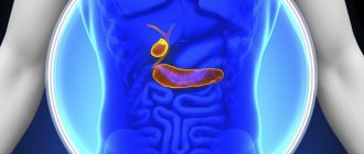
Structure and function of the liver
The liver is one of the largest internal human organs. Its weight at different stages of life is 1.5–2% of body weight. The anatomy of the liver is different in a fetus and an adult; its structure can be seen in the photo. It consists of the right and left lobes. In addition, small lobes are distinguished: square and caudal. To facilitate the description of changes, each lobe is divided into several segments.
The blood supply to the organ is also aimed at ensuring that the liver performs certain functions, in particular detoxification. In addition to the arteries that supply it with arterial, oxygenated blood, the liver receives blood flowing from the intestines. Here it undergoes detoxification, harmful substances and toxins are extracted and destroyed.
In addition to neutralizing agents dangerous to the body, liver tissue performs the following functions:
- participates in metabolism (carbohydrate, fat, protein);
- produces bile necessary for digesting fats;
- synthesizes body proteins, including immunoglobulins;
- destroys bacterial infections and parasites that enter the body with food;
- participates in hematopoiesis, fibrinogen synthesis occurs in it, etc.
The protective function of the liver is possible due to its high repair abilities. But under heavy load, the liver cannot cope, and pathological processes occur in it.
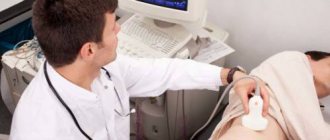
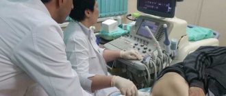
How does the procedure work?
For ultrasound of the liver and gallbladder, equipment is used, the frequency of which is set to 2.5–3.5 MHz. Due to these characteristics, it is possible to consider areas within the range of 1–3 mm. The depth reached by the waves is 24 cm.
The procedure itself is very quick, taking 15–20 minutes. The patient lies on the couch, lies on his back, and exposes his stomach. The doctor applies gel to the skin and begins the examination. First of all, the area of the right hypochondrium is examined. After a thorough study, other areas are studied, the method shows changes.
Unpleasant sensations during the diagnosis of the liver, pancreas and gallbladder are excluded; the patient can feel light pressure from the sensor.
The method is safe and harmless, but it is better if the patient with acute syndrome is accompanied by a friend or relative. When signing up for an ultrasound, it is better to choose comprehensive monitoring of the abdominal organs, then the pancreas will be examined along with other tissues.
Parameters studied
During the examination, the doctor examines the liver in different projections. To conclude, you need to determine:
- the size of each share;
- homogeneity of the structure of the liver tissue, the absence of hypo and hyperechoic areas in it;
- absence of focal inclusions, calcifications, cysts;
- the shape of the edge, normally it is smooth, the surface of the organ is smooth;
- degree of echogenicity, with diffuse lesions there may be a general decrease in density during inflammatory processes or an increase in density during proliferation of connective tissue;
- diameter of large vessels, bile ducts to detect bile stagnation, portal hypertension, Budd-Chiari syndrome, etc.
Changes in the norms of these parameters are considered together with data from laboratory tests and clinical studies to make a diagnosis. The use of Dopplerography and color Doppler mapping (CDM) plays an important role in this. This study allows you to determine the level of blood flow in the organ, areas with increased blood supply, indicating malignant tumors, metastases.
How to prepare properly
In order for the information obtained during an ultrasound to be accurate, you need to properly prepare for the scan. Within 3 days, it is important to start following a special diet and exclude fatty foods, carbonated drinks and alcohol from your diet. Coffee is banned.
In some cases, it is recommended to take enzyme preparations and sorbents that help restore digestion and cleanse the liver. Your doctor will tell you how to properly prepare for an ultrasound examination of the liver, pancreas and gallbladder. Recommendations may differ, but you definitely need to prepare.
Diet
On the appointed day before the ultrasound of the abdominal organs, eating breakfast is prohibited. A light snack is allowed 6 hours before the diagnosis. Preparation for ultrasound examination of the liver, gallbladder and pancreas requires compliance with the following rules and regulations.
They begin to prepare 3 days in advance; before monitoring, the following are removed from the menu:
- bread and other baked goods, cookies and sweets;
- milk and all dairy products;
- vegetables that increase increased gas formation: cabbage, radishes, turnips and radishes;
- whole grain cereals and pasta;
- fatty hard and curd cheese;
- mushrooms and legumes;
- fruit and vegetable juices;
- Carbonated drinks and mineral water are prohibited.
The listed products can increase the release of gases, which complicates the course of ultrasound. The last meal, taken 6 hours before, should be light; overeating is unacceptable. Before examining the liver, pancreas and gall bladder, you can eat oatmeal in water without butter, add a hard-boiled egg.
Taking medications
In order for an ultrasound of the abdominal organs to be successful, it is necessary to prevent bloating and increased flatulence. In case of excessive predisposition to the appearance of these symptoms, even diet does not help. It is better to use medications before diagnosis that will help:
- White carbon, regular activated carbon or Enterosgel. The purpose of these drugs is to cleanse the entire gastrointestinal tract.
- To prevent the formation of gas in the abdomen, use the drugs Espumisan, Simethicone or Bobotik.
- You can minimize gas formation by drinking mint, chamomile tea or a fennel drink.
- Festal, Creon or Mezim helps normalize intestinal motility. You can take other enzyme preparations.
- There is no need to use choleretic drugs. Under their influence, ultrasound will not show the condition of the biliary tract.
You should discuss the possibility of using drugs from this list with your doctor. If the pancreas is additionally examined, you need to be more careful with taking medications. Many drugs interfere with its functioning.
Other recommendations
During the period of preparation for ultrasound examination of the intestine, the following rules must be observed, especially regarding nutrition:
- You must not smoke or chew gum within 2 hours. It's better to increase this time.
- People suffering from type 1 diabetes are usually allowed to eat a small piece of bread the day before and wash it down with a cup of fresh tea.
- Patients taking any medications on an ongoing basis should notify their doctor about this. You can drink them if they are vital. It is necessary to provide him with information before receiving a referral for an ultrasound of the organs.
- You should not exercise before examining the liver, gallbladder and pancreas.
- Overweight patients are prescribed an enema in the morning and evening. The amount of water infused or a special drug is prescribed by the doctor.
The recommendations listed are relative.
Do I need an enema?
An enema should be given if a person is prone to constipation and cannot go to the toilet on his own. For this purpose, you can use drugs like Fortrans and Microlax. If the ultrasound is scheduled for the morning, the procedure should be performed in the evening.
If the patient has emptied on his own, the contents of the intestines cannot be removed using a cleansing enema. The procedure is done carefully. You can use a classic Esmarch mug filled with warm boiled water.
Preparing a child for an ultrasound
Recommendations regarding the preparation of small patients must be given by the doctor. But parents may simply forget something, so the reminder below will help them properly prepare their child for an ultrasound examination:
- 3 days before the procedure, you need to put the child on a diet with a complete restriction of the following foods: dairy products, chocolate, pastries, bread, fruits;
- If an ultrasound is planned for the baby, then the mother should go on a diet so that the child does not have increased gas production;
- If the baby suffers from flatulence, which is important for children in the first year of life, then Espumisan or Bobotik should be given 3 days before the procedure;
- If the child is already taking any medications, then before performing an ultrasound, parents should notify the attending physician about this. He will decide when to conduct an ultrasound and whether the medications taken by young patients will interfere with the successful examination;
- On the day of the study, it is forbidden to give the baby any complementary foods, especially fruit or vegetable purees. It will take a long time to digest, so the results of the study may be distorted. If the baby is hungry, it is better to feed him breast milk or formula. You can also give him some boiled water;
- If the baby suffers from constipation, then the day before you need to give him an enema;
- You should limit your water intake 1 hour before the examination, and you should not eat 7 hours before the ultrasound (for children over 3 years old), 4 hours before the ultrasound for patients under 3 years old, and 2 hours before the ultrasound for infants and children under 1 year old.
What to take with you
When making an appointment for an ultrasound of the liver and gallbladder, listen carefully to the specialist, he will tell you what you need to bring with you. If an ultrasound is performed in a government institution, it is important not to forget and prepare:
- patient's medical record;
- own insurance policy;
- passport confirming identity (adults).
A large towel will come in handy to place on the couch. You will also need shoe covers or slippers. If an ultrasound is performed to determine liver function, the study is first performed on an empty stomach, and then under load. The doctor asks the patient to consume any foods and repeat monitoring. For a snack, you can take a few boiled eggs and sour cream with you.
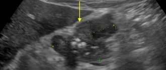
Ultrasound anatomy of the liver
Since the liver is a large organ, it is impossible to image its entirety using ultrasound. A standard scanogram is a series of sections obtained by multidirectional scanning of different lobes of the liver. Having a clear understanding of the features of the anatomical structure of the organ, the doctor must analyze the nature of the sections obtained and mentally restore its shape.
With a longitudinal scan performed through all lobes of the liver, its shape can be compared to a comma located along the patient’s body. A transverse scan of the right lobe allows you to obtain a slice resembling an incomplete circle or an “aged” crescent, and a slice of the left lobe, made in the same direction, looks like the letter “G”. Ultrasound examination allows visualization of all 4 lobes of the liver.
To differentiate all lobes, they rely on anatomical landmarks that are well determined using ultrasound:
Normal liver size according to ultrasound
- the location of the gallbladder (bed) is a hyperechoic cord located between the quadrate and right lobes;
- round ligament or groove of the round ligament - located between the left and quadrate lobes;
- gate of the liver - located between the caudate and quadrate lobes;
- venous ligament - defined as a septum with increased echogenicity separating the left and caudate lobes.
In addition to the lobes of the liver, ultrasound also shows all 8 of its segments. The most easily identifiable segment, commensurate with the caudate lobe - segment 1, has clear boundaries separating it from segments 2, 3 and 4, on the one hand, by the venous ligament, and on the other, by the porta hepatis. The second and third segments are in the left lobe, the second in the lower caudal part of the lobe, and the 3rd in the upper cranial part. The fourth segment is located within the square lobe and is limited by its landmarks.
Segments 5 to 8 are located in the right lobe, and their boundaries can only be determined by focusing on the position of the portal vein and its branches. With ultrasound, the outer boundaries of the organ should have clear outlines, however, the surface contour may have slight irregularities. On the surface facing the abdominal cavity, you can find several irregularities formed due to the tight fit of the kidney, colon and duodenum, stomach and adrenal gland.
Important! When analyzing the echographic picture in obese patients, take into account the fact that accumulations of fatty tissue may look like voluminous neoplasms.
Structure of the liver
Why do you need to follow the training rules?
Doctors recommend that their patients follow the rules for preparing for an ultrasound. If the rules are ignored, the intestines may become overfilled with gases. Because of this, diagnostic results will be inaccurate. The patient may be given an incorrect diagnosis, and the doctor will not be able to fully analyze the functioning of the internal organs and identify any pathologies.
Failure to comply with general recommendations may lead to incorrect determination of the treatment regimen. In this case, the effectiveness of the treatment will be lost.
Diet
Compliance with a three-day diet is a mandatory requirement for routine preparation before an ultrasound scan of the pancreas and liver.
fruits and vegetables during the diet
You will need to exclude foods that promote increased fermentation in the small intestine:
- fresh fruits and vegetables, especially cabbage, beans, grapes;
- dairy products;
- pearl barley and barley porridge;
- sweets;
- Rye bread;
- strong tea or coffee;
- alcohol.
Digestion of such foods leads to the formation of large amounts of gas in the intestines. During examination, it prevents the doctor from determining the size, structure and level of echogenicity of the intra-abdominal organs.
Before an ultrasound scan of the liver, eat a balanced diet that normalizes digestion, reduces gas formation and bloating. Products useful in this situation include:
- carrots, potatoes;
- rice cereal;
- dairy products;
- herbal teas and fruit drinks;
- boiled chicken.
boiled chicken
To improve intestinal function, the dietary regimen in preparation for an ultrasound of the liver is quite strict. But you only need to observe it for three days, so problems usually do not arise.
Products are boiled or baked, without oil or salt. It is advisable to eat a little food every 2-3 hours. This helps the pancreas work.
Last meal 12 hours before the test time. It is held in the morning, so dinner is scheduled at 7-8 pm the night before. A small, light meal is recommended.
Spleen examination
Simultaneously with the diagnosis of the gallbladder and pancreas, the spleen is examined. Most often, complications of this organ occur after injury. During palpation, the spleen cannot be felt. Palpation is possible only when it is magnified 3 times. In this case, the spleen of an adult will weigh about 400 g. A healthy organ weighs 150 g.
Using ultrasound, a slight increase can be seen. Its size depends on the height of the person. Below is a table with normal organ parameters. During diagnosis, it is also determined what structure the spleen has, the nature of its echogenicity, and shape. A healthy organ has a crescent shape. Increased parameters and increased echogenicity are an indicator of serious pathology.
The results are interpreted by a qualified specialist.