To detect pathologies of the gallbladder and biliary tract, various instrumental diagnostic techniques are used, one of which is cholecystography - an X-ray examination of the gallbladder and its ducts.
The essence of this technique is to use a special contrast agent, which is given to the patient before the x-ray.
For these purposes, in the recent past, tetrabromophenolphthalein or tetraiodophenolphthalein was used. Currently, not only they are used, but also other substances with similar properties (for example, biliselentan or bilitrast). They enter the patient's body orally and are available in tablet form.
Once in the body, these substances from the intestine are gradually absorbed into the blood, after which they penetrate into the cavity of the organ being studied through the bile produced by the liver. This makes it possible to clearly distinguish this organ on an x-ray (cholecystogram), which allows a specialist to evaluate the shape of the organ and its functionality.
The degree of functioning of this reservoir is determined by comparing its shape and size before and after the introduction of a special stimulus that causes the muscular walls of the organ to contract. In addition, such a study (using an irritating catalyst) in some cases makes it possible to see the contours of the bile ducts on the cholecystogram and assess the level of their patency.
It should be said right away that cholecystography gives an effective result only if the liver functions normally and hepatocytes (liver cells) produce bile at the proper level, along with which the contrast agent enters the cavity of the organ being examined.
The second condition for the applicability of this instrumental diagnostic technique is the normal patency of both the hepatic and cystic ducts, as well as in the case of a certain ability of the gallbladder to concentrate bile in its cavity. Of no small importance for obtaining high-quality research results is the level of contractility of the organ itself.
Read also: Is a bandage necessary and how long to wear it after gallbladder removal?
If his motility is at a low level, then his cavity is not freed up to accept new portions of liver bile containing a contrast agent.
If all conditions are met, then cholecystography makes it possible to detect congestion in this organ, its atony and the presence of deformations.
What is cholecystography of the gallbladder and bile ducts
This is an examination of an organ using x-rays. The procedure requires the use of a contrast agent consisting of 52% iodine. It is absorbed into the blood, then enters the bladder with bile and illuminates it from the inside under x-rays.
What can be seen in the picture:
- size, shape, position of the gall bladder;
- relief of the inner wall;
- degree of contractility of the smooth muscles of the organ;
- presence of formations.
A distinctive feature of the study is that it is carried out with satisfactory liver function.
Purpose of diagnosis
Cholecystography of the gallbladder helps to detect gallbladder diseases using hardware. HCG reveals:
- features of the bladder (anatomical, functional);
- dyskinesia, tumors, stones, inflammatory processes in the gallbladder.
The method is used for clinical symptoms of the disease for primary diagnosis and to confirm the existing diagnosis (therapy control).
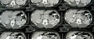
Photo interpretation
The results obtained are the basis for further research or establishing an accurate diagnosis. The attending physician is based on the conclusion of a radiologist, who describes the resulting image in detail (the specialist assesses the degree of darkening of the organ and ducts, analyzes the smoothness of the surface and the volume of tumors). The image shows a violation of the patency of the contrast fluid due to obstruction by stones, scars or neoplasms. Using the resulting image, a reduced concentration of the biliary tract is established.
The resulting image allows us to examine disturbances in the motor function of the excretory bile ducts. The patient does not independently prescribe cholecystocholangiography and interpret the resulting images.
More fresh and relevant information about health on our Telegram channel. Subscribe: https://t.me/foodandhealthru
Specialty: therapist, nephrologist.
Total experience: 18 years.
Place of work: Novorossiysk, medical.
Education: 1994-2000 Stavropol State Medical Academy.
Training:
- 2014 – “Therapy”, full-time advanced training courses at the Kuban State Medical University.
- 2014 – “Nephrology” full-time advanced training courses at the State Budgetary Educational Institution of Higher Professional Education “Stavropol State Medical University”.
Cholecystography is a type of x-ray examination using a contrast agent that enters the patient’s body in different ways at the doctor’s choice. The name of the method combines three words: “bile”, “bubble” and “write” (“depict”). Diagnostics involves a series of images in dynamics, different projections.
Types of techniques
The basis of the method is the ability of the liver to absorb iodine compounds and remove them along with bile into cholecystis, which concentrates trace elements. Types of cholecystography of the gallbladder:
- oral (oral);
- intravenous;
- Cholangiography.
Oral cholecystography
It consists of taking tablets that contain contrast 12 hours before the procedure. The amount of iodine in them is 65-75%. From the small intestine, the drug is absorbed into the blood, accumulates in the liver, and is transported with bile to the gallbladder. The pictures show the configuration of the organ. The patency of the bile ducts and the evacuation activity of the bladder are assessed after eating fatty foods.
Intravenous cholecystography
Characterized by rapid absorption of contrast. The drug can be detected in the common bile ducts after 20 minutes, in the gallbladder after 1.5 hours. X-rays are taken at regular intervals (10-15 minutes), then the patient receives a choleretic breakfast. The goal is to study the functions of the bubble. Intravenous cholegraphy reveals disruptions in the outflow of bile, detects stones, and determines the degree of thickening of the walls.
A subtype of the intravenous method is the infusion scheme. The contrast is administered along with a glucose solution dropwise over half an hour. Gives long-lasting staining of the bile ducts. Used in patients after cholecystectomy.
The advantage of the technique is that the research method is carried out regardless of the condition of the intestines after cholecystography of the gallbladder, with the gallbladder “disconnected” (bile acids do not enter the bladder).
Cholangiography
A method for studying the biliary system in case of abnormalities in the liver. The contrast is delivered directly into the bladder and bile ducts through percutaneous puncture. Blockages, common bile duct tumors, and stones are detected. Laparoscopic cholecystography of the gallbladder is done under eye control. Percutaneous transhepatic cholecystography is performed immediately before surgery.
Modern medicine rarely uses cholangiography due to severe side effects - sepsis, allergies, risk of death (1% of cases).
Indications and contraindications
Cholecystography of the gallbladder is a procedure that uses X-rays and a contrast agent with iodine for diagnosis. This method must be used with compelling reasons for diagnosis.
Reasons:
- confirmation of an existing diagnosis;
- clinical manifestations of gastrointestinal tract diseases.
Indications:
- probable cholelithiasis;
- violation of the motor function of the biliary tract;
- presence of neoplasms;
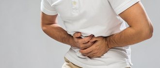
- hypochondrium pain on the right;
- indigestion of fats in the intestines.
Contraindications to cholecystography of the gallbladder are relative and absolute. This is due to the introduction of iodine-containing substances and x-ray radiation.
Relative:
- increased bilirubin levels;
- cirrhosis of the liver;
- inflammation of the intrahepatic ducts (cholangitis in the active phase).
The study can be carried out when the patient's condition improves.
Absolute:
- acute liver diseases;
- renal failure (10% of the contrast is excreted by the kidneys);
- exacerbation of cardiovascular diseases;
- allergic reactions;
- pregnancy and lactation.
In case of contraindications, HCG of the gallbladder is replaced by other types of diagnostics.
Results of the procedure
Best materials of the month
- Why you can't go on a diet on your own
- 21 tips on how to avoid buying stale food
- How to keep vegetables and fruits fresh: simple tricks
- How to curb your sweet cravings: 7 unexpected products
- Scientists say youth can be extended
Cholecystography is prescribed to study the anatomical structure and functional activity of the gallbladder and bile ducts. On the resulting image, you can evaluate the shape and position of the area under study, the displacement of its position, which is deviated from the norm. The size of tumors and stones is assessed from several photographs taken in different planes. The two-dimensional image allows you to evaluate large abnormal formations, tumors and polyps that interfere with the functioning of the gallbladder or ducts.
Cholecystocholangiography provides a clear image of the internal organ: the gallbladder is pear-shaped with smooth outlines and a thin contour. Any deviations from the norm are recorded by a radiologist and are the reason for prescribing additional research methods. The shape of the gallbladder may differ from the norm due to the design features of the body. In hypersthenics, the bladder is round in shape, while in asthenics it is elongated upward: the structural features and position of the organ are assessed by a doctor who writes a conclusion on the cholecystocholangiography performed.
Features of preparing a patient for cholecystography of the gallbladder
Preparation for cholecystography begins 2 days in advance with a diet. Products that cause fermentation (fresh fruits and vegetables, milk, legumes, rye bread), and fatty foods are prohibited. The study is carried out on an empty stomach.
The patient preparation algorithm includes:
- last meal on the eve of the procedure - before 8 pm (water without restrictions);
- An enema is given the day before and in the morning.
These are general requirements that do not depend on the type of cholecystography. For oral cholecystography of the gallbladder, you need to take a drug with contrast 12 hours before the procedure. The regimen and dose are prescribed by the doctor.
For intravenous cholecystography of the gallbladder, contrast is used before the procedure by injection. During cholangiography, iodine is injected directly into the ducts using a puncture in the X-ray room.
24 hours before cholecystography, an iodine tolerance test is performed. Side effects from taking contrast include nausea and diarrhea.
How to prepare for the procedure
Preparing the patient for an x-ray examination of the gallbladder involves cleansing the intestines with enemas. They are performed at night before bedtime and in the morning no later than two hours before the examination. The lack of effect from enemas with the presence of pronounced flatulence is an indication for CT.
Cholecystography should not be performed if there are barium residues in the body. On the day before the study, starting from the second half, you should limit your intake of fatty foods, alcohol, and preferably not smoke
If an oral method is planned, the evening before the study the patient takes a dose of radiopaque contrast agent, calculated individually according to the formula 1 g per 20 kg of weight, following the instructions for use. The period of time before the study should not exceed fifteen and be less than twelve hours. It is recommended to take the medications used with water, sweetened tea or fruit juice.
Intravenous cholecystography involves asking for negative reactions to iodine and conducting a sensitivity test to the drug in a hospital setting.
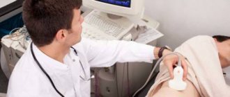
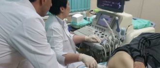
Examination technique
Preparing a patient for cholecystography involves explaining the essence of the procedure, the rules of behavior during it and possible side effects. Carrying out an intravenous injection of a contrast agent requires a horizontal position of the patient; the solution must be heated to 38 degrees in a water bath and injected slowly.
The procedure is carried out on an empty stomach in the morning in a specially equipped room by an x-ray technician under the guidance of a radiologist. Initially, a panoramic photograph of the right side of the abdominal cavity is taken in a vertical projection. Next, targeted images with compression of the gallbladder tube are performed in a horizontal position.
To improve gallbladder motility, the patient is offered breakfast in the form of egg yolks, sorbitol, mannitol, and a piece of butter. This is followed by a series of pictures at 15 minute intervals. Within 20-40 minutes after a test breakfast, the images can reveal polypous formations and small stones that were not previously diagnosed.
The motor function of the gallbladder can be enhanced by injecting cholecystokinin into a vein or muscle. Remains of the contrast agent are excreted in the feces, partially reabsorbed. This feature allows, in the absence of an expressive shadow on the first day of cholecystography, to perform it again the next day. The saturation technique involves taking one or two additional doses of a contrast agent.
Technique
Before examining cholecystitis, an x-ray of the abdominal cavity is taken. The patient takes a vertical position. The goal is to exclude the presence of gases that indicate perforation of the intestinal wall (contraindication to cholecystography of the gallbladder).
The study is carried out with a full organ. The patient lies on his stomach on his left side. The radiograph is then taken in a standing position. This is how the anatomy of the gallbladder and common bile ducts is assessed. The final stage is to study the motor and evacuation ability of the bladder. To do this, the patient drinks 2 raw eggs. The study is done every quarter of an hour for 60 minutes.
There are a number of negative factors that affect the result of cholecystography:
- the contrast agent does not completely enter the bile as a result of impaired liver function, vomiting, diarrhea;
- poor absorption in the small intestine, residual barium on examination of the gastrointestinal tract;
- movement of the patient during radiography;
- improper preparation for cholecystography.
The result will be distorted results and low-quality images. They can cause errors in diagnosis.
The procedure for diagnosing the gallbladder
Before the examination, the diagnostician takes a survey x-ray of the right hypochondrium to assess the degree of preparedness of the digestive organs. Also, the image taken will help identify shadow formations that may indicate the presence of stones, gas, or lime deposits in the bile ducts.
The procedure algorithm consists of two stages: first, an image of the filled organ is recorded, then pictures of the bladder are taken after emptying.
To take contrast images, the patient is placed on a special couch, first in the “prone” position. Next, the doctor asks the patient to move to the left side, stand up straight, while paying attention to the separation of the contents of the bladder or the presence of movable filling defects.
The shadow of the contrasted gallbladder is displayed in the center of the screen, and in the absence of a contrast image - in the area of its projection. The projection point of the organ is located at the intersection of the outer edge of the right rectus abdominis muscle and the arch of the ribs. Given the differences in size and position of the gallbladder and liver, the location of the projection may differ.
In the photographs, the bubble may be reduced or enlarged, deformed, wrinkled, located in the upper part of the right hypochondrium, or extending into the pelvic cavity. The organ may also deviate to the left or right, with its shadow cast on the right kidney or vertebrae.
If the subject has a blockage of the gallbladder (cystic duct obstruction), its shadow will not be visualized on the image. In addition, the lack of contrast in the organ can be explained by functional disorders, for example, deterioration of the excretory function of the liver.
The second stage of diagnosis begins after the patient is given choleretic food or medicine - this is how the phase of contraction and emptying of the bladder begins. During this stage, the doctor can evaluate the so-called evacuation function of the organ, and as the contrasting bile disappears from it, determine the size and location of stones, scars or neoplasms in the walls.
The common bile duct is visible on images taken after 15 and 30 minutes. If the emptying function is slow, the last image recording is made after 60 minutes.
What this study shows
Cholecystography lasts on average 1.5-2 hours. It allows you to consider:
- structural features of the organ, common bile ducts: size, shape, contours, thickenings, scars;
- stones in the gallbladder: number, size, mobility;
- patency of bile ducts;
- neoplasms, polyps (location).
The images are interpreted by a radiologist. Based on them, a medical report is issued .
Norm and deviations
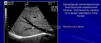
If in the photographs the gallbladder has smooth, thin, even walls, the structure is clearly visible, the shape of the organ is pear-shaped, and it is as full as possible - we can talk about a healthy state of cholecystis.
Deviations
With normal filling, the following are visible: moving contrast elements - a sign of stones, fixed defects - possible benign neoplasms, polyps. If bile is partially filled or absent in the organ, we can talk about inflammatory processes in the bladder (cholecystitis). As a result, obstruction of the bile ducts occurs, and the concentration function of the mucous surface of the organ is disrupted. When studying the evacuation capacity after a choleretic breakfast, insufficient contractility of cholecystitis indicates inflammation of the organ, blockage of the common duct.
Consequences and complications
Negative effects during cholecystography have subtle manifestations. The most common symptoms are:
- dizziness;
- fear;
- nausea, vomiting, diarrhea;
- metallic taste in the mouth;
- feeling of heat during injection.
Complications are caused by the toxic effects of the contrast agent and increased nervous excitability of the patient. Rare deaths are associated with allergies to iodine-containing substances and hypokalemia.
HCG examines the gallbladder using an x-ray combined with contrast. This method is used as an additional, primary study for cholecystis diseases. The value of diagnosis lies in the detection of radiopaque stones, which are visible only on the image.
During surgical interventions on the liver, this method is used to visualize the bile ducts if malignant neoplasms are suspected.
Cholecystography of the gallbladder is highly informative. The complexity of implementation, lengthy preparation for the procedure, and side effects contribute to the fact that this method of studying the gastrointestinal tract is not used as often as we would like.
Is the method dangerous?
Cholecystography is a fairly safe method and can be used without age restrictions, including in pediatric practice. The side effects that occur during the procedure are moderate and disappear quickly. Commonly encountered ones include:
- dizziness;
- nausea;
- loose stools;
- pain in the stomach area.
Vomiting and abdominal pain are rare side effects. Often this is a reaction to the administration of cholecystokinin. Symptoms of iodism are a more serious phenomenon, but quickly resolve with proper correction of the condition. They are treated with alkaline solutions (soda, milk) and medications. The patient and physician should note such a reaction.