Magnetic resonance imaging (MRI) of the liver is a method of non-invasive examination of the gland and its ducts, which is based on a change in the polarity of hydrogen atoms upon contact with radio waves and a constant magnetic field.
When scanning, accurate data is obtained, since photographs of given sections with a minimum step make it possible to construct a three-dimensional model of the organ, but due to the high cost of the study, MRI is prescribed after simpler diagnostic procedures (ultrasound), when there is a need to supplement and clarify the data.
During an ultrasound examination, a specialist receives a low-resolution image, but on it one can still see the inflammatory process and neoplasms, and it is possible to assess the general condition of the organ, its size and morphology.
MRI allows one to judge the type of formation and structure of the parenchyma; with its help, one can distinguish between oncology, hemangioma, fatty degeneration, and find out what is inside the cyst. The main advantages of ultrasound are its availability and the safety of the procedure when carrying a child, and the use of MRI of the liver during pregnancy is limited.
Anatomy and functions of the liver
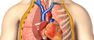
The liver is an organ located in the right upper quadrant of the abdominal cavity, that is, on the right in the area of the lower ribs. The liver is classified as a gland because it produces bile necessary for digesting food. But the functions of this organ are far from limited to this; they include detoxification, protein synthesis, participation in metabolism, regulation of glycogen reserves, and hormone production.
The liver is capable of regeneration, it restores its volume while maintaining only one quarter of its original size. In this regard, in case of organ formations, surgical removal of the affected tissue is possible. Currently, the generally accepted division scheme proposed by Claude Quinot, in which the liver is divided into eight segments. This division is based on the internal features of the branching of blood vessels and bile ducts, so that the removal of certain segments makes it possible to completely preserve the functioning of the remaining part of the organ.
The gallbladder is an organ that stores bile produced by the liver. When eating food, the gallbladder contracts and bile is released through the duct into the intestines to digest food.
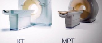
MRI or CT scan of the liver?
When ordering a liver scan, your doctor will usually determine the best test method based on the nature of the possible pathology. World practice describes the superiority of MRI for liver diagnostics over CT due to the absence of ionizing radiation, high tissue contrast, and excellent time resolution when using contrast. In addition, MRI allows you to examine the structure of an organ in more detail due to the use of several MR scanning modes. Therefore, to the question of whether they do MRI of the liver, there is a clear answer - they do it, and with excellent results!
What test is an alternative to MRI?
Alternative methods for examining the liver include ultrasound or CT. However, these methods do not provide complete information about the examined organ and do not allow identifying pathology at the very beginning of its manifestation. In addition, when performing a computed tomography scan, the patient is exposed to x-rays. Of course, the dose of radiation exposure is lower than with standard radiographic examination. If the patient has a neoplasm of an oncological nature, then this effect will be extremely undesirable.
Therefore, you do not need to prescribe a diagnostic method for yourself; it is important to follow all the recommendations of the attending physician, who, before referring you for the procedure, weighs the advantages of a particular method and its risks, and gives preference to the safest diagnosis. MRI of the biliary tract and liver has a number of advantages, which include:
- high accuracy of the result, allowing you to make a diagnosis;
- painlessness;
- no exposure to x-rays.
The only disadvantage of this scan is its high price. However, the high efficiency of this method completely justifies this drawback. In addition, the price factor is influenced by the location of the study. For example, the usual price for an MRI in Moscow is about 7-10 thousand rubles. In the capital, MRI, carried out on Miklouho-Maclay Street, is used not only by state-run clinics, but also by private ones. MRI of the biliary tract and liver is the most effective technique that allows you to identify pathology at the initial stage and begin treatment in a timely manner.
MRI (magnetic resonance imaging) is considered the most objective and informative form of studying the condition and function of the organs of the hepatobiliary system. MRI of the liver provides specialists with detailed information about the structure of the organ, allows them to see abnormalities in the associated gallbladder and bladder, and so on. Diagnostics are prescribed based on the results of a patient examination, ultrasound, and radiography in order to clarify the preliminary diagnosis and detect pathologies.
Indications and contraindications for MRI of the liver
Indications include:
- Assessment of diffuse liver diseases such as hemochromatosis, hemosiderosis, fatty degeneration
- Diagnosis of foci of metastasis, adenomas, areas of nodular hyperplasia
- Differential diagnosis of identified lesions (cysts, fat accumulations, hemangiomas, hepatocellular carcinoma)
- Evaluation of known or suspected organ abnormalities
- Assessment of dynamics during therapy
- Cirrhosis
- Jaundice
- Pain, heaviness in the right hypochondrium
- Palpable enlargement of the liver
- Additional examination in case of detection of pathology by ultrasound, CT
- Monitoring after surgery on the liver/biliary tract in the presence of persistent clinical symptoms
Indications for MRI of the biliary tract:
- suspicion of cholecystitis with unclear ultrasound data or to clarify them
- searching for the location of migrated stones
- diagnosis of dilation of the bile ducts in cholelithiasis to establish indications for surgery
Contraindications to MRI examination of the liver and biliary tract do not differ from contraindications to MRI itself, these include:
- The presence of metal implantable devices for medical purposes (information about this must be reported to the doctor or clinic administrator to clarify the possibilities of conducting the study)
- Possible presence in the body of parts of other metal products (shrapnel, metal shavings, etc.)
- Pregnancy (the degree of possible benefit and risk must be assessed)
- A relative contraindication is the patient’s overweight; the exact acceptable weight figure must be clarified with the administrator in advance.
Alternative diagnostic methods
If a patient has strict contraindications to MRI, then the selection of diagnostics will be based on the proposal of alternative methods. The possible options are described as follows:
- If there are persistent signs of oncology, the patient is prescribed computed tomography as a backup diagnostic method.
- For some types of vascular pathologies with obvious circulatory disorders, MSCT - multislice computed tomography - may be offered. The diagnostic method, like CT, is based on x-rays, which allows you to clearly see hard, bone tissue. These examination methods are also not suitable for pregnant women and diabetics.
- In infants with a large non-ossified area of the cranial vault, neurosonography (ultrasound diagnostic method) is offered as an alternative.
- If you have claustrophobia, it is possible to perform an MRI on an open device.
Preparing for an MRI examination of the liver
Preparation for the examination is generally the same as for an MRI of the abdominal cavity - you should not eat for about 6 hours and drink for 4 hours, however, these requirements may vary; it is better to check with your doctor in advance about the specifics of preparation.
At the medical center you will be asked to fill out a standard informed consent form and your questions will be answered. If it is necessary to contrast the liver, an intravenous catheter will be installed. You must be prepared to hold your breath for a short time. You will be provided with headphones or earplugs to protect your hearing aids from the noise accompanying the MRI examination, and will be explained how to use the intercom and alarm device to communicate with the operator.
Features of the methodology
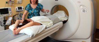
During the examination, the patient lies on a special table, which is pushed into the tomograph machine. It is important to lie still so that the data obtained is not distorted. For this purpose, the patient's limbs are usually fixed.
The procedure is absolutely painless and takes an average of 40 minutes. The patient usually feels warm in the area being examined. During the procedure, the tomograph buzzes, so you can wear special headphones. They have a microphone so that the patient can communicate with specialists in the next room.
The scanned data is transferred to a computer where it is visualized into images. The tomograph scans the body layer by layer - such layers are called slices. The higher the technical capabilities of the device, the thinner the sections obtained. MRI is usually used for a child after 7 years of age, when he can consciously remain motionless. This diagnosis is usually not performed on young children, but if necessary, it is performed after anesthesia or a sedative.
MRI - diagnosis of liver lesions
The size of the liver varies from person to person, even normally. The following linear dimensions of the organ are most often assessed on an MR image, which usually do not exceed 16 cm (the determination of these indicators is important mainly for monitoring treatment during therapy):
- On the coronal image, the perpendicular distance from the dome of the liver to its lower edge, passing through the middle of the organ.
- Maximum craniocaudal size.
Hemangioma
Liver hemangioma is one of the most common benign tumors of this organ. Detection of a mass by ultrasound is usually an indication for further examination, since hemangioma does not have specific differential signs from an oncological tumor on ultrasound.
Malignant tumors
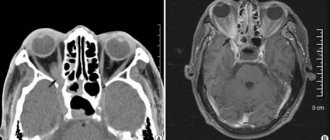
The malignancy of the formation is also clearly determined by MRI (Fig. 3).
Where can you get an MRI of the liver in Moscow and St. Petersburg
An MRI of the liver can be done at various medical centers that provide this service (not all MRI centers perform this test). Before the study, you must inform the administrator if the study will be contrast-enhanced. There are usually no problems with registration in private centers.
If you suspect cancer, it is better to perform an MRI in a medical center that has a doctor on staff who specializes in performing punctures or biopsies of the affected areas. It must be remembered that with the best diagnosis, the final diagnosis of cancer is made only by a histologist when studying biopsy data.
MRI of the liver is a modern examination method that allows you to obtain reliable information about the structure and functioning of the liver, gall bladder, bile ducts, duodenum and pancreas. The ability to examine the functioning of all organs that function in close interaction in one procedure is the main advantage of MR imaging of the liver. The fact is that the listed organs are anatomically and functionally connected in such a way that disruption of the functioning of one of them inevitably entails disruption of the functioning of the entire system.
During an MRI of the liver and gallbladder, layer-by-layer images of sections of the desired anatomical area are obtained. The thickness of the sections ranges from a couple of millimeters to 1 cm and is set manually by the doctor, depending on the purpose of the examination. Thanks to the capabilities of modern tomographs, it is possible to obtain three-dimensional images of organs and individual anatomical structures to make it easier for the doctor to identify pathology.
Reviews of liver MRI with contrast indicate a significant reduction in the time to make a diagnosis. The sooner treatment is started, the better the results.
Diagnostics with contrast
The dye is injected into a vein and distributed throughout the body. It soon reaches the organ being examined. Today, there are different forms of contrast administration during MRI. In the first option, a single intravenous injection occurs immediately before the diagnosis itself. The coloring preparation is calculated from the following proportion: 0.2 mg to 1 kg of patient weight.
How does ultrasound differ from MRI?
In the second option, a drop of contrast is introduced, with the help of which you can control the level of the dye directly during the study period.
Most often, this method is used when conducting dynamic studies. The dye allows you to more accurately determine the size of the tumor, its structure, and outline. As a result of its introduction, the liver segments are more clearly detailed.
The contrast does not pose a danger to the patient and is eliminated from the body through the kidneys within 2 days. Based on this, this procedure is not performed on people with kidney failure. Allergic reactions to the introduction of a dye are quite rare. However, if there is a suspicion of a predisposition to hypersensitivity, then it is better not to perform this type of examination.
A study with Primovist allows you to identify pathologies in the initial stages
In the modern world, there is a growing trend in the success of surgical interventions to eliminate liver tumors. This happens due to its early recognition. A significant role in this was given to innovation in colorants: “Primovist”. After injection into a vein, the drug quickly reaches the liver and makes it possible to examine not only this organ, but also the bile ducts.
An examination with Primovist allows determining the presence of tumors, whether they are malignant or benign, detecting metastases, distinguishing a primary disease from a secondary one. This examination is the most preferable and takes the quality of diagnosis to a new level.
What does an MRI of the liver show?
The following pathological changes can be seen on the images obtained during the examination::
- liver injuries: capsule ruptures, subcapsular hematoma, the presence of free fluid (blood) in the abdominal cavity;
- features of the development of the liver and bile ducts;
- an increase or, conversely, a decrease in the size of the liver;
- accumulation of fluid in the abdominal cavity (ascites);
- focal formations in liver tissue: cyst, adenoma, lipoma, cancer, metastases;
- changes in liver tissue: fibrosis, fatty degeneration;
- changes in liver vessels: thrombosis, aneurysms;
- obstruction of the biliary tract (obstructive jaundice);
- narrowing (stricture) of the bile ducts and pancreas.
The use of a contrast agent significantly increases the information content of images. Thanks to this, it is possible not only to identify a focal formation, but also to determine its type (hemangioma, carcinoma, hepatocellular adenoma, etc.), which makes it possible to make a diagnosis without puncture and other invasive examination methods.
Indications for examination
You can get a referral from your doctor for an examination in the following cases::
- the patient has characteristic complaints and symptoms of liver disease: pain and a feeling of fullness in the right hypochondrium, loss of appetite, yellowing of the sclera, darkening of urine, increased sweating, nausea, vomiting, etc.;
- abdominal trauma with suspected liver damage;
- cholelithiasis;
- fatty hepatosis, liver cirrhosis;
- liver tumors;
- low information content of instrumental examination methods that were used previously, difficulties in diagnosing liver diseases;
- monitoring the effectiveness of cancer treatment and assessing the durability of the achieved remission.
MRI of the liver with contrast is mandatory if cancer is suspected. On regular photographs, the tumor may not be clearly visible, so it may be difficult for the doctor to determine its exact location and size, and to distinguish the primary tumor from the site of stasis. The contrast agent selectively accumulates in the tissues of neoplasms, which makes tumors noticeable and their boundaries clear.
Decoding the result
MRI images show more than just malignant tumors. The most common option when this diagnostic method is prescribed is to confirm the presence of hernias or protrusions in any part of the ridge. Using MRI, affected atherosclerotic vessels and abdominal organs are examined. The method recognizes stones, polyps, adhesions, plaques and narrowed vessels.
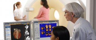
Usually, after an MRI of the liver, a specialist deciphers the result and explains it. After the conclusion and examination, photographs are attached. It is not recommended to try to decipher the result yourself, as only trained people can do this.
Contraindications for examination
MRI is not performed on patients who have:
- there are metal objects in the body (prostheses, pins, knitting needles, metal fragments, etc.);
- a pacemaker or other electronic devices are implanted.
Contrast-enhanced MRI is contraindicated in:
- intolerance to gadolinium-based drugs in the past (these drugs are used as a contrast agent for MRI procedures);
- chronic renal failure;
- pregnancy and breastfeeding.
Application of contrast agent
A contrast agent is used in several cases. The first is the detection of a neoplasm. To understand whether a tumor is malignant or benign, the method of applying contrast helps. The accuracy of clarifying diagnostics is almost 100%.
The dye used in MRI can detect the disease if signs of a malignant lesion are present, but other diagnostic methods do not detect structural changes. Contrast reveals tumors of the smallest sizes.
Preparing for an MRI of the liver
The quality of the resulting images directly depends on the filling of the intestine with gases and intestinal contents, as well as on the activity of peristalsis. The better the intestines are prepared for examination, the more informative the MRI will be.
The preparation algorithm for the procedure is as follows::
- three days before the examination, it is recommended to avoid foods that provoke active gas formation in the intestines (fruits, vegetables, legumes, carbonated drinks, sweets);
- one day before the procedure, you must start taking activated carbon or another enterosorbent;
- on the eve of the procedure, you should empty your bowels or do a cleansing enema;
- on the day of the examination, it is advisable to refuse breakfast if the MRI is scheduled for the first half of the day, or plan a meal 6 hours before the procedure;
- immediately before the procedure (about 30 minutes before it starts), it is advisable to take any antispasmodic, for example, no-shpa.
If MRI is performed for emergency reasons (for example, in case of abdominal trauma), then no preparation is carried out.
If liver MRI with contrast is performed, additional tests are recommended in patients with a history of kidney disease to rule out chronic renal failure.
For examination at the clinic, it is advisable to take with you extracts from your outpatient card or epicrisis from the hospital, doctors’ reports and the results of previous examinations. All this will help the doctor performing the MRI scan to give a clearer and more detailed conclusion. This is especially true in cases of MRI in liver cirrhosis and cancer, when the purpose of the study is to assess the rate of progression of the pathological process, the effectiveness of treatment or the durability of the achieved remission.
Alternative examination methods
Along with MRI, ultrasound and computed tomography are common diagnostic methods. It is not entirely correct to compare them, since the studies have different goals and objectives.
MRI is the most informative, but expensive procedure, which is often performed to confirm the results of ultrasound or CT
Ultrasound examination is a screening procedure, usually prescribed at the initial stages of diagnosis. And MRI is done to confirm or clarify the results of ultrasound. Which is better - MRI or ultrasound, the doctor must decide in each case. The main advantage of ultrasound examination is its absolute safety and the possibility of performing it on pregnant women and children. However, ultrasound does not recognize the initial stages of the oncological process, and the results of the study directly depend on the qualifications and professionalism of the doctor.
The question of which method is more informative - MRI or CT - cannot be answered unequivocally. Computed tomography with contrast is also quite informative. However, with CT, the patient is exposed to radiation, which is extremely undesirable in the development of oncology and can contribute to the aggravation of pathological processes. However, if MRI is not possible, computed tomography becomes an alternative.
CT gives the most accurate results in case of volumetric organ damage - significant enlargement, massive cirrhosis, extensive neoplasm.
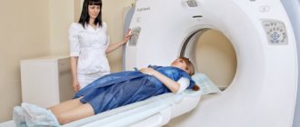
How is the examination carried out?
An MRI scan of the liver takes from 30 to 45 minutes. All this time, the patient remains inside the tomograph and tries to remain motionless. Inside the device it is light, warm and there is a constant flow of fresh air. The operation of the device is accompanied by quite intense noise, so it is better not to refuse the headphones offered by the medical staff.
Examinations performed with contrast enhancement take longer. To administer contrast, a special intravenous catheter is used, which is located in a vein in the patient’s arm the entire time the MRI is being performed.
After the examination procedure is completed and the medical staff checks the quality of the images obtained, the catheter is removed from the patient’s arm.
Reviews
Magnetic resonance imaging is a popular examination, despite its high price, as it allows one to obtain the most complete and reliable information about the onset of the disease in one procedure.
Patients who have undergone MRI note its high efficiency and accuracy of the information obtained. Thanks to the results of the examination, their attending physicians established the correct diagnosis and prescribed competent treatment.
How safe is the MRI procedure?
To scan the body, MRI machines do not use X-rays or other effects harmful to the human body, so MRI is an absolutely safe examination method.
MRI can be performed as often as needed. There are no restrictions on the number of areas surveyed at one time or on the frequency of surveys.
If the patient has been referred for an MRI of the liver, preparation should begin two days before the procedure. Magnetic resonance imaging is one of the most effective ways to study the liver. The study is often carried out together with an MRI of the gallbladder and its ducts, since pathologies may be associated with disturbances in the outflow of bile
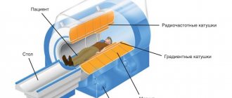
Operating principle of the device
The operating principle of the MRI machine is based on the formation of a magnetic field saturated with radiofrequency pulses. Back in 1946, scientists at Stanford and Harvard universities discovered the phenomenon of nuclear magnetic resonance. Its essence lies in the fact that substances containing hydrogen molecules in their structure, when placed within the range of a magnet, are capable of absorbing and reflecting radio frequency pulses.
The effect is observed only when the frequency of the pulses emitted by the magnet coincides with the frequency of rotation of the nuclei of the substance. It is these impulses that the MRI machine generates and sends them to the area to be diagnosed.
As you know, the human body consists mainly of water - oxygen and hydrogen atoms. Under the influence of a magnetic field, hydrogen acquires the ability to perceive and reflect radio pulses. The signal reflected by hydrogen molecules is sent to special sensors and recorded by the program.
Based on the information received, the program calculates the exact ratio of molecules and diagnostic points, creates a detailed image of the liver as a whole and its individual segments.
The program displays the results in the form of images of layer-by-layer sections. Horizontal visualized sections of the liver are made about 1 cm thick; for a more detailed study of the tissues, the sections are made even thinner - about 5 mm.
Why do you need to prepare for an MRI of the liver?
Preparing for an MRI of the liver has several goals. It is carried out both to improve visualization of the organ and determine the unevenness of its structure, and to identify violations of the outflow of bile through the hepatic ducts.
Liver MRI with preparation is prescribed to detect:
- cirrhosis;
- fatty degeneration due to alcoholism;
- developmental anomalies;
- parasitic infestations (echinococcus);
- benign tumors (hemangiomas, fibromas, adenomas);
- space-occupying formations (cysts);
- injuries (hematomas);
- malignant neoplasms (hepatocellular carcinoma);
- metastases to the liver, for example from the stomach or lungs;
- hepatitis A;
- causes of unknown jaundice.
MRI of the liver is also done in preparation for surgery (liver transplant, removal of a pathological process) or after it to monitor the treatment.
Preparation for MRI of the liver is carried out with the aim of:
- reducing gas formation - for this purpose, a diet is prescribed, which includes foods that do not cause the formation of gases in the intestines; they also use drugs that improve enzyme formation and enterosorbents (activated carbon);
- reduction of metabolic processes, which is important if there is a suspicion of a tumor - since metabolic processes in tumor cells proceed faster, which is associated with increased blood supply, when using a contrast agent they will be visible before the surrounding tissues (therefore, before the examination, a low-carbohydrate diet is prescribed, which reduces the processes metabolism in liver cells);
- sufficient filling of the hepatic ducts - this is necessary to better examine the presence of pathologies in them; You should not eat or drink 5–6 hours before the procedure, which will ensure normal filling of the gallbladder with bile.