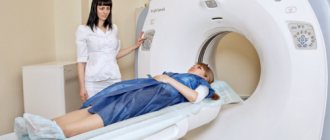
Description of the procedure
The principle of MRI is based on the effect of electromagnetic waves on protons (hydrogen nuclei), which are found in tissues and organs. A proton, absorbing energy from the outside, changes its spatial structure (excites). After cessation of irradiation, the nucleus returns to a calm state, emitting excess energy.
Magnetic waves emitted by an electromagnet or permanent magnet are localized to the area of interest using gradient coils. Radio frequency sensors located on the scanner transmit signals to a computer, where data is processed and an image is formed on the monitor.
Mechanism of action of the tomograph
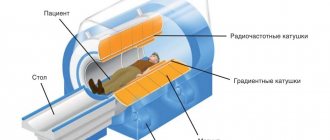
A tomograph is a complex device consisting of magnets, gradient coils, a radio pulse generator and receiver, a power source, a Faraday cage, a data processing system and a cooling unit. To conduct the study, the patient is placed on a special retractable table in a narrow tunnel inside the device.
After switching on, the device processes magnetic pulses reflected from body tissues. Using a computer program, he converts them into images and displays them on the screen. In this case, the organ under study is viewed in various projections and planes. The thickness of the sections depends on the research objectives and can start from 0.8 mm.
Preparation and methodology
No specific preparation is required before MRI. The only exception is contrast diagnostics. Contrast is administered on an empty stomach to minimize side effects such as nausea/vomiting and general worsening of the condition.
Before the procedure, the patient must refrain from eating food for 5-6 hours. Children are allowed to reduce this figure to 3-4 hours. Before and after administering the contrast agent, strictly follow the instructions of the diagnostic technician. If you feel pain, discomfort or any other negative symptoms, be sure to report this to a specialist.
Often, contrast MRI is prescribed if cancer is suspected. For other indications, standard magnetic resonance imaging, and in some cases CT or X-ray, will be sufficient. Get tested only as prescribed by your doctor to avoid side effects and waste of money.
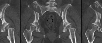
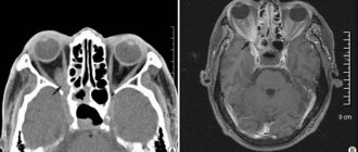
MRI of the paranasal sinuses is carried out according to a standard procedure, but with the use of special programs that help detect defects and pathologies of the paranasal region. What is the reason for this? A lot of mucous, cartilage and bone tissues are concentrated near the nose. The equipment may simply not recognize the necessary areas, reducing the information content of the diagnosis. The tomograph itself is a massive closed chamber in the shape of a cylinder. In the middle of this cylinder there is a retractable table on which the patient is placed during therapy.
The laboratory assistant helps the patient climb onto the extendable table and take a comfortable position. Then the medical worker sets up the operation of the tomograph, correctly installs the scanning ring, reports the location of the call button for communication and retires to the next room.
From this moment on, interaction between the laboratory assistant and the patient is carried out through a speakerphone installed in the room.
The patient should be prepared for the MRI machine to make a lot of noise during operation. If he feels pain/discomfort or wants to stop the diagnosis for any other reason, he must press the button to call a laboratory assistant, and not desperately shout for help.
After starting the device, the scanning ring begins to spin quickly around the examined area. For each turn, the technique cuts the paranasal sinuses at a different angle in order to then form a complete three-dimensional picture from them. During the procedure, the patient must lie still.
Even the slightest careless movement will distort 1 or 2 scans, which will affect the final result and reduce the overall information content. The duration of an MRI scan is 10-25 minutes.
When using a contrast agent, the procedure time increases.
Upon completion of the scan, the patient can return to their usual rhythm of life. No adaptation or special preventive actions are necessary. Remember that the MRI machine uses a magnetic field, which is completely harmless to the human body.
You can pick up your results on the day of your scan. The patient is asked to wait outside the door while the machine processes the information and the doctor makes a preliminary diagnosis.
This usually takes 30-40 minutes, but the time may vary depending on the workload of the medical staff.
Preparatory stage
If you plan to diagnose the nose and nasopharynx without using a contrast agent, special preparation for the procedure is not required.
Immediately before scanning, you will need to remove metal items (glasses, watches, jewelry, etc.). It is recommended to visit the toilet.
In case of contrast MRI, you should refrain from eating and drinking 6-8 hours before the scan. Patients with neurotic disorders are recommended to take sedatives several days before diagnosis.
Which is better: CT or MRI of the paranasal sinuses?
Each method has a uniqueness, thanks to which computed tomography and magnetic resonance imaging complement each other. During a CT scan, you can better examine bone tissue for integrity and the presence of pathological changes. When viewing the sinuses through a computed tomography scan, it is possible to detect tumors that have penetrated the adjacent walls.
The frontal and maxillary sinuses are located superficially, so for diseases of frontal sinusitis and sinusitis, an MRI appointment will be an unnecessary luxury. The best research option would be the least expensive x-ray of the paranasal sinuses. However, only MRI can reveal the cause and type. The sinuses of the ethmoid labyrinth and the canals of the sphenoid bone are located deeper and are more complex, because of this it is possible to identify changes using layer-by-layer research methods - CT and MRI.
An experienced otolaryngologist prescribes a current sinus examination based on the nature of the disease. By contacting a good specialist, you will save not only money, but also your precious health.
During a magnetic resonance imaging study, you can view the mucous membranes of the nose. It is also good to diagnose nasal polyps and the labyrinth of the sphenoid void. The test helps specialists distinguish which bacteria are causing the disease and rule out sinusitis caused by fungi.
What are the sinuses and where are they located?
The paranasal sinuses are bony cavities connected to each other by the nasal passages. They are located near the nose, covered with a mucous membrane, the air passing through them and the cavity of the nasopharynx enters the lungs warm and moist.
Types of sinuses:
- basic;
- frontal - located in the frontal bone;
- maxillary (maxillary);
- ethmoidal - located between the main and frontal sinuses.
Diagnosis of diseases of the nasal cavity and diagnosis of the throat begins with a medical examination of pathologies in this area; if the effectiveness is insufficient, other procedures are prescribed.
Question of price
MRI, in comparison with other diagnostic methods, is a rather expensive technique. The average cost of the procedure in the country is 2500-3000 rubles.
If you plan to use a contrast agent, you will have to pay 2000-3000 rubles more.
The price tag for medical services varies depending on the region, clinic, and equipment used.
MRI of the nasal sinuses is an informative diagnostic method when it is necessary to identify pathologies of the local area in the early stages of their development.
Among the diseases likely diagnosed during tomography are: cysts, neoplasms, injuries, structural anomalies, inflammation.
The procedure is contraindicated for persons with metal objects and electronic devices in the body, and for pregnant women (in the 1st trimester).
List of contraindications for MRI
The limitations to magnetic tomography include the following circumstances:
- metal objects (bullets, fragments, implants, clips) and other medical devices (the presence of insulin pumps, pacemakers, etc.);
- first trimester of pregnancy;
- the patient’s body weight is more than 120 kg (the use of specialized equipment is not excluded);
- pathologies that do not allow the patient to remain motionless inside the tomograph.
Which method is better
Various types of sinusitis are not the only group of pathologies for which the use of MRI of the paranasal sinuses is indicated. There are such types of diseases:
- malignant and benign neoplasms, metastases in the bones of the facial part of the skull;
- injuries to these bones;
- tumor processes in the mucous membranes of the nose;
- polyps that interfere with the release of secretions from the nasal cavities;
- infections in deep-seated junctions of the nasopharynx with the nasal ducts.
In the diagnosis of these processes, radiography and tomography of the nose are used.
1. X-ray is the most accessible and inexpensive method. For injuries and fractures of the facial bone, it is used primarily. Radiography can:
- quickly determine the nature of the bone injury, which will allow urgent treatment measures to be taken;
- see the presence of foreign bodies;
- diagnose congenital anomalies of the nasal bones;
- see inflammation during sinusitis, since these sinuses are located superficially; MRI of the maxillary sinuses is done only in cases where X-ray diagnostics are contraindicated for the patient;
- diagnose frontal sinusitis, since the sinuses of the frontal bone have a simple structure and are convenient for visualization.
X-ray gives a planar image and is not able to reflect changes within the scanned object, which is possible with tomography of the nose and paranasal sinuses.
Tomography is the process of layer-by-layer photographing an object. A series of images of the organ is taken sequentially on one section, then the scanning beams penetrate to greater and greater depths, creating an image of each layer. The result is a three-dimensional image of the studied area with all the features of the internal structure. A CT scan is performed using X-rays, and an MRI uses the effect of the resonant response of tissues to magnetic radiation. It is believed that the information content of both methods is approximately the same, however, with a competent approach to diagnosis, one of them is preferable.
2. Computed tomography is cheaper than MRI of the paranasal sinuses, and, other things being equal, the patient can choose the budget option. Given that X-rays are better at seeing solid structures with little fluid content, doctors prefer CT in the following cases:
- determination of bone tissue integrity;
- congenital abnormalities of bones or nasal septum;
- penetration of metastases through the walls of the nasal cavities.
In addition, a CT scan instead of an MRI of the sinuses is performed if the patient has metal objects, electronic organs in the body, or is claustrophobic. To diagnose relapses of already detected sinusitis or sinusitis and in cases where the diagnosis does not cause difficulties, CT is mainly used. This expensive method is used only in cases where X-ray diagnostics are contraindicated for the patient. The main disadvantage of computed tomography is the possibility of radiation exposure and the risk of developing cancer with its frequent use. For contrast in CT scans, preparations containing iodine are used. And it causes allergies more often than gadolinium, a contrast agent in magnetic scanning.
3. MRI of the nose provides the most information when examining soft and hard nasal tissues: it shows their structure and differentiates detected pathologies.
- For sinusitis of deep cavities - ethmoiditis and sphenoiditis - MRI of the paranasal sinuses is the only possible diagnostic method.
- Deep-lying tissues of the nasopharynx often become a source of chronic infections, especially where the nasal cavity, together with the adenoids, passes into the pharynx. Oropharyngeal examination is only available for sinus MRI.
- This method, in combination with a microbiological study (culture), is able to determine the cause of sinusitis and distinguish inflammation of fungal etiology from bacterial. CT images show only mucosal thickening and darkening.
- Contrast MRI facilitates the study of neoplasms on the mucous membrane: cysts, polyps, cancer cells.
How is the examination carried out?
The procedure is performed on a regular tomograph with standard settings. MRI of the maxillary sinuses is aimed at diagnosing disorders of their functioning. The patient will be placed in a supine position on a special couch, which slides inside the tunnel; the image is displayed on the screen thanks to a magnetic coil placed inside. During the diagnosis, the patient is kept in a closed space; if he is prone to claustrophobia, he is given sedatives, if desired, before scanning. Under standard conditions, scanning takes no more than half an hour, and the patient must remain motionless.
The essence of the method and scope
When performing an MRI of the paranasal sinuses, a strong magnetic field is applied to the patient's head area. During the examination, high-intensity radiofrequency pulses are applied. The result of their influence is a change in the internal magnetization of the tissues of the subject’s body. A nuclear magnetic resonance effect occurs with a subsequent return to the original state. These changes are repeatedly recorded separately for each point of the studied area of the patient’s body. The device allows you to obtain images in various projections.
The resolution of MRI makes it possible to obtain high-quality layer-by-layer images, which makes it possible to separate healthy tissues from pathologically altered ones. A special feature of MRI is the high accuracy of imaging soft tissues, vascular and other formations filled with fluid. It is advisable to study the condition of the bone tissue of the paranasal sinuses using other methods (x-ray).
Additional use of a contrast agent is also used. A paramagnetic substance is injected intravenously, the presence of which in small vessels makes it possible to obtain a more enhanced image compared to surrounding tissues (for example, during tumor growth, bone destruction). This allows us to determine the boundaries and extent of the pathological process. In a number of tumors, there is an increased accumulation of contrast agent in the area of the neoplasm, which indicates the proliferation of the vascular network in the pathologically altered tissue.
MRI is performed if there is suspicion of:
- benign, malignant neoplasms;
- inflammatory changes in the sinuses of an infectious, allergic nature;
- damage to the bone structure due to tumor growth;
- birth defects;
- cavitary cystic formations with liquid contents;
- hemorrhage;
- foreign bodies that do not contain metals;
- the presence of purulent contents in the sinus cavity.
Indications and contraindications
MRI of the nose and paranasal sinuses is performed for the following indications:
- chronic rhinitis, loss of smell;
- pain in the frontal region that occurs regularly;
- difficulty breathing through the nose;
- inflammatory process in the nasal sinuses;
- suspicion of a neoplasm in the area of the paranasal sinuses;
- recurrent nosebleeds.
In addition, a magnetic resonance imaging study is performed if the patient has congenital or acquired defects in the structure of the nasal cartilage and bones due to trauma. MRI for sinusitis is not the main diagnostic method, but can be used if the doctor suspects the allergic nature of the disease. Contraindications to magnetic tomography:
- metal plates, vascular clips or fragments in the body;
- first trimester of pregnancy;
- weight exceeding 150 kg (this limitation applies to most tomograph models);
- a disease that does not allow a person to remain motionless, for example, Parkinson's disease;
- The patient has a pacemaker, hearing aid, or insulin pump.
Intolerance to confined spaces makes research difficult. However, in such patients, MRI can be performed using medicated sleep. Scanning with the introduction of contrast cannot be used to diagnose patients who are allergic to the components of the staining composition or have severe liver and kidney dysfunction.
Indications for use
The reasons for referral for magnetic resonance imaging depend on the area of the lesion.
An MRI of the head is prescribed for symptoms:
- headache;
- fainting;
- noise in ears;
- nosebleeds;
- disorders of nerve conduction (sensitivity, coordination).
See also
Psychosomatic causes of nosebleeds and treatment methods
Read
The nasal cavity is functionally connected to the paranasal sinuses:
- two maxillary ones;
- two frontal;
- basic;
- lattice.
The bone cavities are lined with mucous membrane, which can become inflamed and become a source of infection. The formation of cysts, polyps, and malignant tumors worsens health and is a threat to life.
Examination of the paranasal sinuses is necessary if the patient complains:
- for frequent nosebleeds;
- headache in the frontal and temporal parts for no reason;
- difficulty swallowing, breathing through the nose;
- deterioration of smell and vision;
- noise in ears;
- nasal injury;
- birth defect.
Continuous pain in the back, head, loss of skin sensitivity in the arms and legs indicate the need to check the functional state of the spinal cord and nerve endings.
Signs of joint disease for examination using MRI: swelling, pain.
Pathologies of internal organs have different symptoms. The basis for a more specific diagnosis is most often the results of clinical tests, ultrasound, as well as deterioration of the general condition: weakness, loss of appetite, weight, nausea, localized pain.
Soft tissues include muscle, nervous, glandular, fatty varieties, tendon sheaths, and joint capsules. The infectious or traumatic nature of the injury is indicated by redness, swelling, soreness, and fever. Using MRI, the area and cause of pathology are determined with a high degree of accuracy.
Which is better: CT or MRI?
The paranasal sinuses are costocavity formations lined from the inside with an epithelial membrane. All of them communicate with the nasal cavity through small holes in the bone, through which the secretion produced by the mucous membrane flows out.
Lesions of the paranasal sinuses can have a different nature: a tumor process in the bone or mucous membrane, polypous formations that cover the natural anastomosis and interfere with the outflow of secretions, inflammatory processes (sinusitis, frontal sinusitis) or bone trauma. In complex cases, complete and detailed information about all changes is required.
The nasopharynx is a continuation of the nasal cavity, passing at the level of the lower edge of the velum into the oropharynx. The wall of the pharynx consists of muscle tissue; on their surface are the nasal (adenoids) and pharyngeal tonsils. The surface of the mucous membrane of the nasopharynx can be examined visually or using an ENT endoscope. Only MRI can show the condition of deep-lying tissues; X-ray methods are not informative here.
MRI is designed to study soft tissues, so with the help of this study you can detect benign or malignant soft tissue tumors of the paranasal sinuses, diagnose sinusitis, frontal sinusitis or ethmoiditis, cysts and polyps of the sinus mucosa.
Identified neoplasms can be measured, their relationship with nearby structures can be determined, and, using contrast, even the intensity of blood flow in them can be assessed and the topography of blood vessels can be determined. While a CT scan only shows darkening of the maxillary sinus, which suggests sinusitis, an MRI will show its type and cause.
At the same time, bone tissue, poor in water, is not clearly visible on magnetic resonance scans. Therefore, abnormalities in bone structure and other damage are best examined using an X-ray computed tomograph. A CT scan may show thickening of the mucous membrane or darkening of the sinus, but it will not be possible to determine exactly what caused the changes.
MRI of the paranasal sinuses shows the smallest changes in the soft tissues and allows you to examine them in as much detail as possible, but it is better to use CT to assess damage to bone structures. Only MRI is suitable for examining the nasopharynx
When is diagnostics needed?
MRI of the paranasal sinuses is always carried out as prescribed by a doctor; he can recommend this examination to a patient with the following symptoms:
- the patient complains of frequent pain in the frontal bone;
- the patient has been trying unsuccessfully to get rid of a runny nose for a long time;
- a person is worried about nasal congestion, difficulty breathing, the causes of which have not yet been identified;
- the development of an inflammatory process in one or both nasal sinuses was diagnosed;
- the patient cannot cope with allergies, including allergic rhinitis.
Medical indications for the procedure:
- The patient has frequent headaches, despite the fact that similar research methods did not reveal any abnormalities, and the cause of migraines has not yet been established.
- The doctor suspects that the patient is developing sinusitis or hematosinusitis (accumulation of blood in the sinuses).
- If a person has previously had broken nasal bones or they have an abnormal structure from birth, the person experiences difficulty breathing.
- Fresh nasal injuries, when it is impossible to fully diagnose them without magnetic CT.
- In the event that during a medical examination a cyst was discovered in the nasopharynx area.
- Magnetic CT is prescribed when any tumors appear in this part of the body, especially if there is suspicion of their malignancy.
What you need to know about the scanned area
There are only 4 types of paranasal sinuses: maxillary, frontal, sphenoid and ethmoid labyrinth. Their names indicate the location and structural features of the sinus. Sinuses begin to form in utero. The final period of organ development occurs during puberty. The inside of the nasal sinuses is lined with a specific epithelium, which is capable of producing mucus and having a bactericidal effect.
Content:
- What you need to know about the scanned area
- Specifics of operation of a magnetic resonance imaging scanner
- Contrast MRI
- When can you scan?
- Features of MRI of the paranasal sinuses
- Indications and contraindications for the procedure
- Preparation and methodology
- Evaluation of results
Normally, the sinuses are filled with air, but under certain conditions or during illness, accumulations of mucus/pus/bloody elements are possible.
Protection from pathogenic environmental influences is not the only function of the nasal sinuses. The scientific community still cannot reach a consensus on this issue.
The most likely functions of the organ include humidification and warming of inhaled air, insulation of sensitive structures (eyeballs/teeth roots) and additional protection from injury.
It is also popularly believed that the paranasal sinuses increase vocal resonance, respond to atmospheric pressure and harmonize the shape of the skull. In any case, defects and pathologies of the paranasal sinuses can significantly affect the patient’s quality of life.
Contrast enhancement in MRI of the nose and paranasal sinuses
Usually, for standard diagnosis, coloring enhancement is not required, but if doctors suspect the development of neoplasms in a given area, the introduction of a coloring solution will be planned and mandatory. Any oncological formation is powerfully supplied with a vascular network, which becomes clearly visible when stained.
Experts pay attention to such accumulations.
Contrast therapy is safe in the absence of individual contraindications; the drug does not accumulate in tissues for more than one to two days and is eliminated without residue within 48 hours. The price for an MRI of the nose and paranasal sinuses in Moscow also depends on the use of enhancing dyes. The total amount will include the cost of the required amount of the drug, which is calculated based on the patient’s weight category. A table with the calculated volume of the substance can be found both on our website and at the reception of the clinic that you have chosen.
Doctor's report
After conducting an MRI of the frontal, nasal sinuses and other cavities of the respiratory system, some period will be required so that the diagnostician can carefully examine the areas being examined. Usually the process takes about an hour, but in some cases it may take several hours, up to 1-2 days.
The results are delivered to the client in the form of printed photographs or digitally on removable media or e-mail. The resulting image comes with a written transcript. With this package of documents, the patient is sent to the attending doctor, who, based on the conclusion, makes a final diagnosis and prescribes the necessary treatment measures.
If the examination was carried out in an emergency or on your own initiative, and you do not know where to turn next, consult with the specialist who performed the screening. He will tell you which specialized doctors are competent in your case and which clinics they accept in Moscow. The price for an MRI of the sinuses if an urgent consultation is required will not change.