Types of endoscopy
Examination of the mucous membrane of the nasopharynx can be done in several ways. It all depends on the nature of the symptoms and the age of the patient.
Front
Rhinoscopy is performed by inserting an endoscope to a depth of no more than 2 cm. Local anesthetics can be used to relieve discomfort.
Rear
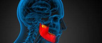
The examination is carried out through the mouth. The device is inserted deeply, up to the wall of the pharynx. Despite the discomfort of the manipulation, this type of examination makes it possible to identify adenoids, tumors and polyps in the early stages. This method is used only in rare cases and when dangerous diseases are suspected.
Average
This method of examination makes it possible to diagnose the condition of the anterior paranasal sinuses. Manipulation is carried out using an elongated instrument through the nasal passages. In this case, local anesthetics and vasoconstrictor drops are often used, which eliminate swelling of the mucous membrane.
Straight
Laryngoscopy is performed using a movable instrument that is inserted into the laryngeal cavity. The procedure can be unpleasant for the patient and often provokes vomiting, so before the procedure the pharynx is irrigated with Lidocaine. The use of microlaryngoscopy makes it possible to identify a wide range of diseases of the larynx.
Indirect
The procedure is carried out using a special mirror, which is placed in the larynx area. In this case, a frontal reflector is attached to the doctor’s head, which reflects light. The manipulation lasts no more than 5 minutes, but does not provide such detailed information as the direct research method.
Surgical
The surgical method is used not only for diagnostic, but also for therapeutic purposes. The manipulation may be accompanied by minor incisions and punctures. Often, using this method, pathological foci are eliminated and tissue biopsies are performed for histological examination. This method involves the use of anesthesia.
What shows, description of results
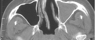
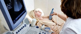
Swelling that occurs with sinusitis causes a change in the density and thickness of the nasal mucosa. In this case, ultrasound of the nasal sinuses shows the homogeneity or heterogeneity of the echostructure of the area under study.
An ultrasound of the maxillary sinuses during sinusitis will allow you to accurately determine the presence of pathology - echo signs of fluid accumulation are visible on the monitor. It is recommended to begin treatment of sinusitis as soon as possible, avoiding a further increase in inflammation and the formation of pus. Also, with the help of ultrasound of the sinuses (price from 1000 rubles), cysts, polyps and malignant tumors are clearly visualized.
In order to sign up for an ultrasound of the sinuses in Moscow and Moscow Region, leave a request on the main page of the site or read the reviews, choose a suitable medical clinic yourself and sign up for an echosinusoscopy on any day convenient for you.
Indications for the procedure
The procedure is used for symptoms and pharynx. An endoscopic examination is carried out if neoplasms are suspected: benign and malignant. Additional indications:
- the presence of inflammatory processes occurring in the nasal cavity and pharynx;
- polyps;
- enlarged adenoids;
- difficulty breathing through the nose;
- pain in the sinuses;
- change in voice, appearance of hoarseness;
- sensation of a foreign body in the larynx while talking or when swallowing food.
The examination shows the presence of purulent foci, the amount of altered tissue and other transformations of the mucous membrane, including microdamages.
Ultrasound of the maxillary sinuses: 7 indications and 4 advantages of the method
Experts say that diagnosing all types of sinusitis is a complex process that requires the right approach and the use of professional equipment. Examination of the maxillary sinuses using ultrasound guarantees accurate results, since the symptoms of this disease are similar to rhinitis, and making a diagnosis without additional examination can be quite difficult.
Modern professional equipment is used to conduct ultrasound examinations. With its help, the maxillary sinuses, which are located under a layer of soft tissue, are clearly visualized, which significantly complicates diagnosis in some cases. Thanks to a special sensor on the device, the doctor is able to detect the presence of fluid in the maxillary sinuses and swelling of the mucous membrane. Considering that inflammation can develop not only in the maxillary paranasal sinuses, but also, for example, in the frontal (frontal) sinuses, then for an accurate diagnosis it is still best to use radiography, which makes it possible to fully examine all paranasal sinuses, which is not always possible with ultrasound.
The specialist conducting the ultrasound examination must have appropriate training and be certified, therefore you should carefully choose a clinic for diagnostics. By seeking advice from a specialist of a different profile or a doctor who is not certified to perform ultrasound, you risk subsequently receiving ineffective treatment that will not give the expected results and can, in rare cases, lead to complications.
The main indications for ultrasound examination of the paranasal sinuses are:
- inflammatory process in ENT organs;
- severe runny nose, which is a symptom of an allergic or other inflammatory process;
- injury to the nasal septum;
- the presence of a chronic inflammatory process;
- tumors (malignant or benign) found in the nasal passages;
- the presence of a foreign body in the nasal passages;
- frequent headaches that occur for unknown reasons.
Ultrasound for sinusitis
Specialists often prescribe an ultrasound examination of the paranasal sinuses for sinusitis or when it is suspected. But most doctors prefer x-ray research methods, which give more accurate results in acute or chronic forms of the disease.
Ultrasound is most often used to diagnose sinusitis in children and pregnant women, for whom radiography is not recommended due to its negative effect on the body. The main advantages of ultrasound are:
- absolute safety for the human body. The use of professional equipment is especially important for diagnosing pathologies. Ultrasound gives accurate results and does not harm the body. That is why it is possible to conduct examinations using it in children of different ages and pregnant women at any stage;
- The frequency of the examination is not limited (which is typical for x-ray examinations, which can be carried out no more than 2-3 times a year). This technique is indicated for sinusitis, when it is necessary to monitor the dynamics of changes and the effectiveness of the chosen treatment;
- ease of use. The equipment is easy to use, the results are visible immediately. To obtain accurate results, a good device and a highly qualified specialist are sufficient;
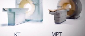
- cheapness. Many other procedures are expensive (for example: CT, MRI), but ultrasound of the paranasal sinuses is available to everyone.
When choosing a medical institution engaged in conducting this type of research, give preference to trusted organizations, since in some private clinics and medical centers the specialists are not properly qualified. An experienced doctor will quickly conduct an examination and provide you with accurate results, which will allow you to immediately make a diagnosis and begin treatment.
Ultrasound examination technique
To make an accurate diagnosis, an ultrasound examination may be required, since based on the results of an objective examination of the patient, it is not always possible to distinguish sinusitis from rhinitis. Inflammation is characteristic of both diseases, which often complicates the diagnostic process. That is why an additional X-ray or ultrasound examination is most often performed; CT, MRI or other diagnostic methods may also be recommended to study the condition of the paranasal sinuses.
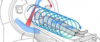
To begin with, the doctor applies a special acoustic gel to the area of the paranasal sinuses. After which the sensor is installed, and the device itself is adjusted to the simplest frequency A-mode. During the examination, the specialist turns the patient's head in different directions to achieve the highest quality image and see the presence of fluid or other pathological contents in the sinus. In this case, the examination time is about 10 - 15 minutes, which is quite enough to make an accurate diagnosis.
When using the A-mode of the ultrasound machine, the usual gray picture is not displayed on the screen. Instead, the specialist sees a simple graphic image in the form of a curve, after studying which the doctor makes an accurate diagnosis. The formation of a curve occurs due to the registration of a beam of rays that are reflected from some media and absorbed by others. If necessary, the results can be printed and, by evaluating the diagram, the doctor will understand whether there is fluid or a foreign body in the nasal passages or paranasal sinuses.
It is difficult to carry out this type of diagnosis of frontal sinusitis (inflammation of the mucous membrane of the frontal sinus), because The frontal bone closes the sinus cavities, which prevents the penetration of the beam of rays from the ultrasound sensor and the making of an accurate diagnosis. In such cases, an additional x-ray of the paranasal sinuses is performed in three projections, so the doctor can see the inflammation and make an accurate diagnosis. But this method is not used during pregnancy and for examining children. Most often, the child is shown an ultrasound, which is safer for the body.
What can an ultrasound of the paranasal sinuses show?
For diseases of the paranasal sinuses, ultrasound, as mentioned earlier, is a safer method than radiography. Echosinusography is used to diagnose the following diseases:
- polyps, cysts or foreign bodies in the nasal cavity and paranasal sinuses;
- in cases where there is a suspicion of accumulation of exudate in the paranasal sinuses;
- for the purpose of more accurate diagnosis or assessment of the dynamics of the disease after the start of treatment.
In most cases, this method is recommended for patients for whom X-ray radiation is contraindicated. These are pregnant women and children of all ages, for whom it is dangerous to conduct an X-ray examination, since its rays can negatively affect their health and even worsen it.
The main thing is to consult a specialist when the first symptoms of the disease occur in order to avoid its becoming chronic or developing complications.
Remember that in addition to the symptoms listed earlier, sinusitis may be indicated by: malaise and headaches that do not go away even after taking strong painkillers. That is why you should not delay visiting a specialist, since only he will be able to quickly and accurately make a diagnosis and select the most effective method of treating sinusitis.
Previously published articles indicate that sinusitis is one of the consequences of a prolonged runny nose, but in most cases a person becomes ill with it only if the immune system is reduced. Under various conditions, innovative or traditional methods are used in the treatment of sinusitis, but all of them are aimed at eliminating the inflammatory process and increasing the patient’s body’s defenses.
Conclusion
Ultrasound examination is one of the methods for diagnosing sinusitis, determining the presence of exudate in the paranasal sinuses, or assessing the dynamics of the pathological process during therapy. If there are no positive changes in the patient’s condition, the doctor adjusts the treatment and selects other methods to achieve a quick recovery. The patient may be prescribed medications and traditional medicine to reduce swelling, remove accumulated pus, and stop the inflammatory process.
Of course, if any serious pathological process is suspected, the patient will be recommended a more informative examination - radiography, computed tomography or magnetic resonance imaging, which are not recommended for pregnant women and children. In the latter case, an ultrasound of the head and neck organs is indeed indicated.
Article rating:
(1 ratings, average: 5.00 out of 5)
Rules for conducting the examination
It is recommended to conduct the study on an empty stomach. There are no special rules, the procedure is quick and painless. If there is a mucous secretion in the sinuses, then it is important to blow your nose so that nothing interferes with the examination. The doctor carries out the procedure wearing gloves, having previously disinfected the endoscope. Diagnosis is carried out with the patient sitting or lying down.
Preparation
The doctor notifies the patient that on the eve of the test it is prohibited to instill any solutions into the nose, use nasal ointments or other means that may complicate the procedure.
It is important to refrain from smoking. The child must be set up for endoscopy, explaining to him how the study will take place. It is important that the person remains motionless during the diagnosis.
Technology
Most often, the patient sits in a special chair during the procedure. Each type of research is carried out differently. When using the direct method, 2 thin and closed jaws are used. The patient is asked to tilt his head back, and the instrument is inserted a few centimeters into the nasal passage. Then the branches are slightly moved apart and the sinuses are examined using special optical equipment.
The posterior method of examination is carried out using a spatula, which is used to move the tongue away from the larynx. Then the device is inserted as deeply as possible, reaching the wall of the pharynx. To reduce the likelihood of vomiting, you need to breathe only through your nose. Before the procedure, you are prohibited from eating or drinking.
The average type of technique involves inserting jaws through the nasal passages and examining using an optical device. Before manipulation, the nasopharynx is irrigated with an anesthetic solution, and a vasoconstrictor is instilled into the nose.
The surgical method requires longer preparation. In this case, various types of pain relief can be used. Often, during the manipulation, an incision is made into the tissues of the nasal mucosa in order to get rid of chronic rhinitis. If polyps are present, a small fragment of material is sent to the laboratory for a more thorough diagnosis.
The indirect type of research is used in any clinic. The patient sits on a chair, slightly throwing back his head and sticking out his tongue. The doctor inserts a mirror into the larynx and examines the tonsils and pharynx. At the same time, the slightest deviations from the norm are clearly visualized.
The direct method is often performed using a moving laryngoscope. A rigid technique with a rigidly fixed apparatus is used during surgery. Before starting the procedure, the sequence of steps is explained to the patient. This method is carried out using general anesthesia.
The laryngoscope is inserted through the larynx and advanced deeply. This method is considered the most informative.
Advantages and disadvantages of the ultrasound method
Of course, the diagnostic method of ultrasound has a number of positive characteristics, due to which it is prescribed almost more often than all other types of examinations.
Best materials of the month
- Why you can't go on a diet on your own
- 21 tips on how to avoid buying stale food
- How to keep vegetables and fruits fresh: simple tricks
- How to curb your sweet cravings: 7 unexpected products
The procedure usually takes place in 10-15 minutes, while other examination methods may take longer than ultrasound. In cases where the doctor urgently needs to obtain information about the condition of the patient’s paranasal sinuses, any delay is inappropriate and can be dangerous.
Other scanning techniques do not allow determining the presence and location of foreign bodies in cavities - this is only possible with the help of ultrasound. Considering that this problem often occurs in young children who love to explore the world in such a traumatic way, the value of an ultrasound examination for them cannot be overestimated.
Scanning with an ultrasound sensor is absolutely painless and does not cause discomfort to the patient; no special preparation is required before it begins.
The safety of ultrasound examination ensures that the procedure can be performed regularly every time it is needed. X-ray or computed tomography cannot boast of this property.
However, the method also has known disadvantages. Echosinusoscopy is an inaccessible type of ultrasound, and not every ultrasound diagnostician has a sufficient level of qualifications to perform it. Some doctors with decades of experience have never encountered the need to perform echosinusoscopy.
In the process of interpreting and deciphering the results, the doctor must remember that ultrasound scanning can produce an image with a certain level of error, and in some cases there may even be such a phenomenon as overdiagnosis, that is, a situation when the sensor detects areas with particular echogenicity, although there is no There is no pathology in this place. Often, ultrasound results must be supplemented with information obtained from other examinations, which requires additional time and expense.
Ultrasound examination of the paranasal sinuses, as a diagnostic method, is highly valued by doctors. Among its advantages are the speed of implementation, sufficient accessibility, in addition, patients note the complete painlessness of the method. Based on the results of the ultrasound, the doctor can detect only pathologies or destructive processes in the images - in the normal state the sinus cavity is not visualized. However, in the presence of polyps or tumors, sinusitis, sinusitis, rhinitis, sinusitis, labyrinthitis, foreign bodies, hemorrhages, the diagnostician will notice characteristic changes in the images, and then, if necessary, prescribe additional examinations, establish or confirm the diagnosis, and propose treatment regimens.
Features of endoscopy in children
An examination of the child's nasal cavity and pharynx is carried out in the presence of parents. Often the manipulation is complicated by the fact that it is most difficult for children to remain motionless for 5-10 minutes. For diagnosis, the most painless methods are chosen, which are practically devoid of discomfort.
If the procedure does involve additional surgical intervention, the child should be carefully prepared. First, determine whether there is an allergy to any medications. Special tests are carried out. To ensure that the procedure itself does not cause shock in the child, he is told and shown what instruments will be used during the study and what they are needed for.
It is important to pay attention to pain relief. Therefore, even when using the most minimally invasive techniques, local anesthetics are used. It is important for a child, like an adult, to refrain from eating and drinking. Children are explained the rules of behavior during the insertion of an endoscope. If this is not enough, then in extreme cases they resort to general anesthesia.
For manipulation in children, an endoscope no more than 2 mm in diameter is used. It does not create discomfort, moves easily through the nasal sinuses and does not injure them. The specialist tries to insert the instrument very carefully so that there is no sensation of a foreign body. At the end of the procedure, it is important to ensure that the child does not pick his nose.
Ultrasound of the maxillary (paranasal) sinuses: technique
Echosinusoscopy or ultrasound of the sinuses - examination of the frontal and maxillary sinuses using ultrasound. It is used in otolaryngology, dentistry, and facial surgery.
Indications and contraindications
The study may be useful for diseases of the nasal cavity, paranasal sinuses, dentofacial apparatus, skin and subcutaneous tissue. Ultrasound is prescribed in the following cases:
- acute sinusitis (sinusitis, sinusitis);
- sinus polyp;
- cyst;
- mucocele;
- frequent nosebleeds;
- rhinitis of unknown origin;
- deformation of the nasal septum;
- purulent discharge from the nasal passages;
- pain in the nose and paranasal sinuses;
- foreign body;
- adenoids;
- injury;
- neoplasm;
- pathology of soft tissues (furuncle, carbuncle);
- performing puncture of the nasal sinuses, hematomas;
- monitoring sinus pathology over time.
The method has virtually no contraindications. It is not recommended for acute mental disorders, acute failure of internal organs (heart, lungs, liver).
How does the examination take place?
There is no need to prepare for an ultrasound of the maxillary sinuses and frontal sinuses. The examination can be performed lying down or sitting. The research methodology is as follows:
- It is necessary to remove jewelry, dentures, and glasses.
- A conductor gel is applied to the nose and paranasal area.
- The sensor moves along the surface of the skin in the projection of the paranasal sinuses. It records the signal from tissues located at different depths. Thanks to the reflection of the echo signal, an image is formed on the screen.
- If it is necessary to determine the fluid level, the patient is first placed on his back, then on his stomach.
The study takes only 5-10 minutes. At the end of the procedure, a conclusion is issued on paper or electronic media. The diagnosis is made by an otolaryngologist after examination and all tests.
What does an ultrasound show?
Ultrasound diagnosis of diseases of the nasal cavity and sinuses is quite informative. It allows you to study the structure of formations and see the speed of blood flow. During the examination, the following indicators can be assessed:
- The thickness and condition of the subcutaneous fat layer.
- Integrity of the mucous membrane.
- Condition of vascular septa.
- Parameters of nasal cartilage.
- Size of cyst, tumor.
- State of blood flow.
- Fluid level in the sinus.
- Condition of bone structures.
Advantages and disadvantages of the diagnostic method
Ultrasound examination has the following advantages:
- availability;
- low cost;
- harmlessness;
- a small number of contraindications;
- quick results;
- lack of preparation;
- painlessness;
- non-invasive;
- information content.
X-ray of the sinuses is the first test that is prescribed if sinusitis is suspected. It should not be performed during pregnancy due to possible effects on the fetus. The method of choice in this case is echosinusoscopy. The examination can be carried out on a stationary or portable device. This makes it possible to diagnose diseases not only in a medical institution.
Ultrasound of the paranasal sinuses has some disadvantages. The echo signal cannot penetrate deeply located objects (ethmoid sinus). The second disadvantage is the lack of awareness among specialists about the methodology. This is because sinus ultrasounds are rarely performed.
Also, the study does not always allow for good visualization of anatomical structures. This leads to the need for additional examinations, which increases the cost of diagnosis. This method is characterized by overdiagnosis - signs of diseases are often detected, which are then not confirmed. This can lead to unnecessary treatment (use of antibacterial drugs, sinus puncture).
Price
The examination can cost from 700 to 1500 thousand rubles, depending on the clinic and equipment.
Echosinusoscopy should be performed only if radiography is not possible. Diagnostic equipment must be modern, and the ultrasound doctor must have the necessary experience.
Author: Natalia Nikulina, doctor, especially for Moylor.ru
Useful video about paranasal sinuses
What are the contraindications?
The main contraindications are persistent nosebleeds. If the vessels are too thin and weak, then the risk of damage is high. Therefore, before resorting to endoscopy, it is important to check the condition of the venous apparatus, as well as donate blood to check the rate of platelet aggregation.
An additional contraindication is an increased gag reflex. However, methods based on deep insertion of the instrument into the larynx are often not used. The procedure is not performed during pregnancy. Endoscopy is contraindicated in infancy, since the sinuses are easily injured.
If the tonsils are very enlarged, manipulation is not performed, since such a clinical picture makes visualization of the tissue difficult. A contraindication is an allergic reaction to painkillers. The procedure is not performed during treatment with anticoagulants, since if a vessel is accidentally damaged, there is a high probability of bleeding, which will be difficult to stop.
For a deviated nasal septum, a pediatric endoscope is used, which reduces the risk of unpleasant sensations. If a person is allergic to local anesthetics, and endoscopy is necessary, then an easy option is chosen, which can be performed without the use of anesthesia.
Contraindications are the patient’s unstable mental state, the presence of schizophrenia, and disorders of the central nervous system.
An endoscopic examination of the nasal cavity in a child helps to examine the nasopharynx and auricles for the presence of pathology and diseases.
Thanks to a long tube no more than four millimeters thick with a camera and a lighting device at the end, the image is displayed on the monitor in an expanded form.
The procedure helps to identify an accurate diagnosis and prescribe comprehensive treatment to eliminate the disease.
Advantages and disadvantages of the procedure
The procedure is quick and safe, and the level of x-ray exposure is minimal. Due to such features, this method is chosen for examining the paranasal sinuses in children and pregnant women. There are no age restrictions.
The significant advantages are:
- simplicity of implementation and absence of the need for preliminary preparation of the patient;
- the possibility of repeatedly conducting research and assessing the course of the disease over time;
- low cost of research;
- the ability to obtain a diagnosis within a few minutes;
- saving the patient’s examination results on the computer’s hard drive, which allows the results to be reused, including by specialists in related fields.
Ultrasound allows you to quickly obtain information about the condition of the frontal and maxillary sinuses, show the presence of air, liquid secretions, thickening of the mucosa, and prescribe surgical treatment within a short period of time. With the help of such a study, doctors are able to monitor the dynamics of pathological changes during drug treatment and, if necessary, make adjustments.
Similar results from ultrasound of the nose and sinuses can be obtained using computed tomography or radiography. In the first case, the patient receives a significant dose of radiation, and the second method is characterized by high cost. Ultrasound examination of the sinuses is not characterized by such shortcomings.
The only drawback can be considered the possibility of obtaining in some cases an incomplete picture of the course of the disease. In this case, repeated studies will be required, which is fraught with additional financial costs for the patient.
For what diseases and symptoms is endoscopy of the nose and nasopharynx prescribed for children?
Nasal endoscopy is prescribed for diseases of the ENT organs, the main ones of which are: sinusitis, tonsillitis, acute respiratory disease, rhinosinusitis, frontal sinusitis.
The procedure makes it possible to identify various pathologies at an early age, the treatment of which in the early stages will be more effective than in an advanced state. Endoscopic examination can detect benign and malignant neoplasms, inflammation of the adenoids and in the sinuses. Polyps are identified through the endoscope. You can also view anomalies in the structure of the nasal passages and septa, and the peculiarities of the nasal mucosa during the disease.
Photo gallery:
Endoscopy of the child’s nose is prescribed for a number of symptomatic features:
- Difficulty breathing, forcing children to breathe through the mouth
- With a decrease or complete disappearance of the sense of smell
- If you have mucus or pus coming from your nasal passages
- Unreasonable headaches in the frontal lobe, or temporal region
- With a decrease in taste buds
- For throbbing pain in the sinuses
- With hearing loss, prevalence of tinnitus
- If you snore during sleep
- Frequently recurring nosebleeds
Advantages of ultrasound examination
Ultrasound of the sinuses is highly informative and safe even for pregnant women and infants. In addition, ultrasound diagnostics are significantly lower in cost than similar procedures:
computed tomography of the nasal sinuses;
- Magnetic resonance imaging.
In the case of small children, the advantage of ultrasound is that parents can be present next to the child. Ultrasound diagnostics are carried out in a special room, without placing the patient in a machine (as with CT or MRI) and without the use of harmful radiation (as with x-ray examination methods).
Preparation for nasal endoscopy in a child
Before the endoscopic examination, a conversation is held with the child about the upcoming procedure.
Parents should explain:
- With the assistance of a doctor, the procedure will take a few minutes.
- Don’t twitch or struggle, then the doctor will quickly and painlessly perform an endoscopy and prescribe treatment.
During the procedure, children use a gel containing lidocaine as an anesthetic. The tip of the device tube is treated with it. When exposed to the gel, numbness occurs in the nasal area, due to which the endoscope painlessly passes into the nasal passage for examination.
An anesthetic spray is also used as a pain reliever. When exposed to it, a tingling sensation occurs in the nasal passages, which irritates the mucous membrane and facilitates painless insertion of the tube.
Preparation for the procedure for adults and children
Echosinusoscopy does not imply any preparation, unlike ultrasound of the pelvis, bladder, abdominal cavity, and prostate gland. There is also no difference in this procedure between adults and children.
When prescribing this study for a child, all aspects of an ultrasound of the nose and sinuses should be explained to him: why it is happening, why he needs it.
All this is done by parents or relatives in advance, to psychologically calm the baby. You should explain to the child that he will not experience any pain, since the examination is truly painless and cannot cause discomfort.
How is nasal endoscopy performed on a child? Features of the event
This is what nasal endoscopy looks like for children
If the child behaves calmly, the endoscopic examination lasts several minutes. Then the doctor takes the tube out of the nose, prints out pictures of the problem areas and makes a conclusion, which includes a description of what he saw, making a diagnosis and prescribing qualified treatment.
When examining ENT organs through an endoscope in young children, during the procedure parents hold the child so that he does not break free and harm himself.
Indications for ultrasound
The main advantages of ultrasound diagnostics are quick results and absolute safety of the procedure for the patient. Such a study is often prescribed to a child, and it is also allowed to be carried out during gestation.
Ultrasound of the paranasal sinuses has a fairly wide range of indications. These include:
- diseases of ENT organs in the chronic stage;
- inflammation of the maxillary sinuses – sinusitis;
- inflammation of the frontal sinuses - frontal sinusitis;
- allergic rhinitis;
- causeless nosebleeds;
- inflammation of the inner ear;
- formation of polyps in the nose;
- purulent-necrotic diseases of hair follicles – furunculosis;
- deviated nasal septum;
- foreign bodies entering the nose;
- various neoplasms.
Is it necessary to do an endoscopy?
Nasal endoscopy can help establish an accurate diagnosis and examine organs for the presence of foreign bodies. It is carried out before and after ENT surgery. During the procedure, the doctor can perform surgery with precision, thanks to the display of the image on the monitor screen.
- Through an endoscope, the doctor examines inflamed areas, deviated nasal septum, neoplasms, and the condition of the adenoids. Endoscopy should be done for a child of any age to accurately identify the cause of the disease and make a final diagnosis.
- Endoscopy is mandatory for children with pathological features of the auditory tubes, enlarged adenoids and swelling in the nasal passages.
- The procedure is financially accessible. Thanks to modern technologies, it allows you to reduce the time and costs of diagnosing the disease by conducting only an endoscopic examination.
If bleeding is frequent, endoscopy should be performed carefully.
Endoscopic examination is painless. Thanks to the flexible hose, it allows you to examine hard-to-reach areas of the ENT organs. It is worth warning your doctor about the presence of nasal and sensitive mucous membranes. With such symptoms, the procedure is carried out with extreme caution.
A contraindication to endoscopic examination of ENT organs is an allergic reaction to the anesthetic. Parents should warn the doctor about this feature before the procedure begins.
What does ultrasound of the sinuses show and where to do it?
Recently, ultrasound examination (ultrasound of the sinuses) for sinusitis has become popular. Ultrasound has become a tool for diagnosing acute sinusitis over the past two decades.
Ultrasound of the nose and paranasal sinuses
Ultrasound of the sinuses is a safe, fast, non-invasive, inexpensive and repeatable method of diagnostic research, which is also commonly called echosinusoscopy.
Echosinusoscopy is very sensitive in detecting fluid in the sinuses. It was registered in otolaryngology (ENT) - the accuracy of determining sinusitis was more than 90%. Ultrasound examination of the sinuses is carried out quickly and painlessly. This procedure is not expensive and is available to every patient.
Accurately diagnosing sinusitis is challenging because the signs and symptoms of sinusitis are nonspecific and distinguishing between rhinitis and sinusitis is not always easy. Tests such as CT and MRI provide an accurate diagnosis; they are not usually used to diagnose uncomplicated sinusitis in an outpatient setting due to the additional cost, time, and radiation risk. That is why ultrasound of the sinuses is relevant today.
Indications for ultrasound examination
Using an ultrasound machine, the maxillary sinuses are clearly visible. They are located under a layer of soft tissue, through which the sensor can easily see the inflammatory process and the presence of fluid in them. To diagnose the frontal sinuses, it is better to use x-ray examination. They are located under the frontal bone, through which the ultrasound sensor of the device will not be able to conduct an examination.
Indications for ultrasound of ENT organs:
- Acute and chronic form of ENT disease.
- Runny nose as a manifestation of an allergic reaction.
- Injured nasal septum.
- Polyposis disease.
- Malignant and benign neoplasms in the nasal passages.
- The presence of a foreign body in the nasal passages.
- Furunculosis.
- Other nasal injuries.
- Frequent headaches.
- For the purpose of observation during the treatment of ENT diseases with medications.
Ultrasound for sinusitis of the maxillary sinuses
A specialist may prescribe an ultrasound if a patient suspects a disease - sinusitis. However, most often doctors use the radiographic method of examination for acute and chronic forms of ENT diseases.
Carrying out an ultrasound for sinusitis
Ultrasound examination in the presence of sinusitis can be prescribed to patients in childhood or pregnant women, a category of people for whom frequent X-ray exposure is undesirable.
Read how x-rays are performed for sinusitis here
Ultrasound examination technique of the paranasal sinuses
Ultrasound of the paranasal sinuses is performed both on stationary equipment and on mobile equipment.
- The convenience of a mobile echo sinuscope is that the doctor can come to the patient’s room or home, conduct an examination and issue a conclusion. A popular echo sinuscope among doctors today is Sinuscan (Sinuscan – 201)
- The advantage of ultrasound equipment in a hospital is its information content. The diagnostician sees the examination results on a large monitor and makes a diagnosis much more accurately. In medical slang, this equipment is called ENT-Combine.
Ultrasound of the sinuses in the hospital
Step-by-step examination of the paranasal sinuses using ultrasound in the hospital:
- The specialist conducts a conversation with the patient and explains to him the progress of the upcoming ultrasound.
- The patient is seated in a specialized chair for further examination.
- The doctor lubricates the skin in the paranasal sinus area with a specialized gel. This is necessary so that the device can better view the sinuses.
- The specialist moves a linear sensor over the skin in the area of lubricated areas.
- During the examination, the doctor tilts the patient's head to the sides to accurately examine the sinuses for the presence of fluid or purulent contents.
- After a complete examination, the doctor writes a transcript of the resulting curved line.
- The patient cleanses the skin of a special gel and awaits the result of the study for a further visit to the doctor and the appointment of qualified treatment, if necessary.
Features of using the echo sinuscope Sinuscan (Sinuscan – 201)
Echosinusoscope Sinuscan 201
This ultrasound machine is manufactured using the latest technology. It is small in size, which allows it to be used in the patient's room. The examination takes a few minutes. The rays penetrate to a depth of eight centimeters. They allow you to scan the frontal and maxillary sinuses for the presence of accumulated fluid. A scale is visible on the built-in small screen. The image changes to a graphical curve if there is fluid or pus in the sinuses.
The echo sinuscope has a built-in memory for four studies. You can operate the device with your left or right hand, according to the desire and convenience of the specialist. The kit includes a charger, a specialized gel and a case for carrying and storage.
Progress of the procedure:
- The patient is informed about the progress of the upcoming study.
- He takes a lying or sitting position at the request of the doctor.
- The specialist moistens the sinus area with a special gel.
- Starts the Sinuscan machine..
- Applies it to the sinuses and moves the echosinoscope within their boundaries.
- Based on the results displayed on the screen, the doctor makes a decision on the diagnostic result.
Examination using the Sinuscan 201 echo sinuscope is fast, painless and effective.
The cost of a Sinuscan 201 examination is estimated at 300-500 rubles
Interpretation of echosinusoscopy
What does the ultrasound examination report show?
- The presence of foreign bodies in the nasal passages and sinuses.
- Malignant and benign neoplasms.
- Cystic disease.
- Accumulated fluid or purulent contents in the maxillary sinuses.
- Examination of the course of the disease and the effectiveness of the treatment prescribed by a specialist.
Echosinusoscopy is most often prescribed to women during pregnancy, children and nursing mothers. In the absence of contraindications to radiographic diagnostics, patients will be prescribed this type of examination. A good picture of the disease is visible during computed tomography and magnetic resonance therapy, but these procedures are expensive and are not prescribed to all people with diseases of the ENT organs.
Photo gallery:
Ultrasound or x-ray of the sinuses
Type of examinationPositive diagnostic criteriaNegative diagnostic criteria
| Ultrasonic | Ultrasound examination does not have a harmful effect on the human body; it can be performed on women during pregnancy and during breastfeeding. The device is also equipped with Doppler ultrasound, which allows you to assess the condition of large vessels in the nasal passages. This diagnostic method can be performed at a low cost in almost every clinic or private clinic. The result is given by the doctor immediately after the examination. | Ultrasound examination is rarely used to diagnose ENT organs. Most practitioners are not properly qualified to conduct examinations using this device. Ultrasound helps to view the maxillary sinuses for fluid accumulation in them; the frontal sinuses are protected by a wide bone; it is almost impossible to view them through equipment. Often an ultrasound examination shows the presence of an inflammatory process that actually does not exist, and the patient is prescribed antibiotic therapy. To get an accurate picture of the disease, it is necessary to conduct an ultrasound examination several times, which takes time and increases the cost of money. |
| X-ray | X-ray examination is carried out for diseases of the ENT organs more often than ultrasound. Most doctors trust this device more. | Frequent x-rays may expose the human body to radiation. This diagnosis cannot be carried out during pregnancy, as it can provoke abnormal development of the intrauterine fetus. |
Ultrasound of the child’s sinuses
Ultrasound of the nose is possible from 2 years of age
Ultrasound of the paranasal sinuses has no contraindications and therefore this type of diagnosis is prescribed to young patients suffering from diseases of the ENT organs.
The study is not suitable for children under two years of age.
The doctor may prescribe a diagnosis of the frontal sinuses and maxillary sinuses, since they are already formed and ready for examination. The rest are formed before the age of twelve, and it is almost impossible to examine pathological changes in them.
The procedure is painless, which is something parents and children need to talk about when going to the doctor. The diagnostic process is carried out in a sitting or lying position at the discretion of the specialist. The duration of the examination, taking into account the wait for results, is no more than thirty minutes.
Clinics and prices where they do ultrasound of the sinuses
Ultrasound of the sinuses is performed both in public clinics and in private clinics in large cities. The price of diagnostics depends on the location and qualifications of the specialist. Approximate cost from 500 to 1550 rubles.
City and clinicCost
| Moscow "Miracle Doctor" | From 1240 rubles |
| St. Petersburg “Dr. Pel’s Clinic” | From 700 rubles |
| Krasnodar "Krasnodar Balneotherapy Hospital" | From 500 rubles |
| Yeysk "Territory of Health Clinic" | From 550 rubles |
Is the endoscopy procedure painful?
Thanks to local anesthesia, nasal endoscopy is performed painlessly. An unpleasant feeling as the tube passes through the nasal passages is present in children with pathologies in the structure of the ENT organs and a deviated nasal septum.
The procedure itself lasts no more than 2 minutes.
Allows you to accurately examine the internal structure of the nasopharynx or ears in an enlarged form. Also, during an ENT examination, the doctor may take biological material for analysis.
During the diagnostic procedure, most patients experience discomfort during pain relief. After a few minutes they pass and the doctor begins an endoscopic examination.
Modern office equipped with advanced equipment for nasal endoscopy
Endoscopy of the nose and nasopharynx for children can be performed by an experienced otolaryngologist in a treatment room equipped with all the necessary equipment. Endoscopic examinations are carried out in clinics and specialized medical centers.
Parents prefer modern clinics with a license, qualified doctors and modern equipment.
Before performing nasal endoscopy, the otolaryngologist prescribes blood tests to rule out allergies to painkillers in children.
Endoscopy of a child’s nose video:
There are many methods for diagnosing diseases associated with the nasopharynx. The main methods are: rhinoscopy, palpation method, x-ray and, of course, endoscopy of the nasopharynx. Nasopharyngeal endoscopy is a more modern, more accurate, less traumatic and painless type of examination of the nose.
What is ultrasound, its advantages
This diagnostic method is based on the different ability of tissues to reflect ultrasound waves (echogenicity). Dense muscle or connective tissue structures, liquid or air behave differently under the influence of ultrasound, which is displayed on the monitor screen in the form of a two-dimensional image. The result is ready immediately.
This feature of the study is very important in the serious condition of the patient, as well as in the need for dynamic monitoring during endoscopic puncture or surgical intervention on the sinuses.
An ultrasound examination carried out for the purpose of diagnosing inflammation of the nasal sinuses has the highest accuracy (up to 100%) and information content. On the monitor screen, the doctor sees a thickened, swollen mucous membrane of the sinus, the presence of air and liquid in the cavity. He immediately diagnoses perforation of the maxillary sinus floor after dental operations and the presence of foreign bodies in it.
Various polypous growths, bone and soft tissue cysts, deviated nasal septum, injuries of the nose and facial bones - all these conditions are clearly and quickly visualized using ultrasound. This method is invaluable in emergency cases when there is a threat to the patient’s life due to serious injuries or developed complications of sinusitis.
Compared to traditional radiography of the nasal sinuses and computed tomography, the ultrasound method has the main advantage of the absence of ionizing radiation. This allows ultrasound to be used for all pathologies without exception and in every patient, regardless of his condition. It can be given to a child at any age, a pregnant or lactating woman, or a person who has recently received radiation exposure.
Ultrasound is more informative and safer than radiography, comparable in importance to computed tomography, but cheaper and more accessible than the latter. The absolute non-invasiveness and painlessness of the method is used in cases where the patient refuses diagnostic punctures of the maxillary sinuses using a needle or endoscope.
This research method can be repeated as often as required by the process of therapy and monitoring its effectiveness.
The procedure itself takes very little time, about 10 minutes, and does not require any preliminary preparation. This is also an important advantage of the method.
Progress of the procedure
The procedure is performed in a sitting position. The patient is seated on a chair (akin to a chair in dentistry), and his head is slightly tilted back. After this, the nasal cavity is anesthetized. This could be a gel that is used to lubricate the endoscope, based on lidocaine, or it could be a spray - an anesthetic that is used directly in the nasopharynx. After the anesthetic, a tingling sensation occurs in the nose, which causes some discomfort. Next, the endoscope is inserted into the nose.
The picture is displayed on the monitor and the doctor, assessing the situation, makes a decision on treatment.
The procedure, as a rule, takes no more than twenty minutes (anesthesia, direct endoscopic examination, printing of photographs and drawing up doctor’s reports).
Surgical endoscopy allows you to remove formations without affecting important mucosal structures. They are removed within healthy tissue. During such an operation, bleeding is minimized, there are no scars, cicatrices or incisions on the face, and there is no postoperative rehabilitation period. The patient is under the supervision of doctors for only a day after the operation.
Where is the procedure performed?
The procedure is carried out in municipal public medical institutions and in private paid research medical centers. The main criteria for choosing a place for examination:
- the establishment has all the necessary documents and permits to carry out this examination;
- satisfactory sanitary condition of the premises where the entire process will take place;
- availability of a full set of medical equipment necessary for diagnostics - sensors, monitor, gel;
- The procedure should be carried out by competent and experienced specialists whom patients trust.
You need to be careful if you choose a paid institution, and this is not a large medical center that has long earned a good reputation.
Experienced diagnosticians with a diploma do not always work in private offices - before doing an ultrasound, you should make sure of the specialist’s competence!
Description of the procedure
The ultrasound technique is based on the ability of ultrasonic waves to pass through the tissues of the human body and be reflected at the border of areas of different densities. This is called a reflected echo. The ultrasound machine “catches” him.
Waves are unable to travel through a mass of air. Since in a healthy state the sinuses are filled with it, the ultrasound signal is completely reflected and is not recorded on the equipment. If there is mucus, liquid inside the paranasal sinuses, or there are seals - signs of the development of the disease - then ultrasound penetrates inside without obstacles and is recorded by an ultrasound machine.
Scanning of the internal contents of the nasal sinuses occurs using a high-frequency linear sensor. A gel contact is first applied to the surface of the skin of the examined area. The gel is needed for a better visual display of the study results on the monitor, since the bone prevents this.
The sensor is fixed motionless in one place. It collects information about the state of the sinuses and displays it on the screen in graphical form. The specialist interprets the data obtained and draws conclusions about the existing diagnosis.
What does the doctor evaluate during an ultrasound?
As mentioned above, a doctor can use ultrasound to determine not only the state of the human body at the time of the examination, but also how the disease progresses. Thus, it becomes possible to understand how effective the prescribed treatment is.
When performing an ultrasound, the specialist identifies the following indicators:
1. Presence of any shadows.2. What is the size of the sinus, as well as its volume.3. How thick are the mucous membranes of the nose.4. Shape of the sinuses of the nose.5. Internal protrusion.6. Amount of liquid.7. Area of distribution of pathology.8. Shadows.
Indicators are assessed quickly, no longer than ten minutes. Next, the patient is prescribed a treatment regimen. It can be conservative or surgical.
Contraindications
Contraindications to the procedure are as follows:
- phobias (before visiting the ultrasound specialist’s office, take sedatives for several days);
- state of psychosis and exacerbation in severe mental illness;
- wounds and abrasions on the area of skin that is to be treated with gel;
- acute cardiac, pulmonary, liver failure;
- the presence of serious facial injuries in which the person experiences severe pain from touch.
If there is a metal plate in the skull or a pacemaker, you must inform your doctor about this and consult with him about the possibility and conditions of ultrasound diagnostics. In general, the procedure has no special contraindications.
Examination algorithm
In the ultrasound room, a person sits in a chair facing the diagnostician. Glasses or nose piercings must be removed in advance. When examining sinuses in adults, the doctor tells you when you should not move and gives other instructions.
Methodology for examining the paranasal sinuses using ultrasound:
- The diagnostician applies a thick layer of gel to the skin in the examined area.
- The doctor places the sensor perpendicular to the cheek near the nose or on the bridge of the nose.
- The sinus area is scanned on the right, left in a vertical and horizontal position. The patient sits upright, then lies down or tilts his head slightly forward, to the side, or back.
- After the examination, the gel is wiped off the face, and the diagnostician draws up a conclusion.
Ultrasound of the sinuses and echosinusoscopy are performed according to the same research algorithm with minor differences. Both procedures last no longer than 20 minutes.
Features of examination of children
It is not advisable for a child under 3 years of age to have an ultrasound examination of the paranasal sinuses. They are scanned if defects of the upper respiratory tract, facial trauma, or foreign body are suspected. Newborns or infants lie down during the procedure, older children sit. For medical reasons, they may be put into medicated sleep.
Ultrasound of the nasal sinuses in children over 14 years of age and in adults is performed using the same technique.