Radiography is a relatively quick, inexpensive and non-invasive study that allows us to judge not only the state of the bone structure, but (mostly indirectly) also about changes in soft tissues. It is not surprising that the method remains relevant even after the advent of more accurate (but also more complex and expensive) examinations, such as MRI and CT.
X-ray of the paranasal sinuses is one of the most informative diagnostic techniques in otorhinolaryngology, allowing to identify both inflammatory processes and neoplasms, fractures and other pathological changes.
The paranasal sinuses (sinuses) are cavities in the bones of the skull, lined with mucous membrane and communicating with the nasal cavity. The paranasal sinuses not only reduce the weight of bone structures, but also warm the air passing into the lungs, moisten it, serve as a resonator for the voice and a “buffer” for injuries.
Indications for x-rays of the nasal sinuses
Indications for which an X-ray photo of the maxillary sinuses is taken:
- Difficulty breathing, constant runny nose and increased tearing, copious discharge from the mucous membranes and sweat glands.
- Clinical or indirect signs of sinusitis. A referral for the diagnosis of sinusitis is issued by a doctor after an initial examination and testimony of the patient.
- Severe nasal congestion and constant runny nose. The congestion does not go away and is not relieved by nasal cleansing products. The mucus output is weak, the temperature of the facial part is increased.
- Severe facial pain, facial swelling that forms in the cheeks and eyelids. When the maxillary sinuses are completely blocked, the accumulated mucus begins to put pressure on the skull and soft tissues of the nose. This causes severe, unrelenting pain.
- Benign neoplasms. Tumors with benign symptoms make it difficult to breathe through the nose and cause a feeling of constantly stuffy nose. It is determined during the initial examination and, for confirmation, a referral is issued for the diagnosis of sinusitis on an x-ray.
- Malignant neoplasms. Tumors are accompanied by severe pain and a feeling of constantly stuffy nose. Yellow mucus is constantly secreted, which may be the first sign of meningitis. On x-ray it appears as an area with ragged and sharp edges.
- The presence of foreign bodies in the patient’s nasopharynx. To fully check the condition of the object and its position, fluoroscopy is prescribed out of turn. After assessing the condition, surgery is performed.
- Frequent nosebleeds. They may accompany benign and malignant neoplasms in the maxillary sinuses, which may be an indirect sign of acute meningitis. The development of this disease cannot be allowed.
- Nasopharyngeal injuries. In case of mechanical damage to the skull, an x-ray is prescribed to determine the presence of foreign body fragments and assess the extent of damage to the area.
- Severe and acute headaches for no reason. Acute attacks of headache accompany acute sinusitis or the initial stage of meningitis. An x-ray will help diagnose the disease.
- Suspicion of cysts. Neoplasms can lead to complications and loss of organ functionality. A maxillary cyst can lead to loss of smell and partial vision. X-ray will show the presence of a cyst and the possibility of its puncture drainage under ultrasound guidance.
- Development of caries of the upper teeth. The X-ray method of the nasal sinuses will determine the degree of damage to the teeth and the possibility of their removal without complications.
- Dental implant surgery. To determine the location of the dental implant and the presence of pathologies, an x-ray of the maxillary sinuses is taken.
- Polyps in the maxillary sinuses. They can form as a result of complications and relapse of chronic sinusitis. The disease affects about 4% of the population. Men are more susceptible.
- Decreased performance and general fatigue, periodic dizziness, constant sleep disturbances, lack of appetite.
Symptoms
Acute sinusitis has the following symptoms:
- Fever, chills, fever, temperature rise to maximum levels.
- Headache that comes and goes suddenly.
- Thick discharge from the nose mixed with pus.
- Dull pain in the location of the sinuses.
- Severe nasal congestion, runny nose.
- The voice becomes rough, nasality and hoarseness appear.
- Fatigue, weakness, deterioration in general health.
Pansinusitis in children is characterized by the presence of cough during night rest. In addition, the child has abundant purulent mucus from the nose.
Signs of pansinusitis appear suddenly. It all starts with fever, excessive sweating, chills, body aches and increased body temperature. Further, the clinical picture is complemented by headache. The patient’s skin also turns pale, sleep is disturbed, and appetite disappears. There may be congestion in the ears and impaired sense of smell.
If treatment is not started on time, the consequences can be unpredictable. The disease can become long-lasting or affect the meninges.
Chronic pansinusitis is similar in symptoms to acute, only it manifests itself weakly or is completely absent. Pain in the head and projections of the sinuses rarely appears, the increase in temperature is insignificant - up to 37.5ͦC.
X-ray for sinusitis in children
X-ray of the nasopharynx in children is performed if other diagnostic methods do not provide a sufficient picture for diagnosis. The procedure is low-risk for the child's health. Most often, X-rays are used in children if there are symptoms and suspicion of serious illness. The referral is issued after the initial examination, tests and testimony of the patient.
The maxillary sinusoids of children are formed and will look similar to those of adults on an x-ray photo by the age of 16. Until this age, sinusoids in children are displayed, for the most part, in the form of light spots. They have a dense structure and do not appear hollow.
Using an x-ray of sinusitis in the photo, you can obtain information about the following pathologies:
- accumulation of masses of pus;
- excessive enlargement of pharyngeal tonsils;
- the presence of sinusoid and sinusitis in acute stages.
Mainly used to more accurately determine the degree of enlargement of the nasopharynx with adenoids.
You can find out about the need to do an X-ray examination in a video from the author “Doctor Komarovsky”.
Treatment
If there is at least one sign of illness, you should consult a doctor. If the diagnosis of “acute pansinusitis” is confirmed, then the patient is recommended to go to the hospital. Therapy at home is prohibited due to the high likelihood of complications.
Treatment should begin with taking antibiotics with an extended effective mechanism - Azithromycin, Amoxiclav, Ciprofloxacin. The duration of therapy is on average 7-10 days. In addition, antibacterial drugs are prescribed in the form of sprays and drops (Isofra, Polydex).
If the cause of the disease is a fungal infection, Flucanozol, Ketocanozol are prescribed, viral - Kagocel, Ingavirin.
For nasal congestion, vasoconstrictor medications are recommended. The products will quickly ease breathing, eliminate a runny nose, and reduce mucus production. Nazivin, Noxprey, Tizin, Rinonorm are suitable for these purposes. Rinofluimucil aerosol has vasoconstrictor, anti-edematous, anti-inflammatory and mucolytic effects. In addition, nasal rinsing is prescribed with solutions based on purified sea water - Dolphin, Aquamaris. In the hospital, the procedure is carried out using an electric suction, or the Proetz method.
Among anti-inflammatory drugs, preference is given to Diclofenac, Nurofen, Paracetamol. Suprastin and Zyrtec are indicated as antihistamines (if the disease is caused by allergic rhinitis).
The patient must undergo physical therapy:
- UHF;
- magnetic therapy;
- ultra-high frequency therapy;
- laser therapy.
If conservative therapy does not have a positive effect, they resort to surgery. During surgery, the surgeon punctures the sinus to create a hole through which accumulated mucus will be pumped out. Puncture is also carried out to introduce antibacterial and anti-inflammatory drugs into the sinus cavity. Polypous pansinusitis is also treated exclusively with surgery.
After the acute process has resolved, you can supplement the main treatment with folk remedies. Breathe over the steam of freshly boiled potatoes or a hot decoction of chamomile and sage for 20 minutes. The head is covered with a towel.
To strengthen the immune system, relieve inflammation, and eliminate pain, take linden tea.
The nose can be instilled with aloe and Kalanchoe juice.
A saline solution is used as a home remedy for rinsing the nasal cavity - 1 tsp per glass of warm water. salt.
For pansinusitis, it is recommended to eat a lot of vegetables and fruits, especially those with a high content of vitamin C (black currants, parsley, bell peppers).
Pansinusitis is a disease that requires urgent action. If treatment is not started in a timely manner, then there is a risk of serious complications - the pathology becoming chronic, the inflammatory process spreading to the membranes of the brain. You can get rid of the disease only after taking antibacterial drugs.
Where to go? ENT clinics in Moscow:
SM-Clinic on Klara Zetkin Moscow, st. Klara Zetkin, 33, bldg. 28 Dobromed on Timiryazevskaya Moscow, st. Yablochkova, 12 Medical center Family clinic on Izmailovskaya Moscow, st. Pervomaiskaya, 42 Family Doctor Clinic on Usacheva Moscow, st. Usacheva, 33, building 3 Medical center SM-Clinic on Volgogradsky Prospekt (metro Tekstilshchiki) Moscow, Volgogradsky Prospekt, 42, building 12 Tatyana Vladimirovna Korotkova Moscow, 9th Sokolinaya Gora street, no. 12 All ENT clinics in Moscow on the map
How is a sinus x-ray performed?
X-ray of the nasal sinuses includes two stages - preparation and execution.
Preparation for the procedure
Before the x-ray procedure of the nasal sinuses, you must take off your outer clothing and change into a robe, if necessary. Remove all metal jewelry, earrings, and piercings. If the patient uses dentures, they must be removed.
Before the procedure, the patient must tell the doctor:
- how many procedures were performed before this one;
- are there dental implants and where exactly?
- about facts of a fracture of the nose or facial bone.
Methodology
The procedure for radiography of the nasal sinuses for sinusitis goes like this:
- First, the radiologist will explain to the patient in detail the essence of the diagnostic process. The patient should not move during the image and strictly follow the instructions.
- The patient will need to rest their nose and chin on the X-ray machine stand. It is adjusted to the patient’s height in advance or during preparation.
- The doctor will leave the room and take the first x-rays.
- From the next room, the specialist will report further actions, while observing the condition of the sinuses on the monitor. The radiologist will ask the patient to hold their breath for a short period of time. The duration of holding your breath will be no more than 10 seconds.
- The process will take place in two projections. The patient will have to face the X-ray stand in the direction indicated by the radiologist. Sometimes it is necessary to check the condition of the sinuses while lying down. Then you will need to lie down on a special table for examination.
The procedure lasts no more than 10 minutes.
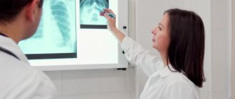
Decoding the result
Upon receipt of the X-ray results, the interpretation is carried out by the attending physician - an otolaryngologist and a radiologist. You cannot decrypt images yourself.
Photos of the sinuses will be taken and labeled indicating potential pathologies and darkened areas.
By analyzing the images, the doctor can determine:
- The presence of pathogenic foci. They appear as darkened or sub-darkened areas in photographs.
- Presence of sinusitis. In the pictures it is indicated by lightened areas of the sinuses, which is an indirect sign of the presence of mucus. It can be purulent and catarrhal.
- Nature of the disease. The doctor carries out this diagnosis only after a course of therapy prescribed earlier and if the problem has not been eliminated. They differ in sluggish, stopped, chronic and acute forms. All of them have a different nature of the course of the disease and are determined by the doctor.
- The presence of cysts and tumors in the nasal sinuses. They have clear shapes and are easily visible on an x-ray. When taken again after a while, they may enlarge, spreading to the sides.
- To compare color saturation in normal conditions and in pathology, comparisons of the color of the eye sockets and maxillary sinuses are used. Normally, their colors should match or be close in color. With pathology and the presence of disease, the colors differ sharply. This effect is called “milk in a glass.”
Types of pansinusitis
Pansinusitis can be acute or chronic. In acute sinusitis, symptoms increase quickly, within a few hours. In the case of a chronic form, the disease has a wave-like course in the form of remissions and exacerbations; symptoms are less pronounced in contrast to the acute form. Based on morphological characteristics, they are distinguished:
- Hypertrophic pansinusitis. Thickening of the mucous membrane of the sinuses, without changes in the morphological structure of the cells.
- Catarrhal. The discharge is mucous in nature, the face is swollen, the mucous membranes are hyperemic (have a pronounced red color).
- Purulent. The mucous membrane swells, due to which the evacuation of the sinus contents is blocked, secondary flora joins and a purulent process develops.
- Exudative. A mucous secretion is observed in the sinuses.
- Polypous. Develops with chronic pansinusitis and is characterized by the presence of polyps in the sinuses.
- Hyperplastic pansinusitis. There is a proliferation of fibrous tissue with the appearance of atypical cells, dysfunction of the lacrimal glands.
Only a doctor can determine the morphology of the pathological process after an examination. Depending on this, the principles of treatment will differ.
Diagnostics
Doctor — Young female consultant or ENT specialist — with a patient in her practice, examining the throat with a spatula
During examination, you can see pale skin, puffiness of the face, swelling of the nasal septum, nasal discharge. When pressing on the projection of the sinuses, pain is noted. The doctor pays attention to the nasal septum, the presence of spines, polyps, adenoids, and examines the teeth. In the CBC there is an increase in the number of leukocytes and an increase in ESR.
An endoscopic examination is carried out to examine the sinuses - with inflammation there is swelling, hyperemia, and purulent discharge in the nasal passages. CT or MRI allows you to assess the condition of the sinuses, note the level of their filling with discharge, and the presence of formations. A study of the discharge is being carried out. A swab is taken from the nasal mucosa or a biopsy obtained during endoscopy and a culture is done. This will help determine the type of pathogen and prescribe the correct treatment.
Sinus puncture is a diagnostic and treatment procedure. With its help, the contents of the sinuses are evacuated, followed by sowing, and the patient’s condition is alleviated. Along with fluid removal, antibiotics can be administered into the sinus cavity.
Is it necessary to take an x-ray to diagnose sinusitis?
An x-ray is the most informative method for examining and diagnosing sinusitis, sinusitis and related diseases. However, there are other ways to diagnose sinus diseases.
Diagnostic methods without x-ray examination
In addition to radiographs, the following methods are also used to diagnose sinusitis and related diseases:
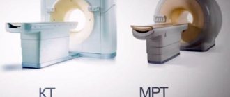
- Tomography. Magnetic resonance and computed tomography techniques allow you to examine the entire facial area from different sides. This is an effective, but rather expensive procedure with a high radiation exposure. Thanks to this diagnostic method, it is possible to visualize the size of the sinus canals and their condition. It is also possible to determine the amount of purulent accumulation and identify complications or pathologies.
- Endoscopy of the nasal and sinusoidal sinuses. Allows you to examine organs from the inside and make a conclusion. The procedure carries some inconvenience for the patient, since the diagnosis will be carried out through the natural respiratory tract. It is carried out in a stationary position and requires highly qualified specialists performing the procedure.
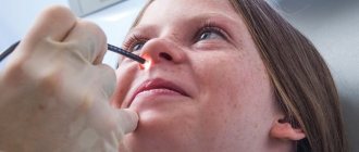
- Diagnostic puncture. To extract a sample of accumulations from the maxillary paranasal sinuses and determine the material, it is necessary to carry out a therapeutic and diagnostic puncture. The procedure is invasive and inconvenient for the patient. The procedure involves puncturing an area with a special needle that has previously been subjected to local anesthesia. The doctor pierces the wall of the sinus with it where it is considered the thinnest. The procedure can be performed on children only after reaching 6 years of age.
- Ultrasound examination allows us to determine whether pathological changes have occurred or have not yet occurred in the maxillary sinuses. If there is no pathology, then we need to look for the cause of the disease in other directions. This type of study precedes an x-ray for sinusitis. The study is of an intermediate nature and does not have sufficient effectiveness.
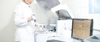
- Bacteriological diagnostics. They collect mucus from which pathogens are isolated. These can be bacteria, viruses and even fungi. It is necessary to determine their sensitivity to antibiotic medications. Microbiological procedures for the nasal and throat samples are carried out separately and using different methods. The drugs are selected separately, taking into account the microflora of the sample collection site. If the attending physician suspects the development of an allergic process, it is necessary to conduct skin tests.
- Additional consultations. It is possible to prescribe additional medical consultations if it is not possible to accurately identify the cause of the inflammatory process. It is worth considering that not only infection can cause inflammation in the maxillary sinuses. This could well be a dental disease, allergy, or intracranial complications.
- General inspection. In addition to X-ray photos, symptoms of sinusitis in adults can be detected during a general examination. While examining the patient, the ENT palpates the area of the maxillary sinus. If a person suffers from sinusitis, he will feel very painful and unpleasant sensations.
Reasons for development
Chronic pansinusitis is a complication of ordinary sinusitis. But the inflammatory process in this case spreads to the paranasal sinuses. When several sinuses are affected, polyposis pansinusitis develops. The process can be hypertrophic or purulent.
Factors causing the disease:
- Bacterial or viral infection;
- Allergy;
- Nasal polyposis;
- Trauma to the nasal septum;
- Fungal infection.
This type of runny nose requires contacting a qualified specialist, correct diagnosis and appropriate treatment. Most often, experts call the cause of the disease – infection.
How often can an x-ray be taken for sinusitis?
During such a procedure, the radiation dose to the body is 0.1-1.2 mSv, which is quite small. The equipment used during the examination greatly influences the radiation dosage.
To minimize exposure to radiation, you can visit private clinics; they are often equipped with the latest devices for this type of diagnosis. The procedures can be done quite often and in a short period of time (3-4 times of diagnosis will not cause significant harm to health).
Before scheduling an X-ray examination, the healthcare provider will review the medical record and find out the current level of radiation exposure. If the dosage exceeds the normal value, an x-ray examination will not be performed.
Photo gallery
Sinuses before and after treatment
Bilateral purulent sinusitis
Degree of filling of sinuses with fluid
Video
The video from the author of “Treatment of ENT Diseases” talks about x-rays of the nasal sinuses.
Do you have any questions? Specialists and readers of the HROMOSOMA website will help you ask a question
Was this article helpful?
Thank you for your opinion!
The article was useful. Please share the information with your friends.
Yes (100.00%)
No
X
Please write what is wrong and leave recommendations on the article
Cancel reply
Rate the benefit of the article: Rate the author ( 1 vote(s), average: 5.00 out of 5)
Discuss the article: