The inner ear is located deep in the temporal bone and is responsible for perceiving sound signals, maintaining balance and identifying the body in space. Symptoms of inner ear diseases are not always specific, and it is difficult to make an accurate diagnosis.
To understand whether the inner ear or another organ is the source of the malaise, a thorough examination of the patient should be performed.
The best method for making an accurate diagnosis is an MRI of the ear.
Advantages of the method:
- high image quality, the possibility of detailed study of the structure of the organ;
- health safety, no radiation exposure;
- no damaging manipulations are performed during the procedure;
- Due to layer-by-layer scanning, there is no effect of overlapping images on each other.
MRI clearly shows the soft tissues located in the bony labyrinth of the temporal bone pyramid. The inner ear consists of the vestibule (a small cavity bordering the middle ear), from which the cochlea extends anteriorly, and the semicircular canals of the vestibular apparatus extend posteriorly. With the help of the inner ear, a person not only perceives sounds, but also orients himself in space, instantly determining the bottom and top with any movements of the head and torso.
What can you see on magnetic resonance imaging of the ears?
What does a magnetic resonance imaging scan of the ears show:
- ear structure;
- anatomical features;
- foci of inflammation, tumors;
- condition of the vestibulocochlear (VIII pair of cranial nerves) and facial (VII pair) nerves;
- neighboring structures of the head - eyes, temporal bones, cerebellopontine angle, blood vessels.
MRI of the inner ear allows you to detect minor changes, the initial forms of diseases. Such information makes it possible to carry out timely treatment and stop the disease at the very beginning of its development.
MRI of the ear with contrast allows you to look at the inner ear in more detail and assess the extent of its damage. The method is based on the ability of the metal gadolinium, which is part of contrast agents, to give an enhanced signal from the tissues in which it accumulates. The inflammatory focus has a stronger blood supply than the healthy ear, so it is clearly visible. MRI of the ear with contrast clearly shows areas of insufficient blood flow and tumors.
Indications for the procedure
If the slightest suspicion of disease in the jaw and face area is detected, it is recommended to conduct an examination of the temporal bones or the extended protection - X-ray according to Schüller. Mayer's X-ray is usually recommended if it is necessary to view the mastoid cave or antrum. This method is a narrower method of examination. The examination is performed at an angle of 45 degrees, since the analysis represents axial protection of the auditory organ.
We recommend reading: Phlegmonous appendicitis - causes, symptoms, diagnosis and treatment
When is it necessary to conduct an X-ray examination of the temporal bones (X-ray according to Schüller):
- inflammatory process of the middle auditory region in an advanced phase;
- acute otitis with consequences;
- trauma to the auditory organs;
- cholesteatoma;
- benign or malignant formations;
- inflammatory process of mastoid cells.
Doctors advise carrying out this research method after surgical resolution of diseases of the auditory organs. The procedure will help to identify changes in the ear and assess their condition after the intervention.
Purulent otitis.
When is an MRI of the inner ear prescribed?
The examination is carried out if there are symptoms that indicate damage to the ears or surrounding tissues, so not only the ear, but also the brain is often checked. MRI of the ear allows you to confirm or exclude its participation in the formation of symptoms and conduct an accurate diagnosis.
Symptoms for which an MRI of the inner ear is indicated:
- progressive hearing loss, partial or complete loss;
- congestion, tinnitus;
- headache;
- dizziness at rest and when changing position, accompanied by nausea, vomiting, headache;
- distortion of sound perception;
- unstable balance at rest and when walking;
- head injury;
- signs of damage to the facial nerve - asymmetrical facial expressions, immobility and weakness of one half of the face;
- elevated body temperature of unknown origin;
- suspected tumor of the ears or surrounding structures.
The symptoms that form the basis for indications for MRI of the ear may be similar in diseases of the brain, blood vessels, or the ears themselves. An MRI will show the cause of the ailment with high accuracy and allow you to begin the correct treatment. The examination allows us to identify the initial manifestations of pathology, which increases the success of treatment and allows you to preserve your hearing.
An important indication for MRI of the inner ear is preparation for surgery. Depending on what the image shows, the surgeon decides on how to access the inner ear and draws up a surgical plan. In the postoperative period, the MRI method is most optimal for monitoring the stages of healing and the condition of the ears and implant.
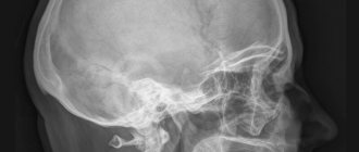
Interpretation and description of ear x-ray
In cases where a visual examination does not provide a satisfactory assessment of the patient’s health, he is recommended to undergo an x-ray examination. Since a survey X-ray of the skull will not provide the opportunity to obtain the necessary results about the condition of the middle and inner ear, radiology doctors use targeted images of the temporal bone.
Ear structure
Radiography makes it possible to project images of organs and tissues onto film using special rays. Improved digital devices allow for more accurate diagnosis and recording of studies on digital media. Since the temporal bone is quite complex, it allows you to assess the condition of all its parts.
What will an ear x-ray show? Ready-made images make it possible to assess the condition of the temporomandibular joints and understand whether there is skeletal deformation.
The benefits of the study include:
- Speed of execution, which is very important in vital situations with severe skull injuries;
- Visibility of the results, which is expressed in a picture reflecting the problem.
- General accessibility, which is ensured by the presence of an X-ray machine in every district hospital.
- The procedure is painless, which is important when examining small children.
- Economic benefit associated with low cost.
Ear examination
Cases requiring the procedure
Ear x-rays are done at any age. Does it make it pregnant? No, just like research in this way of other parts of the body. The presence of a metal implant in the patient's temporal lobe is considered a contraindication. Studying the placement of ear implants made from other materials is permitted.
X-ray of the temporal bone is used in the following cases:
- Otitis and other diseases. Suspicion of otitis media, its transition from the acute to the chronic stage. Other diseases of the middle area, such as mastoiditis. Suspicion of pneumatization (formation of air cavities) in the mastoid processes.
- Dental pathologies. It is performed before cutting out ulcers formed on the jaw.
- Tumor processes. X-rays of the middle ear are performed to diagnose tumors and other space-occupying processes.
- Injuries. Injury to the temporal region and ear structures is an indication for the procedure. Based on the results, the integrity of the skull, the presence of debris and exudate, and the degree of damage to soft tissues are assessed.
- ENT diseases. Used as an additional diagnostic method.
Methods of conducting
There is no need to prepare in any special way for radiography. Pictures are taken with the patient sitting or in an upright position. All jewelry must be removed.
X-ray according to Schuller
Extraoral (extraoral) diagnostics can be performed with portable or stationary devices. Extraoral visualization of the temporal bone is carried out using three main techniques:
- Schuller projection. Performed at a thirty-degree incline. The result makes it possible to evaluate and examine the structure of the mastoid process, understand the distribution of cells, and identify the degree of its airiness. Is a Schuller projection done when a person is standing? No, only when lying on your side. The walls of the external meatus and the areas of the sigmoid sinuses will also be reflected on the film. X-ray of the ear for otitis is carried out only according to Schüller.
- Mayer's projection. An x-ray shows the condition of the auditory bones, the space above the drum, and the mastoid cave. The picture is taken in a special projection, when the rays are directed at a forty-five-degree angle.
- Stenvers projection. The most difficult method to perform, as it is performed with a ten-degree tilt. The film shows the diagnostic results of the apex of the pyramid, temple, and internal auditory canal.
Skull shot
Decoding the final results
It is quite difficult to obtain high-quality and informative images. It all depends on the correct installation, the possibility of applying shadows and projection distortions.
Only an experienced doctor can correctly interpret the results obtained.
- with otitis media in the acute phase, the transparency of the cavities of the mastoid process and middle ear decreases;
- in case of mastoiditis, the level of airiness of the cells and the degree of destruction of the internal bone partitions are considered;
- Symptoms of otitis media in purulent form are darkening of areas of the mastoid process, sclerosis and destruction in bone tissue;
- Choleastomy is manifested by an enlargement of the mastoid cave and cavities formed in the bones. It is worth noting that small tumors may not be detected during a routine examination, while the diagnosis of tumors of a larger diameter does not cause any difficulties;
- skull injuries show up well on film. Micro damage is indicated by dark spots formed due to the accumulation of blood in these areas.
Two X-ray projections
Today, X-rays are significantly inferior in information content to the results of computed tomography and magnetic resonance therapy. These methods are more accurate and less harmful. However, due to their high cost, they may be available only to a certain category of patients.
Source: https://rentgenovski.ru/vidy/opisanie-rentgena-uha
What does an MRI scan of the ears show?
Good quality images allow you to thoroughly study the structure of the anatomical structures of the area under study, note pathologies and developmental anomalies, and determine the stage and extent of the lesion.
MRI of the inner ear helps in diagnosing the following diseases and pathological conditions:
- neuritis of the vestibulocochlear (auditory), facial nerve;
- inflammation of the inner ear (labyrinthitis) in acute or chronic form;
- acoustic neuroma;
- congenital and acquired deformations of ear structures;
- Meniere's disease;
- meningioma of the cerebellopontine angle;
- otosclerosis of the bone labyrinth;
- vascular pathologies of the temporal region;
- complications of purulent labyrinthitis - meningitis, encephalitis, abscess;
- traumatic injuries to the ears and the area next to them.
Since the examination does not expose the body to radiation, it can be performed an unlimited number of times. Treatment of diseases is much more effective if it is possible to control the body’s reaction and, if necessary, make adjustments or change tactics.
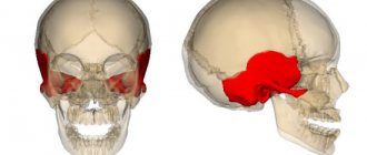
Neuroma of the vestibulocochlear nerve
The most common benign tumor of the cerebellopontine angle, its development may be due to a hereditary predisposition. Acoustic neuroma develops from Schwann cells surrounding the nerve fiber and is an irregularly shaped formation surrounded by a capsule. The disease practically does not become malignant, but as it increases in size, the tumor affects the inner ear and brain, causing clinical manifestations.
The disease is characterized by decreased hearing, impaired balance, tinnitus, headache, and numbness of the face. Less commonly observed are pain in the ears, paresis of the facial nerve, double vision, and involuntary frequent eye movements (nystagmus). Most often, symptoms are observed on one side.
In most cases, acoustic neuroma grows slowly, and symptoms do not always appear clearly. MRI of the auditory canal allows you to detect a tumor and begin its treatment, preserve hearing and avoid compression of brain structures and intracranial hypertension.
Labyrinthitis (internal otitis)
Inflammation of the inner ear occurs as a result of infection entering from the middle ear or from other sources through the circulatory system. Infection can get into the ears due to injury to the eardrum or temporal bone. By nature, labyrinthitis can be serous or purulent, acute or chronic.
With the development of internal otitis, hearing decreases (impairments can be irreversible, especially with purulent labyrinthitis), attacks of dizziness with nausea and vomiting, nystagmus occur, balance and coordination of movements are impaired. Internal otitis is dangerous due to complications when the infection spreads to the brain and meninges with the formation of an abscess and meningitis.
Severe forms of internal otitis may lead to removal of the labyrinth on the affected side and complete hearing loss. If symptoms of damage to the auditory analyzer appear, an MRI of the ear should be performed - this will identify otitis media and stop its spread.
Meniere's disease
The disease is characterized by attacks of dizziness in combination with tinnitus and progressive hearing loss. The symptoms are caused by increased pressure in the inner ear due to an increased amount of endolymph, the fluid that fills the labyrinth cavity. The causes of Meniere's disease are unknown.
The disease is characterized by three main symptoms:
- sudden dizziness, the attack lasts 4-5 hours, accompanied by nausea and vomiting;
- unilateral low-frequency noise in the ear, intensifies during dizziness;
- periodic unilateral hearing loss.
An MRI of the inner ear in Meniere's disease is performed primarily to exclude other diseases that may present with similar symptoms.
Sensorineural hearing loss
The disease occurs when the ears are affected or is a consequence of other diseases. In this case, there is a loss of the sound-perceiving function of the nervous apparatus of the ear with a gradual decrease in hearing acuity until it is completely lost.
The causes of hearing loss may be the following:
- infectious diseases - influenza, measles, tick-borne encephalitis, mumps, meningitis, syphilis;
- acute intoxication with ototoxic substances at home and at work;
- taking certain antibiotics, anti-inflammatory drugs, diuretics, antidepressants;
- cerebrovascular accident due to arterial hypertension, atherosclerosis;
- genetic predisposition;
- long-term exposure to industrial noise exceeding maximum permissible levels.
MRI of the ear canal will allow us to establish the true cause of hearing loss and make a differential diagnosis. An examination is necessary to exclude or confirm ear diseases as a cause of hearing loss.
X-ray of the ear and temporal bone: what will show according to Stenvers, Schuller and Mayer
Radiography is a time-tested diagnostic technique. It is used by otolaryngologists, dentists, and surgeons. X-rays of the ear and temporal bone reflect structural changes to the same extent as modern computed tomography. The diagnostic procedure is in demand for injuries or for adjusting postoperative rehabilitation.
When is an ear x-ray prescribed: indications
X-rays are used in the following situations:
- The medical institution does not have computed tomography or MRI machines.
- An x-ray of the ear for otitis media is performed when complications develop or a suspicion of tumor formation arises.
- When examining the condition of sensors implanted in the inner ear.
- If the injured person has fractures of the temporal part of the skull.
- To adjust the treatment of choleastomy.
- With mastoiditis - an inflammatory process in the cavity of the mastoid branch of the temporal bone.
- When there is a need to exclude or confirm a congenital anomaly.
- To determine the cause of hearing loss due to ear diseases.
When there is purulent inflammation of a tooth, the dentist needs to know whether the exudate has penetrated into the adjacent areas. This is clearly visible on an x-ray. The method allows you to detect cavities filled with air.
How the research works
Preparation for x-ray of the temporal bone consists of removing metal objects. Before the procedure, rings, bracelets, crosses, and dentures are removed. The radiologist is warned about the presence of implants. Electrical conductors distort the image, which leads to an erroneous diagnosis.
If X-rays of the temporal bones have already been performed, the patient should be provided with previous images so that the doctor can trace the dynamics of the pathological process.
Radiographs are taken in the supine position. The following methods are popular:
- According to Stenvers.
- According to Schuller.
- According to Mayer.
Laying according to Stenvers
The method makes it possible to identify destructive inflammatory changes in the hearing organs. The image shows the tip of the cone-shaped temporal bone and shows the internal auditory canal. X-rays of the temporal bones according to Stenvers are performed in a transverse projection.
The patient is placed on his stomach, the cheekbone and eye socket are pressed against the plane of the table. They slip in a cassette. A series of photographs are taken. The doctor guides the patient's head rotation to obtain images from different angles. The quality of radiographs depends on the qualifications of the doctor.
Laying according to Schüller
Used in maxillofacial surgery and dentistry. The shooting is carried out in a lateral, oblique position. A fist is placed under the healthy half of the head. The cassette is placed behind the ear and pictures are taken from several positions. Laying according to Schüller reveals destructive processes in which the bone is destroyed.
Laying according to Mayer
The technique is used to diagnose otitis media of the middle ear. The patient is placed on the table, arms are placed along the body. Mayer's styling consists of a series of shots with a cassette located behind the ear at an angle of inclination of the head relative to the table of 45 degrees.
Important! If internal inflammation is suspected, photographs are taken in the Stenvers position. To diagnose middle ear pathology, the Mayer method is used. If bone tissue destruction is suspected, the Schüller method is used.
What will an x-ray of the ear and temporal bone show?
The radiologist takes pictures, describes the results and gives them to the patient. The patient receives a transcript within 24 hours. Treatment is prescribed by an otolaryngologist, dentist, oncologist or surgeon.
An ear x-ray shows the following symptoms:
- darkening of the cells of the mastoid branch occurs with otitis media, accompanied by suppuration;
- decreased transparency in the medial ear area is a symptom of an inflammatory process;
- if an x-ray of the ear reveals bone tissue cavities, cholesteatoma is diagnosed;
- when melting of the septa of the mastoid cavity is detected, a diagnosis of mastoiditis is made.
X-ray of the temporal bone shows the following pathological signs:
- Ankylosis. The picture shows that the temporal bone has fused with the lower jaw.
- Osteophytes. Articular cartilage is destroyed, bone tissue grows uncontrollably.
- Subchondral cyst. A round dark cavity forms under the surface of the joint.
- Flattening of the articular joints of the temporal bone and jaw.
Important! If X-rays do not provide comprehensive information, additional studies are prescribed.
During an X-ray examination, a person receives a dose of radiation equal to 4% of the annual limit. Therefore, doctors do not recommend more than three sessions per six months. Alternative methods are MRI and computed tomography.
Source: https://DiagnozPro.ru/rentgen/kosti-sustavy/visochnoj-i-uha-ukladki
Contraindications for magnetic resonance imaging
Obtaining MRI images is possible due to the interaction of a high-power electromagnetic field with the structures of the body. If you place objects that have ferromagnetic properties in a magnetic field, they will react in a certain way. The special magnetic susceptibility of ferromagnets forms the basis of contraindications for MRI.
The examination is prohibited for people with implants containing a magnet and prostheses made of iron, cobalt, nickel, and steel. In a magnetic field, objects implanted into the body are displaced and cause pain and internal injuries. Implants can fail and distort the image.
MRI of the ear is not performed on patients who have inside the body:
- pacemaker;
- insulin pump;
- cochlear implant;
- hemostatic vascular clips;
- metal fragments after injury.
Modern prosthetics and implants can be made from MRI-compatible materials. If there are contraindications, it is necessary to study their characteristics to determine the feasibility of the procedure.
There are relative contraindications that, under certain conditions, allow examination. These include pregnancy in the first trimester, serious condition of the patient, age under 7 years, claustrophobia, mental illness, epilepsy.
MRI of the ear with contrast is a safe procedure, but it is contraindicated in people with severe renal failure and at any stage of pregnancy. Breastfeeding mothers can be administered a contrast agent, provided that feeding is stopped for 1-2 days - until the drug is completely eliminated from the body.
Methodology
Ultrasound of the ear is performed with patients lying down, having previously lubricated the area under study with a special solution that acts as a connecting medium.
Preparation
No special preparation is required for the examination. It is better for children to calm down before a planned diagnosis, show or tell them what will happen so that they are not afraid.
Procedure
Decoding the results
When performing an ultrasound, the doctor measures the necessary parameters, which are entered into a special form. An experienced specialist interprets the results and makes a preliminary diagnosis based on the indicators.
Preparing and conducting a study using a magnetic resonance imaging scanner
At the appointment, it is advisable to take the results of all previous tests and studies related to the current state of health, as well as advisory opinions and extracts from medical records. No special preparations are required.
If an MRI of the ear with contrast is performed, a renal function test should be performed first to rule out renal failure. Women should be aware of the possibility of pregnancy - contrast agents have a negative effect on the developing fetus. A contrast agent should not be administered if an allergic reaction to its components has previously been documented. The examination is carried out on an empty stomach.
Metal objects and magnetic devices must not be brought into the room where the research is being conducted. The patient changes into light disposable clothing, having previously removed jewelry, watches, belts, removable dentures, clothing with metal parts and leaving a mobile phone, money, and cards outside the office. The subject is positioned on a table that slides into the body of the tomograph. Most devices are not designed for body weights exceeding 130 kg. When performing an MRI of the ear, a special coil is placed on the area under study to read the signal. The procedure lasts about 20 minutes.
If an MRI of the ear canal is performed using a contrast agent, the drug is administered intravenously after the first scan. After a few seconds, you can rescan. The contrast agent rarely causes side effects or allergic reactions. You may feel warmth or tingling at the injection site.
The examination can be stopped at any time at the request of the patient if he becomes ill during the procedure. People suffering from mild claustrophobia are recommended to take sedatives half an hour before the test. In some cases, a loved one may be present during the procedure.
How is this research going?
Computed tomography, regardless of which part of the body is examined, is carried out according to the following scenario:
- The patient is placed on a mobile table, which is placed in the tomograph machine during the scan.
- The body, and in this case the head, is fixed with special straps and bolsters so that the patient does not make involuntary movements during the scan.
- When the table is placed in the cylinder, the tomograph ring begins to rotate around the patient; its movement may be accompanied by clicks or slight noise.
- The images will be displayed on the screen; after evaluating and studying them, the doctor can draw a conclusion about the presence or absence of diseases.
- The procedure without contrast takes about five minutes, CT with contrast can last up to 20 minutes.
Features of CT with contrast
To carry out this scan, a special contrast agent is often used - a special pharmacological preparation, which can be based on various harmless drugs. For each patient, this drug is selected individually by the attending physician; its choice depends not only on the patient’s health status, but also his weight is taken into account. Most often, an iodine-based drug is used as a contrast agent; it is administered intravenously in advance of the scan. The contrast quickly colors the vessels in a reddish tint, which makes them more visible on the screen. The body is released from this drug on its own within a few days, it is excreted by the kidneys, therefore it is not used for patients with renal failure.
What does the result of magnetic resonance imaging of the ears show?
Preparing a report can take from one hour to a day. MRI images of the inner ear and surrounding brain structures are analyzed by a radiologist. The patient receives a description of everything that the MRI shows and a conclusion. The photographs can be received electronically and the most significant images can be additionally printed on film.
A healthy MRI of the ear is characterized by:
- absence of foci of increased or decreased intensity;
- the ventricles of the brain are not displaced;
- the internal auditory canals on both sides are of a normal shape, not expanded;
- the course of the nerves (vestibulocochlear, facial) is not disturbed, the diameter is within normal limits;
- the configuration of the cochlea and semicircular canals is typical;
- the cerebellopontine angles on both sides are unremarkable;
- The pyramids of the temporal bones are not changed.
The administration of a contrast agent should not be accompanied by pathological accumulation of the drug in any areas of the inner ear.
What research shows in some diseases:
- meningioma looks like a volumetric formation of irregular shape with clear, uneven edges, the structure of the formation is heterogeneous, cysts may appear inside, there is an area of edema around it, the ventricles of the brain are deformed by the tumor;
- neuroma of the vestibulocochlear nerve is characterized by the appearance of a space-occupying formation that extends into the internal auditory canal, the cavity of the fourth ventricle of the brain is deformed;
- Meniere's disease may appear on magnetic resonance imaging as the absence of the endolymphatic duct.
Depending on what the study shows, the patient may need additional consultation or an appointment with a specialist doctor. This could be an oncologist, surgeon, neurologist, otorhinolaryngologist or audiologist - a doctor who treats hearing disorders and specific lesions of the auditory analyzer.
Preparing for a CT scan
If you plan to perform a CT scan without contrast, no special preparation is required. To avoid an adverse reaction to the injected substance, when studying using contrast, the patient must refrain from eating and drinking 4 hours before the scheduled procedure. In the case of a study with contrast, the patient needs to donate blood the day before for a biochemical analysis - to determine creatinine and blood urea; The contrast agent is excreted by the kidneys and before starting the study, you must ensure that they are functioning normally.
Where can I get an MRI of the ears?
Choosing a clinical diagnostic center for magnetic resonance imaging of the ears is not easy to do. Our website contains information about 18 clinics in Moscow where you can undergo an examination of the inner ear at the best prices. Any patient can independently compare and choose a medical center or call by phone to receive a free consultation and make an appointment at a convenient time at any of the proposed clinics.
On the website you can find the following information:
- contact details of the clinic (address, metro);
- detailed price list for services;
- type of equipment used (closed or open tomograph);
- operating hours of the institution;
- availability of free places for registration;
- special offers and promotions;
- possibility of undergoing tomography for children;
- patient reviews.
The average price for magnetic resonance imaging of the ears is 4,400 rubles. The price range is between 3,600-10,500 rubles. When you make an appointment through our website for your first appointment, you can get a discount of up to 1,000 rubles.
Doctors performing a CT scan of the ear
Chervonenko Svetlana Vladimirovna Radiologist Make an appointment
Filippov Vasily Vasilievich RadiologistExperience 38 years Make an appointment
Syubaev Roman Borisovich RadiologistExperience 16 years Make an appointment
Filatov Dmitry Aleksandrovich Radiologist, Head of the Department of Radiation Diagnostics Make an appointment
Advantages and disadvantages
The main advantages of ultrasound examination are high information content and safety. The procedure has no contraindications since it is not invasive. Even age restrictions are excluded. Ultrasound is allowed, including for infants. The advantages of this method of studying the vascular system of the brain include the following:
- painlessness;
- the ability to study the area under study in several projections;
- good visualization of soft tissues;
- possibility of frequent use due to its harmlessness to the body;
- reliability due to real-time implementation;
- low cost.
Some objects are still difficult to visualize due to their complex projection layering. This is one of the disadvantages of ultrasound diagnostics. Other disadvantages of this procedure include the following:
- lower spatial resolution compared to magnetic resonance and computed tomography;
- excess weight can complicate diagnosis, since adipose tissue absorbs part of the ultrasound;
- difficulty visualizing bone tissue.
Decoding the results
X-ray images are prepared within 1 day. If the result is high-quality, the doctor determines the pathological conditions that have arisen.
What diseases are detected using x-rays?
- If the patient has acute otitis, the transparency in the area of the middle ear, as well as the cells of the mastoid process, will be reduced.
- In acute mastoiditis, pneumatization of the mastoid cells will be reduced or completely absent.
- Foci of bone tissue destruction signal advanced otitis media.
- If the mastoid areas are darkened, this may indicate purulent inflammation.
- Cholesteatoma is determined by the enlargement of the cave area.
- Large-scale formations are visible on their own in the image.
X-ray examination is recommended for all patients. This method accurately identifies pathological changes and conditions in the ear.
How to do a vascular ultrasound
Before the ultrasound begins, the patient lies down on the couch and rests his head on a small pillow. At this moment he needs to relax and restore his breathing. Next, the specialist will feel the vessels to determine the degree and depth of their pulsation. After this, the study begins using a special sensor that emits ultrasonic waves.
Dopplerography, duplex scanning and three-dimensional ultrasound have almost the same technology. During the procedure, the doctor may ask the patient to do the following:
- take a vertical position;
- hold the breath;
- blink faster;
- Breathe frequently and deeply (hyperventilation).
Another functional test is pressing the vessels with your fingers. This helps to more accurately diagnose the mechanism of blood flow regulation. Before starting the diagnosis, a special gel is applied to the area under study, which is washed off at the end of the session with warm water or removed with a damp cloth. The ultrasound diagnostic process can take about 45-50 minutes. It does not bring any discomfort or pain. The difference between the types of ultrasound is only in the places where the specialist passes the sensor:
- During a transcranial study (of vessels located in the head), the sensor is placed in those areas of the skull where ultrasound more easily penetrates bone tissue: above the eye sockets, in the back of the head, in the area of the temporal bone.
- With extracranial ultrasound, vessels are studied before they enter the cranial cavity. To do this, the sensor is placed in the neck area.
Anatomical features of the ear
First of all, the ear is a paired organ. It is located directly in the temporal part of the human skull. The main part of the organ is located deep, so only the auricle can be visually seen. Three main components can be distinguished:
- external;
- average;
- and inner ear.
The outer one is made in the form of cartilage, covered with skin in each person. The earlobe is located in the lower part; it consists of adipose tissue. The outer part of the hearing organ is quite sensitive. Any, even the slightest, mechanical damage can serve as deformation. One and two ears are the primary receiver of mechanical vibrations, converting sound waves into sound at a distance of up to 25 meters.
Next is the auditory canal, which produces earwax, which can protect the inner ear from germs and viruses. Vibrations of sounds, passing through the ear canal, hitting the eardrum, enter the middle ear.
The middle ear consists of auditory ossicles; these ossicles convert incoming noise and transmit it to the internal part of the organ. If you carefully examine their arrangement sequence, you will notice a chain through which the incoming sound is transmitted. A violation in one of the links leads to diseases of the auditory organ. The middle ear is connected to the nasopharynx, hence the feeling of stuffiness in the ears, due to an increase or decrease in pressure.
The inner ear is the most complex in structure; it is called a labyrinth, so otolaryngologists must carry out diagnostics. The shape resembles a snail. His condition can be judged from MRI images.
The human ear is responsible for the ability to keep the body in balance and convert mechanical vibrations directly into sound. It is also responsible for a person’s ability to concentrate on any object, which means it helps with visual function.
Now we can conclude that the ear is the most important organ, the condition of which must be monitored and, in case of any violations, immediately go to the doctor for a referral for examination.
Contraindications for CT scanning of the ear
For children under 14 years of age, due to limited radiation exposure, the study is carried out for health reasons. However, a doctor may prescribe this diagnostic method if other methods are not informative enough.
It is not performed on pregnant women, as even a small dose of radiation can negatively affect the development of the fetus.
CT with contrast is prohibited for patients who are allergic to iodine, and is also contraindicated for people suffering from renal and liver failure.
People with mental disabilities and in an unconscious state - because During the examination, the patient should lie still.
what is ultrasound of cerebral vessels?
This is the name of a modern method of assessing the state of blood flow in the capillaries, veins, and arteries that supply blood to the brain. Its ultrasound examination is carried out using different methods. The first experiments with this procedure were carried out back in 1955. Since that time, ultrasound examination has been improved, so it has become safe, accessible and informative. This technique is highly accurate because it helps to study any organs or tissues in detail.
Ultrasound examination of the head can be performed using three different methods. Each is based on an image that is displayed on the screen due to the movement of a special sensor across the human body. In this case, the patient does not feel pain or discomfort. A timely ultrasound of the vessels of the head and neck helps:
- identify the early stages of impaired blood supply to brain tissue;
- prevent oxygen starvation, as well as stroke, heart attack;
- monitor patients with vascular pathologies;
- evaluate the effectiveness of prescribed treatment for diseases associated with arteries or veins.
How is an MRI of the hearing organ performed?
It is recommended to carry out diagnostics on devices with field induction from 1.5 Tesla (high-field tomographs), since less powerful equipment does not provide complete information about possible hearing impairments.
The ear scanning procedure does not require preparatory measures and is carried out similarly to other types of MRI - the patient is placed with his back on the tomograph table, a special coil is put on the area under study, and in the case of a contrast procedure, the drug is injected into a vein through a syringe injector. After this, the table with the patient moves inside the tomograph and scanning begins.
The duration of the study is approximately 20 minutes, with contrast enhancement - about half an hour.
Which is better: MRI or CT
During MRI, the patient is not exposed to ionizing radiation, as with X-ray methods, and there are no restrictions on the number of procedures performed. Computed tomography gives a large radiation dose to the body and is not recommended for children and pregnant women.
In each clinical case, the choice of method remains with the doctor, so it is important to consult a specialist if symptoms appear. After examining the complaints and checking the ear using available methods, the doctor gives a referral for the most appropriate examination.
Computed tomography is more informative for injuries and damage to the skull, damage to bone structures, and accumulation of pathological fluid in the middle ear cavity. MRI will more accurately show the condition of soft tissues, tumors, inflammatory processes, and vascular diseases.
Types of CT ear
There are two main ways of performing computed tomography of the ear:
- without the use of a contrast agent, i.e. native scanning. This diagnostic method is the main one and, as a rule, answers all questions. The scanning step (or the thickness of the resulting slices) is 0.5 mm, allowing you to study the smallest structures of the ear (auditory ossicles, vestibular apparatus);
- with the use of contrast - prescribed for the study of tumor processes in the ear (in order to more accurately determine the nature and extent of the space-occupying formation) and a detailed study of adjacent tissues and vessels. To do this, a special iodine-containing substance is injected into the patient’s body, which passes through the vessels and accumulates in the tissues of the area being examined.