X-rays of the foot are important to prevent various injuries and diseases. Using this diagnostic method, you can determine not only the presence of diseases, but also identify possible complications after injuries and bruises. If a person feels severe pain while walking, he needs to have an x-ray of his foot and leg, which will help accurately determine the condition and identify possible diseases. Most often, two images are enough to establish a diagnosis, but it is best to take x-rays from different angles, as this will give a more detailed picture.
Indications and contraindications
An x-ray of the foot is necessary in the following cases:
- recent injury;
- severe pain and discomfort during movement;
- deterioration of joint mobility;
- increase in bone size;
- severe swelling;
- fractures, bruises.
This diagnostic method also has contraindications. Doctors prohibit X-rays if:
- a woman is carrying a child (this especially applies in the early stages, when the child’s organs and systems are just being formed; in later stages, the procedure is carried out only with the permission of an obstetrician-gynecologist);
- severe bleeding;
- inadequate condition of the patient.
Is special training needed?
This procedure does not require any preparation. It must be remembered that if an x-ray of the leg was previously taken with a load, and after therapy a repeat diagnosis was prescribed, then it is important to take the second x-ray in the same position as the first. To carry out the procedure correctly, you need to remove shoes, jewelry and clothing that have metal parts.
The essence of the method
Diagnostic radiography allows you to analyze the structure, position and structural features of internal organs by passing directed x-rays through them. Due to the different densities of the tissues under study, the flow of electromagnetic waves is unevenly attenuated and scattered, which makes it possible to obtain a shadow image of the object at the output. The radiation passing through the biological medium is recorded on a special photodetector: cassette, electronic matrix. The projection of the organ or its area being studied is formed on an X-ray sensitive film or monitor screen. The object of study is the resulting photograph or digital image. An orthopedic surgeon is involved in X-ray morphometry or interpretation of the examination results.
The reliability of the method is directly related to the accuracy of the resulting projection of the body part being studied, which depends on the quality of the equipment, film and reagents, as well as diagnostic conditions. Random movements of the patient during the examination can also reduce the clarity of the image.
Features of diagnostics
Patients with unpleasant sensations in the leg undergo x-rays of the foot in two projections:
- straight - to obtain such an image, the patient stands on a cassette with X-ray film with one leg (on which he does not lean); a direct photograph is taken;
- lateral – the picture is taken from the side, and the ankle joint is also captured; The patient in this case also rests on only one leg.
Pictures in 2 projections are taken first for one leg, and then for the second. Normally, bones should be homogeneous in structure and intact.
Standard
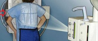
This diagnostic procedure is carried out quickly. The patient places the bare leg in which he feels pain on a special disc-stand. At the same time, the doctor checks that no jewelry or other unnecessary objects remain on the patient’s body. The healthy leg bends at the knee. With this position, the body weight is transferred to the sore leg; An x-ray is taken.
The nurse places the X-ray cassette so that it is located along the leg at the very top and is pressed with a special weight. The central beam hits the center of the cassette; during the procedure, the patient's body is covered with an apron on top. This is important to protect his organs from radiation. Pictures are taken in the following projections:
- anterior-posterior;
- dorsoplantar;
- oblique, which is at different angles;
- lateral.
After receiving the images, the doctor carefully examines them and makes an accurate diagnosis. If a doctor takes an x-ray of the foot to check for flat feet, then in the image he pays special attention to the shape of the leg and the degree of curvature.
X-ray of the foot with weight bearing
This diagnostic procedure is most often used in surgery, traumatology and orthopedics. The test is used to identify diseases and disorders in the foot. Using x-rays with a load, you can determine not only injury and pathology, but also obtain accurate information about the structure and condition. The doctor can also see all the anatomical changes in the foot in the image.
Nowadays, this method of examination is the main one in diagnosis; it helps to accurately diagnose the patient and select more effective treatment. The procedure is carried out using an image in 2 projections. In the clinic, the doctor will independently determine which projections to use, based on the symptoms and distinctive features of the lesion:
- lateral image – helps to obtain detailed information about the condition of bones and joints;
- anterior-posterior, in this case a picture of 2 feet is taken;
- oblique - done at an angle of 45°;
- dorsal-plantar image – performed when the tibia is deviated, the beam passes vertically.
In children
The child is prescribed x-rays of the foot and leg in the following cases:
- falling, injury at birth;
- for problems with the process of bone formation, a lack of minerals in the bones of children or diseases of bone tissue;
- foreign objects entering the respiratory organs or digestive system;
- head injury, cancer, congenital disorders in the structure of the skull bones;
- suspicion of the presence of Koch's bacillus, bronchial asthma, inflammatory processes in the lungs, arthrosis, arthritis;
- before surgery;
- For children under 1 year of age, X-rays are performed to detect dislocation or subluxation of the hip joint with complete displacement of the femoral head.
X-rays are prescribed at a young age only when other diagnostic procedures do not provide the necessary information. Modern examination methods, based on X-rays, use a small amount of radiation. They are not dangerous to the patient's health.
In older people
The accelerated aging process of the body of an elderly person occurs against the background of metabolic disorders and problems with tissue renewal, so old people in most cases suffer from diseases of the musculoskeletal system.
The treating specialist prescribes radiography if there is pain, as well as for preventive purposes. The number of procedures will be determined by a specialist based on the patient’s condition.
During pregnancy
For a child actively developing and growing in the womb, X-rays pose a particular danger, as they negatively affect the formation of organs and systems in the embryo. It is most dangerous to use such a diagnostic procedure in the early stages of pregnancy. An X-ray can be performed on a pregnant woman only after consultation with a treating specialist.
Structure
The skeleton of the foot and joints is considered incomplete without muscle function. The main operating and actively working muscles are located in the ankle, foot, and lower leg. Together, the work of all muscles allows a person to fully move.
- Calf muscles - in the front of the calf is the tibialis muscle, which is responsible for flexion and extension of the feet. Thanks to the proper functioning of these muscles, a person is able to make extension movements with his fingers. This section also includes the following types of muscles: peroneus brevis and peroneus longus. They take on the job that is responsible for performing lateral flexion of the foot. The back of the tibia is responsible for plantar flexion. The triceps, gastrocnemius, and soleus muscles are involved here. It is this part that is subjected to serious daily stress.
- Foot muscles - are a dorsal muscle group that is responsible for the extension of the small toes (all four small toes, except the big one). Additionally, there are several small muscles located on the sole of the foot. They are responsible for abduction, adduction and full flexion of the toes.
What does a foot x-ray show?
Using radiography, a doctor can detect the following diseases in a patient:
- congenital disorders in the formation and structure of the osteoarticular apparatus;
- arthrosis, bursitis, varicose veins;
- foot injuries and dislocations;
- problems with the structure of the limb;
- fractures;
- cracks;
- hallux valgus;
- flat feet;
- gout.
conclusions
X-rays of the foot are used as a preventive diagnostic procedure, as well as to identify the presence of various abnormalities in the patient and monitor the process of his recovery. The radiation exposure provided during this examination does not allow it to be used to assess the structure of the foot tissues often enough without harm to the patient’s health. There are alternative examination methods, but radiography is used most often due to its low cost, information content and ease of implementation.
Decoding the results
A properly performed x-ray of the foot will show the structure of the bones and the condition of the bone tissue. After receiving the image, it first goes to a radiologist for review. He does not make an accurate diagnosis, but only provides a detailed description of all problems in the structure of the leg identified during the procedure. Afterwards, the image goes to the attending physician, who analyzes it, makes a diagnosis and prescribes suitable and effective treatment.
For longitudinal and transverse flat feet
A thorough analysis of the image is especially important if you have flat feet. To identify degrees 1,2 or 3 of longitudinal flatfoot, a specialist determines the angle of the arch of the foot. In the normal state, the angle of the arch does not exceed 130°, and the height of the arch reaches 3.5 cm. If transverse flatfoot is suspected, special attention is paid to examining a direct photograph of the foot. The result is considered normal if only the heads of the 1st and 5th metatarsal bones are adjacent to the support.
Technologies and devices in modern radiography clinics are completely safe and cannot threaten human health. The radiation in this case is minimal and, if all recommendations are followed, does not affect the human body in any way. To prevent possible pathologies and problems with the foot, it is important to carry out X-rays in a timely manner.
When is it appointed?
X-rays of the feet are prescribed by a doctor if there are patient complaints such as:
- Pain in the foot;
- Swelling;
- Deformed lower limb;
- Changed skin color;
- Impaired ankle mobility;
- Calf cramps.
Pathologies for which x-rays of the feet are prescribed
An X-ray allows you to see pathological foci of bone tissue, ligaments, joints, and make an accurate diagnosis. On its basis, a conclusion is given about the nature of the disease, whether it is congenital or acquired. The doctor determines the presence of:
- Deformations resulting from inflammatory processes;
- Heel spurs;
- Damage resulting from injuries of varying severity;
- Flat feet;
- Congenital and acquired bone anomalies;
- Clubfoot;
- Osteoporosis;
- Deformed big toe;
- Heel, hollow foot.
On a note!
To take a control x-ray of the foot after treatment, you need to take the same position as the previous time.
How often can a foot x-ray be taken?
Despite the fact that every person’s body is resistant to ionizing rays, experts identify some standards that are important to follow.
X-ray examination of the feet at an early age is allowed no more than 5 times a year, since the child’s body suffers the most from ionization. Although there are some modern devices that provide minimal radiation, which is almost safe for the child, such equipment is only available in good medical institutions.
In adults, the frequency of foot x-rays will directly depend on their age, individual characteristics, stage of the disease and symptoms of the lesion.
Functions
The human foot performs 3 main functions:
- Support. This function is explained by the ability to easily resist and prevent reactions when vertical loads are applied. When walking, this function is pushing. This task of the foot is the most difficult, since it simultaneously uses both functions - balancing and springing. As this function worsens, a person begins to suffer from pain in the ankle when running or jumping.
- Spring. Aimed at smoothing out shocks during physical activities (running, jumping, walking). With low arches, a person may suffer from diseases of the lower extremities and spine. Internal organs can also be injured.
- Balancing. Aimed at adjusting the posture of the human body during movement. A healthy foot can spread out and embrace the underlying surface, thereby giving a person the opportunity to feel the area where the foot is placed.
All functions of the foot interact with each other during active physical activity. If one of the functions is violated, the remaining two are automatically violated.
Clinical picture of longitudinal flatfoot
There are three types of longitudinal flatfoot, which differ in the severity of symptoms and changes:
- First type. The initial stages of deformation, the pathology is mild, the angle of the longitudinal arch of the foot varies from 130 to 140˚. It is impossible to visually determine the deformation. Patients complain of symptoms associated with a feeling of fatigue after long walking and periodic swelling in the feet. There is no pronounced pain syndrome; mild pain appears when pressing on the area of the distal foot.
- Second type. The deformity is moderate; upon examination, the arch angle ranges from 141 to 156˚. There is a visible flattening of the feet, and with a detailed diagnosis - changes in the arthritic nature of the metatarsal bones. There is an increase in pain even after minor physical activity. In this case, the pain spreads throughout the lower leg, right up to the thigh.
- Third type. It is characterized by pronounced flat feet, where the arch angle is more than 157˚. Clinical picture: constant pain, swelling in the legs, development of arthrosis, difficulty moving. The patient cannot use ordinary shoes; there is an urgent need to wear special orthopedic ones. In advanced cases, deformation of the ankle joints occurs.
Please note that normally, the arch angle should be 125-130˚, which indicates the absence of a deforming process.
Benefits of the study
Modern radiography has the following advantages:
- low radiation exposure;
- possibility of digital recording;
- painlessness;
- Possibility of carrying out for children and pregnant women;
- non-invasive;
- quite high information content.
7 minutes Author: Lyubov Dobretsova 6187
The evolution of man, which ensured walking on two legs, gave him not only additional opportunities and conveniences, but also became the cause of many diseases of the musculoskeletal system. Constant load on relatively small organs, such as the feet, often leads to the occurrence of various pathologies, and due to the nature of their functioning, they are susceptible to multiple injuries.
X-ray diagnostics have been successfully used for many years to identify and differentiate diseases of these areas of the lower extremities. An X-ray of the foot allows the doctor to obtain enough information in a short time to make a diagnosis, without exposing the patient to unpleasant or painful sensations. And if you consider that the procedure does not require preparation and is affordable for everyone in need, then it can safely be considered one of the most irreplaceable.
Types of radiography
Foot X-ray has the following types:
| Variety | Peculiarities |
| Side shot | Can be performed with or without load. The study helps assess the functionality of the foot |
| Oblique shot | Performed in both projections at an angle of 45 degrees |
| Plantar | The photo is taken from the sole side |
| Front shot | The study provides information about the forefoot |
Worth knowing! Despite the fairly high information content, if an oncological process is suspected, this study is not enough. In such a condition, the patient will be prescribed a CT or MRI diagnosis.