Since over the last 2 decades, progressive gastrointestinal diseases have become more common in people, when the corresponding symptoms appear, doctors prescribe their patients a time-tested research method called abdominal x-ray. This type of medical non-invasive manipulation is based on the effect of ionizing rays on the body, which “transparent” a certain area of the body and reflect the patient’s condition at the current moment in time, visualizing the most important structures - organs and tissues.
Main types of diagnostics
At the moment, the study is divided into only three main types, each of which is widely used in medical practice:
| Name | Short description | «+» | «-» |
| Plain radiography of the abdominal cavity | Only one main image is taken, reflecting the general condition of the pelvic bones, organs located below the diaphragm, and major large tissues. This type of diagnosis is often carried out during routine medical examinations. | relatively low radiation dose, speed (the procedure lasts a maximum of 1-2 minutes) | insufficient information content |
| Contrast radiography | Before the X-ray session begins, the patient is asked to drink a special composition, which will quickly spread throughout the body through the bloodstream and color its barely noticeable components. In rare cases, a person is given a substance through a tube | high level of visualization of dense tissues, detection of tumor formations of any stage | duration (a session can take up to 2 hours), rare occurrence of side effects (headaches, weakness, etc.) |
| X-ray | In this case, the image of the organ system is displayed on a special screen - a small sheet of thick cardboard on which special fluorescent particles are evenly distributed. In new generation devices, the image can be reflected on a computer monitor in real time | short duration (up to 7–10 minutes), quick development of the image, the ability to transfer the image to modern electronic media, painlessness, volumetric visualization | increased body radiation |
Types of radiography
There are two types of examination of the abdominal organs using X-rays: contrast and plain radiography of the abdominal cavity.
The introduction of contrast during the procedure significantly increases its information content and diagnostic value. The contrast for the intestines is a barium solution. The patient drinks a glass of this substance (100 ml of water per 80 g of barium), and then a series of photographs are taken over the course of an hour. If the study is carried out to diagnose rectal pathology, the solution is administered using an enema. It consists of 720 g of barium and 1 liter of 0.5% tannin solution. This contrast agent is not absorbed into the blood and is excreted along with feces.
The radiologist observes how barium passes through the intestines, how the loops of the intestinal tube are filled, and after what time barium is evacuated from different sections. This makes it possible to visualize both structural changes in the intestine and disturbances in its motor function.
Diagnosis using x-rays should only be carried out with the direction of the attending physician. After all, only a qualified specialist can comprehensively assess the patient’s condition and find out whether he has contraindications to abdominal radiography.
All contraindications can be divided into absolute (in which it is strictly prohibited to conduct research) and relative (it is permitted only in extreme cases, when the possible risk is less than the expected benefit).
Listed below are only the most basic conditions that are a contraindication to any x-ray diagnostics, not just the abdominal cavity:
- pregnancy, especially the first half, since during this period the formation of the child’s organs occurs;
- the patient’s serious condition, in which it is impossible to transport him to the X-ray room;
- open pneumothorax - a pathology in which air from the environment enters the pleural cavity located around the lungs;
- bleeding.
Progress of the procedure
As soon as the patient removes metal objects and decorative elements, he should position himself near the device, the position of which will be adjusted by the radiologist in accordance with the person’s height. After preparing the device, the specialist goes to his own separate office and adjusts its individual settings. Then the X-rays will begin to have a localized effect on the body, at this moment it is necessary to remain static - this will allow you to obtain the most reliable image.
If you need to hold your breath for a few seconds, the doctor will warn you about this. Since X-rays are a two-dimensional (flat) image, a second scan may be required with the body in a horizontal position. This approach allows you to study a medical problem in several perspectives, which increases the likelihood of a correct diagnosis.
Before the examination, you do not need to take off your clothes, but in some cases you will need to expose your stomach: you must first put on a loose jacket that can be easily lifted
When the examination results are ready, they are transmitted either to the patient or to the clinician. If an X-ray with contrast is required, before the actual start of the study, the person will be given a container with a suspension, which should be drunk to the bottom. Often, this radiopaque substance is barium sulfate, which has excellent throughput. The chemical element is insoluble in water, and therefore does not penetrate into the cellular structures of the gastrointestinal tract and is not absorbed into the blood.
The contrast is painlessly eliminated from the body naturally over the next 12–24 hours. Sometimes barium sulfate is replaced with another substance, for example, nitric oxide. This happens at the slightest suspicion of the presence of perforations (through holes) in the internal organs, since the more common contrast drug, when it enters the abdominal cavity, can cause peritonitis, which can be fatal.
Before taking an x-ray using a contrast solution, you should find out whether there is an individual intolerance to its components.
Methods for eliminating pathology
To get rid of the accumulation of free gas in the abdomen after laparoscopy or in the presence of a disease from which a symptom appears, it is possible only through laparocentesis. This operation is aimed at evacuating pathological contents in the peritoneum. Laparocentesis is also prescribed for administering medications into the abdominal cavity, including after operations. In severe cases, laparoscopic diagnosis is repeated after primary surgical interventions.
It is necessary to understand the difference between the free gas in the abdominal wall and that which is in the intestines and stomach. In the second case, the condition does not require radical intervention and cannot indicate the development of perforations, acute purulent processes and other life-threatening pathologies.
Object of study
X-ray diagnostics allows you to determine the structural features and location of such organs as
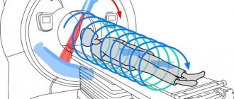
Tomography of the abdominal organs
- spleen;
- prostate;
- ureters;
- pancreas;
- intestines;
- ovaries;
- testes;
- liver;
- uterus;
- kidneys;
- gallbladder and bladder;
- stomach.
An overview X-ray image can also show the diaphragm, heart, lower part of the lungs, and free pairs of ribs. Fluoroscopy, in addition, reveals the dynamics of development - displacement, extensibility and contraction of the heart muscle and other types of organs and tissues.
Indications for use
An X-ray of the abdominal organs is extremely necessary for those who have:
- abscess;
- polyps;
- pain in the lower back or abdomen;
- tumor process;
- suspicion of injury to any organs;
- diverticulitis;
- cysts;
- intestinal obstruction;
- chronic bloating, etc.
Sharp pain in the right quadrant of the abdomen is a reason to urgently consult a doctor and undergo an x-ray: this symptom may indicate inflammation of the appendix
A survey x-ray should be taken at least once a year to prevent gastrointestinal diseases.
Intestinal obstruction: X-ray signs
Among the pathologies of the intestinal tube, X-ray has the greatest diagnostic value in acute intestinal obstruction. Quick results and the presence of symptoms characteristic of obstruction enable the surgeon to make a diagnosis and carry out immediate surgical intervention.
In case of obstruction, a survey radiography is performed in a vertical position. Characteristic is an increase in the airiness of the intestinal tube, which indicates excessive accumulation of gas in the intestines.
The most academic symptom is the presence of fluid levels, the so-called Kloyberg cups. Moreover, by the shape and location of these cups, the level of pathology can be determined. If the cups are high and narrow and are located on the periphery of the x-ray, we can talk about the presence of obstruction in the large intestine. Surgeons also use the term "low obstruction".
If the Kloyberg cups are low and wide, and also located closer to the center, this indicates small intestinal obstruction, or high. Also characteristic is the expansion of the intestine to the site of the obstruction and the collapse of the intestinal section after.
When performing contrast radiography, you can notice that the evacuation of barium is slow or absent altogether.
In some cases, radiography allows one to visualize the obstacle itself (tumor, intestinal torsion, adhesions).
When diagnosing obstruction, it is important to distinguish in which part of the intestine the problem arose: in the small or large intestine. In addition to the difference in the location and shape of the Kloyberg bowls, there are a number of other features.
If it is not clear from the cups which part is involved in the pathological process, contrast radiography of the abdominal cavity can be done. In this case, distended intestinal loops are clearly visualized.
Signs of small intestinal obstruction:
- swollen loops occupy predominantly the center of the image;
- they do not exceed 4-8 cm in size;
- characterized by the presence of transverse striations against the background of swollen loops;
- there are no specific concavities along the edges (haustra).
Signs of large intestinal obstruction:
- swollen loops of larger diameter;
- there are concavities - haustra;
- characterized by the presence of arched folds (symptom of arches).
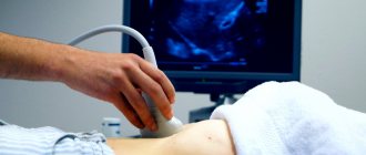
Thus, the value of abdominal radiography in diagnosing obstruction is extremely high. However, there are cases when the x-ray picture is unclear. Then it is necessary to carry out other examination methods: ultrasound, computed tomography.
Main contraindications
The procedure using ionizing radiation is characterized by an almost complete absence of contraindications. X-rays are prescribed even if a person has implants, piercings and tattoos, which is impossible with MRI. However, two categories of people should not visit the X-ray room without special circumstances: pregnant women, minors under the age of 12 years.
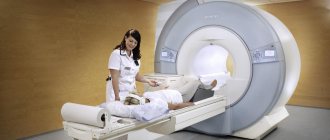
If necessary, expectant mothers will most likely be prescribed alternative diagnostic methods - ultrasound or MRI. After all, X-ray radiation will focus specifically on the abdominal region of the body, where the developing fetus is located. Since the consequences of X-ray exposure on the baby are officially considered unstudied, this diagnosis should be neglected until the end of the gestation period.
If a person suffers from dehydration (dehydration), cystic fibrosis or bronchial asthma, the possibility of undergoing the study must be discussed in advance with the treating doctor. X-rays are not performed on children at all, since the radiation dose from the device significantly exceeds the usual daily norm.
What does an x-ray show?
A doctor with the proper level of qualifications will be able to detect the following diseases and phenomena on an x-ray:
- tumors at any stage of development;
- appendicitis;
- inflammatory process (peritonitis, cholecystitis, etc.);
- nephrolithiasis (formation of kidney stones);
- pancreatitis;
- polyps;
- internal hematomas;
- abnormal structure of hollow organs;
- cholelithiasis;
- polycystic disease;
- diverticulitis;
- intestinal obstruction;
- various injuries;
- biliary and hepatic colic.
In the X-ray images, Kloiber's cups are visualized on the left, and the crescent sign on the right
In a young child, using X-rays, you can determine the location of swallowed foreign objects: coins, buttons, needles, beads, nuts, etc. Sometimes a specialist can detect quite dangerous phenomena on photosensitive film: Kloiber's cups and the crescent sign.
The sickle symptom is an accumulation of free air caused by a violation of the integrity of one of the sections of the gastrointestinal tract. Externally, it is a lightened crescent-shaped spot, which is most often located under the right dome of the diaphragm - the muscular septum separating the abdominal cavity from the chest. Kloiber's bowls resemble bowl-shaped two-layer elements filled not only with gases, but also with liquid.
If the number of vertically oriented spots prevails in the image, then the patient has obstruction of the large intestine, and if horizontal, then obstruction of the small intestine. Both cases considered indicate the presence of extremely advanced pathologies, which after X-rays should be eliminated as soon as possible.
How is an X-ray performed?
A survey radiography of the abdominal cavity is done in two projections: lying and standing. Most often, the first option is omitted and X-rays are taken only while standing; this method is more informative. This allows for better visualization of intestinal obstruction, as well as perforation or perforation of ulcers.
Contrast radiography is more difficult. Here it is necessary to take a series of photographs after a certain time and observe how the barium solution moves through the intestines. This process of moving barium is called passage.
The location of barium as a function of time is as follows:
- after 1 hour - the contrast is partially in the stomach, partially in the small intestine;
- after 3 hours - the contrast should completely leave the stomach and fill the small intestine;
- after 6 hours - contrast in the initial parts of the large intestine (cecum and ascending colon);
- after 9 hours - contrast in the transverse and descending colon;
- after 12 hours - contrast in the descending colon and sigmoid colon;
- after 24 hours - contrast in the rectum.
Immediately before the test, your doctor will ask you to remove items from your pockets that may interfere with the image being displayed on the screen. An x-ray of the abdominal cavity is taken in both vertical and horizontal positions. In some cases, it may be necessary to scan the body in two projections in order to better see the condition and structure of the internal organs. The patient takes a standing or lying position. In order for the pictures to be of high quality, it is necessary to remain still.
Stages of preparation for radiography
If a person is urgently admitted to the hospital, special preparation for an x-ray is not required. In the case of a scheduled examination, it is worth taking care in advance to cleanse the body of toxins, which are reflected in the image in the form of unnecessary shadows that make it difficult to make a diagnosis. 2-3 days before the x-ray, beans, confectionery, raw fruits and vegetables, milk, and fast food should be excluded from the diet.
This product causes gases to appear in the intestines, which also “smear” the image on the film. You should drink enough clean water and simple meals. On the eve of the study, it is advisable to do a cleansing enema. The use of strong laxatives, for example, Guttalax and Bisacodyl, is discussed individually with the doctor.
The diet involves unlimited consumption of slimy porridges and creamy soups
The procedure for performing radiography
- The patient is placed in the starting position in front of the X-ray machine and stands motionless for several minutes.
- The radiologist starts the machine and takes a picture in the original projection.
- If necessary, the patient changes position, and the study is carried out in a lateral or oblique projection.
- At the same time, images can also be taken in a supine position - for this, the patient needs to lie on a couch under the X-ray machine and also lie still, awaiting the doctor's instructions.
Be sure to read:
Sigmoidoscopy and colonoscopy: what is the difference between the studies and which is better?
Where can I get an abdominal x-ray?
After the patient receives a referral for an x-ray from a nephrologist, gastroenterologist, endocrinologist or urologist, it is worth finding out about the availability of specialized centers equipped with the necessary devices at their place of residence. You need to pay attention not only to the price of an x-ray, but also directly to the category of the medical institution, the qualifications of the doctors and the model of the equipment used for scanning.
Also, do not neglect reviews on the Internet and the opinions of familiar people. If the previously chosen clinic is characterized extremely negatively by former patients, you should refuse to visit it. Even if the stumbling block becomes a financial issue (in the case of paid centers), it is better to save the required amount of money and maintain your health than to lose it due to your own stinginess.
Cost of examination
Since each region of Russia has different price indicators, the cost of research in different cities varies significantly. As a rule, a person will need to pay from 350 to 2100 rubles for an x-ray of the abdominal organs. In the Moscow region, the price can reach 3,000 rubles. It is worth keeping in mind that interpretation of the x-ray is also included in the total cost. If the doctor has given the patient a referral for the procedure, then it will be carried out at the expense of compulsory medical insurance (i.e., free of charge).
If a doctor at a private clinic insists on paid x-rays in their institution, without particularly justifying his choice, it is recommended to consult with independent doctors before agreeing to radiation diagnostics
Differences and advantages of CT and X-ray
In fact, computed tomography is also a type of research based on the use of X-rays. Only when scanning does the image reflect the state of the organs in several sections and projections, which allows you to view a particular area of the body from the necessary angles. Although CT is more informative, it has not managed to displace classical x-rays from medical practice, which has some advantages:
- a high level of safety (the radiation dose is 10–20 times lower than with tomography);
- low cost;
- accessibility for the majority of the population.
Moreover, X-rays best visualize bone tissue, and CT scans best visualize the structure of bones, adjacent vessels, organs and soft tissues. In order to make the right choice, you should consult in advance with a specialist of a certain profile, and then adhere to his recommendations, developed on the basis of the patient’s individual indicators.