What does a shoulder x-ray show?
For an accurate diagnosis, a history and physical examination are not enough. The doctor prescribes additional tests, depending on the pathology.
When X-rays pass through tissues of different densities, the beam is scattered and slowed down, resulting in images of different intensities. Radiography is considered not the best way to diagnose soft tissue pathologies. But for diseases of the osteoarticular system, this research method is the main one. In the photographs, soft tissues are darker, and hard tissues are lighter.
An X-ray of the shoulder joint in two projections (frontal and lateral) shows a fairly detailed picture of pathological changes. In addition to the condition of the joint itself, it can be used to assess the condition of the collarbone and scapula. Thanks to the direct projection, the doctor determines the degree of displacement of the fractures (if any); changes in the edges of the acetabulum are clearly visible on lateral (axial) photographs.
Diagnosis and differential diagnosis
The diagnosis is made taking into account characteristic clinical and radiological indicators. X-rays of the shoulder joint reveal dystrophic transformations and marginal bone growths (osteophytes); in the later stages, narrowing of the joint space, deformation and changes in the structure of the underlying bone are determined. The joint space may take on a wedge-shaped shape, and osteosclerotic transformations and cyst-like formations are detected in the bone.
X-ray classification of arthrosis of the shoulder joint:
- Stage 1 – a small number of osteophytes, the joint space has been slightly reduced or not changed.
- Stage 2 – there are noticeable osteophytes, the joint space has been concluded.
- Stage 3 – osteophytes are clearly defined, the joint space is clearly defined.
- Stage 4 – large osteophytes are identified, the joint space is significantly drawn, the underlying bone is deformed, indicators of osteosclerosis are determined.
In highly doubtful cases, especially in the initial stages of the disease, the patient may be referred to a CT scan of the shoulder joint or MRI of the shoulder joint to obtain additional information about the condition of the bone, cartilage and soft tissue structures. If secondary arthrosis is suspected, consultations with relevant experts are prescribed: a surgeon, an endocrinologist, etc.
Differential diagnosis is carried out with gouty, psoriatic, rheumatoid and reactive arthritis, and with pyrophosphate arthropathy. In case of arthritis, blood tests reveal indicators of inflammation; transformations on radiographs are poorly expressed, osteophytes are absent, and there are no indicators of deformation of the articular surfaces. With psoriatic arthritis, along with joint manifestations, skin rashes are often found. In rheumatoid arthritis, a positive rheumatoid factor is determined. With pyrophosphate arthropathy and gouty arthritis, a biochemical blood test reveals corresponding transformations (increased levels of uric acid salts, etc.).
Indications for radiography of the shoulder joint
Pathologies of the shoulder joint are found not only in surgery, but also in orthopedics and neurology. Patients come with various complaints, and the doctor prescribes an x-ray for the following symptoms:
- Sharp, aching or shooting pain in the joint area.
- Stiffness in movement.
- Asymmetry of the shoulder area.
- Swelling in the collarbone and shoulder area.
- Hyperemia of the skin at the shoulder joint.
In addition to symptoms, there are other indications for research:
- If a shoulder dislocation is suspected, x-rays are the primary diagnostic method.
- Fractures of the humerus, clavicle or scapula.
- Diseases of the joints of inflammatory nature (arthritis).
- Pathologies of joints of a degenerative-dystrophic nature with destruction of cartilage (arthrosis).
- Suspicions of the occurrence of various neoplasms.
- Control of congenital structural or functional abnormalities of the joint.
Many pathologies of the shoulder joint have similar symptoms and course features. Diseases need to be differentiated; x-rays allow you to do this quickly and as accurately as possible.
Symptoms
Arthrosis of the shoulder joint begins to develop in people aged over forty-five years. This happens due to degenerative transformations that are observed in the human body over time, in other words, due to aging. This stage of arthrosis is called primary. A big role in the development of the disease is played by what profession a person has, what specific bad/food habits he has, and what kind of lifestyle he leads in general.
The initial circumstance that the cartilage slowly deteriorates is, of course, a lack of fluid (lubricant), in addition, injuries to which the joint was exposed play a key role.
Arthrosis of the shoulder joint can manifest itself in different ways. In most cases, at first small pain or discomfort appears in the shoulder, while it, the shoulder, is in a certain position. Then the pain may subside for a while or disappear completely, but after a while it will show itself again, moreover, the disease will begin to progress. The pain that a person feels with shoulder arthrosis is aching, pulling. It is worth noting that the pain may be constant, and its intensity will worsen in the presence of physical activity or, in addition, due to the weather. From time to time, the course of the disease may be caused only by sharp pain, if there was a lot of physical activity. In a state of calm, the joint will not be disturbed.
It is also worth noting that the localization of this disease itself may also be different: it can be pain all over the shoulder, and often in the arm, the elbow can ache, prick, and the back muscles on the affected side will also ache, or specifically on the affected side. joint.
As a result, there is a certain restriction in the movement of the joint, and the consolidation of the pain syndrome at chronic bases. The joint will deteriorate more and more every day, the cartilage will change into a scar, and the pain will worsen. Be prepared that, without taking measures, you may find yourself in a situation where your arm begins to rise only 90?, and subsequently there will likely be complete limitation in movements.
From all of the above, it is necessary to conclude that treatment for this disease must be applied without fail.
Diagnosis of gouty arthritis link here
Contraindications
Despite the high information content, accessibility and ease of implementation, in a number of situations it is necessary to abandon radiography and diagnose diseases using other methods. The reason for this is the presence of contraindications to the procedure. The shoulder joint is not x-rayed if the patient has the following pathological conditions:
- Pregnancy, especially the first trimester.
- Extremely serious general condition.
- Open pneumothorax.
- Heavy bleeding.
- Severe pathologies of the liver or kidneys.
- Decompensated diabetes mellitus.
- Pulmonary tuberculosis in open form.
In modern X-ray machines, ionizing radiation is minimal. Only a doctor can assess the possible risks and feasibility of the procedure.
Anatomy of a joint
The shoulder joint consists of the head and glenoid cavity of the scapula. The first element has a spherical shape, and the second is presented in the form of a concave bend. The articular surfaces of the elements are congruent relative to each other.
The ends of the bones interact with each other due to the presence of hyaline cartilage. Hyaline is presented in the form of an elastic and hard substance that is responsible for shock absorption. The joint cavity contains synovial fluid. It reduces friction between joints, performing a nutritional function. The pressure inside the cavity is normally negative. With inflammation, it increases, which contributes to dislocation of the limb.
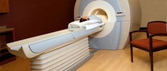
How is a shoulder x-ray performed?
One of the advantages of radiography is that no special training is required. You can undergo the test immediately after seeing a doctor by taking a referral from him. An X-ray of the shoulder joint goes like this:
- Cell phones and other emitting gadgets are left outside the room where diagnostics are performed.
- Remove outer clothing and metal jewelry. The arm and shoulder are completely exposed.
- They lie down on a special table and take a position that is optimal for x-rays.
- The chest, abdominal and pelvic areas are covered with a lead cape.
- The doctor brings the emitter to the shoulder and goes into the next room to take pictures. At the time of its implementation, the body must be motionless.
- After the procedure, the patient gets dressed and waits for the image with explanations from the radiologist in the corridor.
Another advantage of radiography is that the image can be used for consultation at another medical institution or by another specialist. A record of the procedure is made in a special accounting book; it can be requested at any time.
Preparation, carrying out the procedure
There is no special preparation for the patient before the examination. All you need to do is take the directions in your hands and go to the X-ray room.
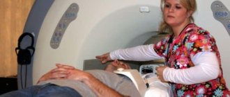
The diagnostic process includes a number of subsequent steps:
- The patient removes clothing in the area of the limb and shoulder, as well as all metal objects (jewelry, watches, etc.).
- The patient is placed on the table, the specialist fixes the arm in the desired position (depending on the X-ray projection).
- The laboratory assistant covers the patient's chest, abdomen and pelvic area with a special lead blanket.
- The specialist leaves the office - the patient remains in a motionless position.
- The device starts up. The laboratory technician may ask the subject not to breathe for a while.
- After 2-3 minutes, the patient gets dressed and leaves the room.
If a child is being examined, the eye organs and thyroid gland are also covered with protection.
If the rules of the procedure are followed, the output should be a high-quality image that provides information about the condition of the joint. Otherwise, the image may be damaged and a repeat x-ray will be required.
It is possible that the image will be distorted if the patient is overweight. A large number of fat cells causes blurred images.
X-ray harm and precautions
Radiation energy, passing through the patient's organ being examined, is redirected to the receiver, transforming into an image on photographic film. Unfortunately, some of it “settles” in the patient’s body, to one degree or another contributing to changes at the cellular and tissue levels.
Ionizing radiation can cause radiation burns, malignant tumors, and radiation sickness.
Of course, it is impossible to make an X-ray of the shoulder joint absolutely safe, but protective measures can reduce the degree of harm from radiation. There are a number of ways to protect yourself from harmful ionizing radiation:
- Maintaining a special record sheet in which data on the frequency of procedures is entered. Accounting allows you to control radiation doses.
- Compliance with all precautions and recommendations by the patient. When X-raying the shoulder joint, a special lead apron is put on the patient, leaving only the area of study open.
- Development and use of new, safer equipment.
- Complete immobility at the time of shooting. If the patient is unable to sit still, the image may be unclear and the x-ray may need to be repeated, which in turn increases the radiation dose.
Causes
The trigger point for changes in arthrosis of the shoulder joint can be either the normal process of tissue aging or damage or disruption of the structure of the cartilage as a result of mechanical influences and various pathological processes. Primary arthrosis is usually detected in older people; secondary arthrosis (developed against the background of other diseases) can occur at any age. The main reasons are considered:
- Developmental defects
. Pathology can be detected with underdevelopment of the head of the humerus or glenoid cavity, capomelia of the shoulder and other anomalies of the upper limb. - Traumatic injuries
. Arthrosis of traumatic etiology most often occurs after intra-articular fractures. A possible cause of the disease may be a shoulder dislocation, especially a habitual one. Less often, severe bruises act as a provoking injury. - Inflammatory processes
. The disease can be diagnosed with long-term glenohumeral periarthritis, previously suffered nonspecific purulent arthritis and specific arthritis of the joint (with tuberculosis, syphilis and some other diseases).
Risk factors
Arthrosis is a polyetiological disease. There is a wide group of factors that increase the likelihood of this pathology:
- Hereditary predisposition
. Many patients have close relatives who also suffer from arthrosis, including other localizations (gonarthrosis, coxarthrosis, arthrosis of the ankle joint). - Joint overstrain
. It can occur in volleyball players, tennis players, basketball players, sports projectile throwers, as well as in people whose profession involves a constant high load on their hands (hammers, loaders). - Other pathologies
. Arthrosis is more often detected in patients suffering from autoimmune diseases (rheumatoid arthritis), some endocrine diseases and metabolic disorders, systemic connective tissue insufficiency and excessive joint mobility.
The likelihood of developing the disease increases sharply with age. Frequent hypothermia and unfavorable environmental conditions have a certain negative impact.
The value of x-rays in pathologies of the shoulder joint
X-rays have been used in medicine for a long time. The study is used to diagnose gastrointestinal diseases, infectious and tumor diseases of the chest, teeth, and female reproductive organs.
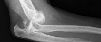
Diagnostics is especially important when identifying pathological changes in various parts of the peripheral skeleton. Using an X-ray of the shoulder joint (photo shown above), fractures, dislocations, diseases of an infectious and tumor nature are detected. Due to the different densities, all structures of the articulation and adjacent soft tissues are clearly visualized, down to the smallest detail. The study is specific; the image clearly shows what kind of pathology arose in the person.
An x-ray is a flat image of a three-dimensional organ. Therefore, when diagnosing, you should take pictures of at least two surfaces. By superimposing images, the localization of the pathological focus can be established. It is almost impossible to do this on your own.
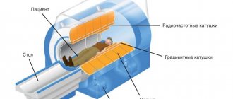
Principle of radiography
After the x-ray is taken, the display will show not only the joint itself, but also other areas, including the scapula and collarbone. In this regard, a specialist can examine these areas in detail and determine possible damage options.
The X-ray image contains all the data on the current state of the tissues, the presence of cysts, inflammatory processes and tumor formations. The technique also makes it possible to detect deformation processes and diagnose arthrosis, osteophyte or osteoporosis.
During a comprehensive examination of the patient, the doctor may prescribe a chest x-ray. This approach allows you to carefully study the characteristics of the joint cavity and then look for ruptures in muscle connections. If such changes are present, surgical intervention cannot be avoided.
Decoding the results
Unlike ultrasound images, x-rays are not operator-dependent. Only a specialist can decipher them.
To understand exactly what abnormalities were revealed in the image, you need to know what the image looks like after an X-ray of the shoulder joint is normal. The doctor distinguishes darkening, sees the presence of gaps in the structure of bone tissue, determines its changes, and also accurately finds the source of the disease. Indicator standards:
- The contours of the bones are clear and even. The bone structure is not changed.
- Full conformity of the shape of the contacting articular surfaces.
- The joint spaces are clearly visible and not narrowed.
- The contours of adjacent surfaces on both sides are clear and even. No bone growths are noted.
- No bone-dystrophic changes, as well as dystrophic or traumatic ones, were detected.
- Soft tissues are not changed.
When viewing the image, the doctor may ask several questions that will help to understand the causes of anomalies and correctly prescribe treatment:
- The doctor may ask for a description of the circumstances and duration of the injuries.
- Ask if there are any changes in symptoms, and if so, what they are.
- The doctor may do a sensory test.
Treatment
Medicine offers many effective treatment methods. To eliminate inflammation and pain, painkillers are prescribed. After the acute inflammation is relieved, the doctor prescribes a course of restorative and regenerating agents. To increase the therapeutic effect, drug treatment is supplemented with diet and the use of alternative techniques.
Medication
For effective and rapid treatment of arthrosis of the shoulder joint, a specialist prescribes medications from different pharmacological groups. They are necessary to restore damaged cartilage tissue, eliminate pain symptoms and improve the general well-being of the patient. You can achieve results by using the following medications:
- Chondroprotectors. These are medicines made from herbal ingredients. They effectively nourish cells. It is recommended to use medications during remission and during exacerbation. The most popular chondrotectors include Artu, Chondroitin, Glucosamine.
- Corticosteroid group. Medications can provide long-term pain relief. This is an excellent solution for a serious stage of pathology. If you follow the scheme of a competent specialist and undergo complete treatment of the diseased shoulder joint, you can reduce the foci of pathology and restore cartilage.
- NSAIDs. The drugs quickly eliminate the pain that occurs with degenerative changes. Relieving pain and inflammation ensures free movement.
Along with medications for internal use, the attending physician prescribes external treatments. We are talking about ointments Voltaren, Ketoprofen, and Ortofen. They are rubbed directly into the inflamed area. Properly structured drug treatment for arthrosis of the shoulder joint relieves inflammation and eliminates pain.
Surgical
This surgical intervention cannot be avoided if the following factors occur:
- Lack of effect from previously prescribed and administered conservative treatment;
- The occurrence of complications;
- Manifestation of degenerative destructive changes.
If irreversible changes in appearance or functioning occur, endoprosthetics is performed. The affected part is replaced through surgery with an artificial one. This is a complex operation that requires ideal professional qualifications from the doctor.
Therapy
When relieving the signs of arthrosis of the shoulder joint, attention is paid to physical therapy. After the main course of treatment, you need to reduce pain and restore natural mobility. The following procedures are prescribed:
- Cryotherapy on the affected area;
- Electrophoresis;
- Magnet therapy.
You can achieve results from this form of treatment if you follow the rules. Physiotherapy is prescribed only in the absence of acute painful manifestations. The procedures must be performed by highly qualified doctors.
Gymnastics
We are talking about physical therapy. Gymnastics with a slight load is prescribed at an early stage of the pathology. This will relieve attacks of exacerbation. Will allow you to quickly cure a sore shoulder joint. A standard training complex looks like this:
- A man sits down on a chair. Secures brushes in a lock. Several stretches are made forward and immediately back. You need to perform this movement twice with short pauses.
- In the same position, you need to put your hands on your hips and relax. Next, you need to do 4-5 shoulder manipulations, moving in different directions.
- In a standing position, you should perform several jerks while simultaneously moving your elbows behind your back. This is an excellent method to give the spine a load and work the entire shoulder area.
Regular exercise will relieve pain for a long time and restore shoulder mobility.
Massage
When deciding how to treat the shoulder joint, doctors often prescribe a course of massage. This will provide results such as:
- Improving tissue nutrition;
- Relieving swelling;
- Toning muscles;
- Eliminate pain.
If you entrust the massage to a professional, after a few sessions you can notice a positive effect.
Traditional methods
Despite the rapid development of modern medicine, treatment of shoulder arthrosis with folk remedies is considered relevant. Here are the most effective recipes:
- Compress with cabbage leaf. Reduces pain due to arthrosis, removes inflammation and improves mobility. The method is easy to use. You need to apply the leaf to the sore shoulder and leave it overnight.
- Decoctions of chamomile, nettle, burdock and calendula should be added to the bath.
- Hirudotherapy. This is an excellent option for combating acute and chronic shoulder arthrosis. The procedure quickly restores blood circulation, removes pain and swelling.
Despite the ease of use and versatility of folk methods, you should not self-medicate. Treatment of chronic arthrosis of the shoulder joint using traditional methods should be prescribed by a doctor after an examination and understanding that there is no allergy to the components present in the recipes.
The most common pathologies of the shoulder joint
All pathological changes are recorded by the doctor in conclusion. The images are the basis for making a diagnosis. By X-raying the shoulder joint, the following pathologies are revealed:
- Anterior shoulder dislocation is the most common structural disorder, occurring in 98% of cases.
- Posterior dislocations are rarely diagnosed
- Dislocation with fracture of the neck of the humerus.
- Old dislocation. Such pathologies are difficult to treat. Scars form on soft tissues, muscle tone changes, and a degenerative process of muscle tissue develops.
- Rotator cuff injuries.
- Arthrosis.
- Arthritis.
- Osteophytes, characterized by compaction of the capsule and the formation of calcifications in it (deposition of calcium salts).
- Osteoporosis is more often diagnosed in older people.
Why does it happen?
The causes of omarthrosis can be divided into several groups:
- Injuries (sprains, sprains and torn ligaments, bruises), heavy physical work and intense sports, which put excessive stress on the joints, forcing them to work “for wear and tear”. This also includes metabolic disorders and the appearance of excess weight in a person because of this. Arthrosis that arises as a result of an injury is called traumatic, and that which develops after its healing is called post-traumatic.
- Heredity. If arthrosis is observed in parents and relatives, there is a high probability of its occurrence in children.
- Genetic abnormalities. Mutation of type 2 collagen (disturbances in the structure of connective tissue fibrillar proteins) leads to rapid wear of cartilage.
- Diseases – rheumatoid and purulent arthritis, psoriasis, systemic lupus erythematosus, venereal pathologies, thyroid diseases, hemophilia, Perthes disease, in which the blood supply to the articular head deteriorates.
- Elderly age. Natural aging of the body, especially if a person makes no attempt to somehow maintain his health, is associated with deterioration of blood circulation and a decrease in physical activity. As a result, the nutrition of the cartilage is disrupted, and it also begins to “age”.
Arthrosis that arises due to a hereditary or genetic factor is often called primary, and that which develops after an illness is secondary. Pathology can be acute or sluggish. In the first case, severe pain occurs without any prerequisites.
Omarthrosis is almost always accompanied by arthrosis of the subacromial joint (acromio-clavicular arthrosis). This is the junction of the acromion process of the scapula and the humerus.
There is also a combined pathology - cervicobrachial arthrosis, which is divided into omarthrosis and uncovertebral arthrosis of the joints of the cervical spine.
Where to take an x-ray of the shoulder joint
The study is included in the list of free procedures. It will be carried out upon presentation of a passport, compulsory medical insurance policy and referral (if any). Stationary X-ray rooms are available in any district clinic.
Private commercial medical institutions rarely provide such a service, with the exception of specialized centers. This is due to the mandatory consideration of the patient’s ionizing radiation. But if we take into account that the study needs to be carried out as rarely as possible, then it is quite possible to get by with the number of inpatient rooms.