What does an x-ray of the elbow joint show?
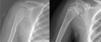
Elbow dislocation
Having received an X-ray, the specialist can see on it not only damage such as cracks, dislocation or fracture. An x-ray of the elbow joint gives the doctor information about the presence or absence of bone growths, the width of the joint space, how well the articular surfaces correspond to each other, in which areas calcium is deposited, and much more.
Using this diagnostic procedure, you can make a differential diagnosis of diseases and injuries such as:
- fracture;
- dislocation;
- cracks;
- arthrosis and arthritis;
- synovitis;
- congenital anomalies;
- metabolic disorders.
Features of the procedure
On any x-ray of the elbow joint, traumatologists will be able to see bone growths that are not characteristic of the normal state of bone structures, discrepancies in the surfaces of the joints, as well as areas where calcium salts have begun to be deposited.
Content:
- Features of the procedure
- Related and Complementary Research Methodologies
- Symptoms for radiography
- Indications and contraindications for radiography of the elbow joint
- Methodology of the procedure
In this case, radiography will be able to help identify all existing anomalies, make a diagnosis, diagnose complications and those signs of this pathology that can affect or are already affecting other organs and systems, prescribe a treatment regimen and monitor its dynamics.
X-ray diagnostics of the elbow area is a research medical method with which you can obtain a complete picture of the condition of the corresponding part of the human body by directing X-rays at it with a special device. This ultimately gives the specialist an objective image of the area being examined. It is important to know that soft tissues are capable of transmitting X-rays, while hard tissues, on the contrary, absorb them.
Because of this difference, in the images, specialists can see bone tissue painted in a very light, almost white color, and all soft tissues will have shades of gray - from lighter to almost black. Based on this change in shades, the doctor can judge what pathologies are present in the area under study.
At the same time, experts believe that X-rays are the best diagnostic technique for analyzing the condition of bone tissue.
In what cases is an elbow x-ray prescribed?
The patient complains of pain in the elbow joint
An X-ray is prescribed if, upon examination, the elbow joint has the following pathological symptoms:
- pain in this area;
- swelling of tissues;
- color change (redness, bluishness);
- high temperature (both general and local);
- limitation of mobility.
If only the soft tissue of the elbow is damaged, then the x-ray will not show such changes.
In this case, computed tomography, arthroscopy or ultrasound are additionally prescribed. X-rays are particularly effective in detecting bone damage resulting from trauma, degenerative disorders, and various tumors.
Where can I get an elbow x-ray?
If you fall or just seriously hurt your elbow, you can find out about the possible absence or presence of damage by contacting the emergency room. The traumatologist will examine the elbow, prescribe an x-ray procedure and issue a conclusion. If there are no queues, it will take about an hour.
If you have prolonged or recurring pain in the elbow joint, you can consult a therapist or orthopedist. They will also order an x-ray examination, establish a diagnosis and prescribe treatment. As a rule, an x-ray of the elbow joint can be taken in all major clinics or diagnostic centers.
X-ray room setup
Ideally, such centers should have modern X-ray equipment and qualified personnel. Before providing a service such as an x-ray of the elbow joint, the radiologist should ask you about the disease, inquire about the presence of contraindications, and also provide brief information about the procedure itself.
Related and Complementary Research Methodologies
Modern digital X-ray equipment provides doctors with images on the screen of a connected computer, on paper or digital media.
At the same time, radiologists say that pathological conditions of the joints cannot always be fully displayed on an x-ray, so other examination methods are used for them:
- computed tomography of joints;
- ultrasonography;
- Magnetic resonance imaging.
Each of the above methods demonstrates to specialists certain tissues, including the soft tissues of patients - muscles, tendons and ligaments. The high accuracy of these diagnostic methods makes it possible to mutually complement information about the pathological aspects of various periarticular tissues.
To analyze the cavities inside the joints, it is necessary to puncture the synovial capsule. The procedure is carried out with the collection of joint exudate. Impurities are easily detected in this joint fluid, which may indicate a number of diseases. These impurities are studied in more detail in laboratories under a microscope, and on the basis of such conclusions, experts draw conclusions about the infectious or non-infectious nature of the disease.
If it is necessary to study the structure of the joints, another diagnostic technique is used - arthroscopy.
Arthroscopy is performed through two punctures, into the first of which an arthroscope with a special camera is inserted, and in the second, the necessary manipulations are performed with special instruments.
In this case, such a procedure is very informative and provides specialists with the opportunity to see firsthand the development of various pathologies.
Based on the clinical manifestations of diseases, doctors choose those diagnostic methods or their complex that can most accurately provide information about all the processes occurring in the human elbow joint in order to make a timely and accurate diagnosis and prescribe the necessary therapy.
How can an x-ray of the elbow joint be taken?
At the moment, there are two ways to conduct an X-ray examination of the elbow joint: analog and digital. The first option involves obtaining images of articular structures on a special film that requires development. In the second case, the specialist sees the damage displayed on the screen. This method allows you to take as many pictures as needed, since the information received can be stored on a digital medium. Today, this is the most preferred method of examination, as it is considered to have greater accuracy as well as less radiation exposure to the patient. In addition, the time required to conduct a digital survey is reduced.
How is the elbow x-ray procedure performed?
An X-ray examination of the elbow does not require special preliminary preparation. The patient is seated and the arm is placed on the table as necessary. Immediately during the acquisition of the image, the limb must be absolutely motionless. To ensure this, use bags containing sand or other aids.
In order to view changes in tissues as much as possible, images should be obtained in three projections.
X-ray technician preparing a small patient for an X-ray of the elbow joint
The position of the patient depends on the degree of damage to the joint; most often, the examination is carried out when the person is sitting and the hand is on the table. In some cases, it is possible to take an x-ray while standing or lying down.
When an AP X-ray is taken, the patient's arm should be fully straightened and elevated. During a lateral examination, the arm will need to be bent 90 degrees and also raised. The third projection in which an x-ray is taken is axial. To do this, the arm is bent to the maximum and then placed on the table, resting on the humerus.
X-ray of a child's elbow
The bones of the shoulder, forearm, and human connective tissue are formed already at the 8th week of embryonic development. The final formation of the joint occurs in the first months of the baby’s life, when the arms and elbows begin to constantly move.
Growing up, an active child can suffer both dislocations and fractures, since childhood elbow injuries are considered the most common among joint injuries. The most difficult thing is the diagnostic process, since bruises are often accompanied by swelling of the adjacent soft tissues, which significantly complicates the examination.
How is it carried out, is it dangerous
An x-ray is done for a child in the same way as for an adult and does not take more than 10 minutes. The doctor describes the images obtained and makes a diagnosis.
To reduce harmful radiation, children are examined only on new, modern equipment using protective aprons. In this case, the thyroid gland, genitals and eyes of the child are especially carefully covered, and infants are covered completely, leaving only the examined area.
Preparation for the procedure does not require any special measures. The only rule for examining the elbow joint is its complete immobility. For this, various weighting agents in the form of sandbags can be used.
The X-ray dose directly depends on the equipment. New modern technology causes much less harm.
Normal indicators
The standards for the elbow joint in adults and children differ significantly. Let's consider three age categories:
- Babies from 3 months - the anatomy of the coronoid bone is not yet very developed. The edges of the block-shaped wall are rounded. The angle between the forearm and the longitudinal axes of the shoulder is 170-175 degrees.
- Children from 7 to 11 years old - the corresponding angle is 175±2 degrees.
- Adults have an angle of 180 degrees.
The anatomical characteristics of the child's bones, joints and joints are formed in the womb, and acquire their final characteristics by the age of 10.
Deviations and consequences
In childhood, due to the slow formation of articular bones and cartilage, deviations from developmental norms may be observed. This applies not only to the elbow bend, but also to the knee. Orthopedists treat the following conditions:
- norm;
- dislocation;
- subluxation;
- high dislocation.
Such deviations in children are often encountered in pediatric practice, but are amenable to surgical and physiotherapeutic treatment. Methods for examining infants are not based on X-ray radiation. For this purpose, ultrasound diagnostics is used.
The manifestation of arthritis in infants is almost always of an autoimmune nature. Infectious and inflammatory processes in the joints of babies can be a consequence of a complicated pregnancy of the mother.
Processing the results obtained
After undergoing the X-ray examination, the patient is asked to wait. As a rule, the images are ready after 15 minutes, and they are brought to the doctor’s office or given to the patient. The specialist carefully studies the results and makes a written conclusion about the condition of the elbow joint. Based on this conclusion, the patient can be diagnosed and treated, and may be referred to other specialists.
Doctor examining x-ray
A diagnostic method such as an x-ray of the elbow joint makes it possible to accurately discern bone pathologies. Studying the images, the specialist clearly sees cracks, displacements, the development of degenerative processes, and the formation of tumors. Much attention is paid to the size of the joint space, the condition of the articular surfaces of the bones, and the olecranon process. At the same time, the doctor examines nearby tissues.
If the data obtained is not enough to make a diagnosis, then additional diagnostic procedures are prescribed.
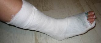
This is usually MRI, CT, arthroscopy or ultrasound. If a traumatic injury is detected, a fixing bandage, plaster splint or splint is applied. In some cases, hospitalization is required. Therapeutic treatment can be carried out both at home and in a hospital or outpatient clinic.
Who should not have an x-ray?
Since X-rays pose a danger to the successful development of the fetus, women are not recommended to have X-rays taken during pregnancy. If it is absolutely necessary to carry out diagnostics using this method, the abdominal area is covered with a protective apron. They should also be used in other cases of radiography.
Contraindications include children's age. It is not recommended to do X-rays unnecessarily for children under 14 years of age. In order to reduce the risk of radiation exposure, children are covered with a protective apron around the pelvic area, abdomen, chest and thyroid area, as well as the organs of vision. If we are talking about a newborn, then only that part of the child’s body that needs to be examined is left open.
The human elbow joint is exposed to stress and external influences every day. Physical stress and habits that are not noticeable at first glance, such as incorrect hand positioning when working at a computer or while driving, can lead to diseases that require radiography for diagnosis.
When X-rays are contraindicated
X-rays are not prescribed for pregnant women. This is due to the fact that radioactive radiation negatively affects the fetus at any stage of its development.
For children under 14 years of age, X-ray examinations are replaced by alternative gentle methods, such as ultrasound and MRI. Exceptions are those situations when the doctor needs to diagnose any congenital pathology at an early stage of its development, for example, radial or ulnar clubhand.
To reduce harm from the procedure, studies in children are carried out only with modern equipment and protective equipment for the thyroid gland, eyes and genitals is used. Newborns are covered completely, leaving only the part to be examined outside.
The dose of X-ray radiation during diagnostics directly depends on the quality of the equipment. Modern devices cause much less harm to the body.
An X-ray of the elbow accurately determines the pathological changes that have occurred in the bone tissue. The photographs clearly show cracks, tumor and degenerative processes, and fractures. An X-ray examination allows us to assess the size of the intra-articular space, the narrowing of which is a diagnostic indicator in identifying arthrosis and arthritis.
X-ray of the elbow allows you to assess the condition of the end surfaces of the bones of the shoulder, forearm and elements of other joints. In addition, the areas adjacent to the joint are examined.
After taking all the images, the radiologist interprets them and makes a description, which, together with the images, is transmitted to the doctor who referred the patient for an x-ray.
Alternative names: X-rays of the elbow joint, English: X-rays of the elbow joint
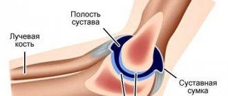
The elbow joint is characterized by a complex anatomical structure. It is an articulation of three simple joints - radioulnar, humeroradial and humeroulnar, which are connected in one articular capsule.
The elbow area is quite vulnerable; it is constantly exposed to stress and negative mechanical influences, which leads to the emergence and development of various diseases of the elbow joint. For their initial diagnosis or clarification of the diagnosis, radiography is prescribed, which allows one to clearly visualize changes in the structure of bone tissue or the presence of pathological processes in other elements of the joint.
Objects of study during radiography
During an x-ray, rays pass through the tissue of the organ being examined and are projected on x-ray film. Bones absorb the least amount of rays, so their image will appear whitish on an x-ray.
The objects of study for the radiologist are the components of the movable joint formed by the bone sections:
- shoulder;
- elbow,
- radial.
Together they participate in the formation of 3 movable bone joints enclosed in a single capsule:
- Humeral-ulnar - it is formed by the capitate eminence of the humerus and the fossa located on the head of the ray. The connection has 1 transverse rotation axis. The shape of the joint is close to spherical.
- Humeral - it is formed by the articulation of the head of the condyle of the humerus with the fossa of the head of the radius. The appearance of the joint is shaped like a ball; it is capable of movement around 2 axes.
- Proximal radioulnar - formed by the articular circumference of the ulna and the radial notch located on the ulna. Thanks to the joint, it is possible to perform the flexion-extension function of the arm, rotational movements of the radius, rotation of the hand inward (pronation) and outward (supination).
The joint is held in place by 2 ligaments - the catarrhal and lateral ligaments, as well as a group of muscles.
When performing an x-ray, the doctor assesses the condition of the bones adjacent to the joint, identifies areas of bone growth, areas with calcium deposits, and measures the size of the joint space.
Diagnosis of diseases
A referral for examination of the elbow joint is given by the doctor to whom the patient turns to identify the cause and treat the disease. This could be a family doctor or a specialist:
- rheumatologist - will diagnose and prescribe therapeutic measures to eliminate inflammatory diseases;
- traumatologist - will help with pain caused by injury;
- orthopedist - will determine the direction of treatment for organ deformation;
- oncologist - will make a diagnosis and choose treatment tactics for tumor formations;
- neurologist - will prescribe treatment if pain occurs due to nerve damage.
If you receive an injury, you must go to the emergency room, where a doctor will examine you and send you for an x-ray. In other cases, the procedure is carried out in diagnostic centers and medical institutions where appropriate equipment and specialists are available.
The average cost of radiography of the elbow joint in 1 projection is 500 rubles, in two projections - 1200 rubles. However, depending on the level of prestige of the clinic, the class of equipment, and the number of projections, the cost of the procedure can reach 9,500 rubles.
Indications for x-rays
The use of radiography is often prescribed if the development of articular pathology is suspected. This procedure is especially recommended for athletes and people whose bodies are predisposed to arthritis.
The joints of the elbows have a complex structure. They involve the articulation of three simple joints:
- radioulnar;
- humeroulnar;
- brachioradialis.
They are all connected by one capsule. This area is very vulnerable because it is subjected to constant loads and various mechanical influences of a negative nature. All these factors lead to pathological changes in the area of the elbow joint.
Using an x-ray of the elbow joint, you can notice changes in the structure of bone tissue and other articular elements.
This primary diagnosis is indicated when the patient exhibits the following symptoms:
- elbow pain;
- poor mobility;
- swelling, redness and increased temperature in the elbow joint;
- club-handedness
X-rays are also indicated for planned surgery and after a course of treatment. It is prescribed when a patient is suspected of having inflammation or degenerative changes in the elbow joint, or with tumor formations in the elbow.
Who to contact
Who to contact
A referral for examination is given by the specialist with whom the patient was seen. With pain in the elbow area, people most often turn to a family doctor or a specialist.
To identify causes and treatment, you can consult a rheumatologist. He will conduct a diagnosis and prescribe a course of therapeutic measures aimed at eliminating the inflammatory process.
Traumatologists will provide assistance if you feel pain due to injuries. The orthopedist will be able to determine the further direction of treatment when the organ is deformed. The oncologist will make the correct diagnosis and then select the necessary course of treatment when diagnosing tumors. For this purpose, a biopsy is also prescribed to make the information more reliable. When pain occurs due to nerve damage, treatment is prescribed by a neurologist.
The procedure is carried out in medical institutions that have the necessary equipment and the necessary specialists.
Research methods
The elbow joint has small joints. Because of this, x-rays are the most effective method for diagnosing pathology. The device shows various deviations from the norm and the development of diseases:
- arthrosis and arthritis;
- bone injuries;
- neoplasms;
- dislocations, subluxations and cracks;
- sprains and ligament tears;
- congenital ulnar and radial clubhand;
- gout and bursitis;
- epicondylitis;
- abscesses and synovitis;
- osteomyelitis.
Pictures are usually taken in three projections: lateral, direct and axial. During the direct posterior projection procedure, the patient stands to the side of the table or lies on its surface. The doctor moves the limb to the side of the table and extends the arm at the elbow joint as much as possible. The hand is turned to the supination position. An x-ray is also taken with the elbow bent, which allows you to get a picture of the size and condition of the joint space. It makes it possible to evaluate structural changes in bone tissue and joint-forming bones.
When taking a lateral projection photograph, the patient sits sideways to the table, and his arm is bent and lies palm down. It is necessary that the limb coincides in the direction of the body with an angle of the elbow joint of 90°. This allows you to see the upper parts of the elbow in the image.
During axial projection, the patient sits sideways from the table or can face it. The back of the hand is pressed to the surface of the shoulder and bent at the elbow. The fingers of the hand should touch the collarbone. The image allows you to evaluate the olecranon process and the trochlear bone of the shoulder.
The whole procedure takes no more than 10 minutes. The radiologist spends the next 15 minutes describing the image. He gives his conclusion, which indicates a specific diagnosis or the most likely definition of the disease. To perform an X-ray of the elbow joint, no preliminary preparation of the patient is required.
https://youtu.be/W5xOzpPJsZQ
Contraindications for radiography
There are certain contraindications for x-rays. This is due to the effect of X-ray radiation on the human body. With the use of more modern equipment, the dose received is much less during such a procedure. The newest equipment makes it possible to take photographs with minimal harm to the health of adults.
However, X-ray machines are harmful to children's bodies. After such a procedure, radiation exposure negatively affects the child’s future health. This can manifest itself in delayed development and growth of children.
For this reason, X-rays should not be taken for children under 14 years of age and pregnant women during breastfeeding. This examination is also not recommended for patients whose condition may worsen after it is performed. Sometimes, in the case of diagnosing serious diseases, it is necessary to take an image for some patients. For this purpose, whenever possible, only high-quality equipment is used. During the examination, the specialist must provide maximum protection for the eyes, genitals, and thyroid gland of the subject.
X-ray diagnostic results
On an x-ray, the elbow joints of an adult and a child are very different. The age of a person is influenced by certain normative characteristics that differ for each age group.
In healthy adults, the X-ray of the elbow joint is normal: 180°. This is the value of the angle between the forearm and the longitudinal axes of the shoulder. In a healthy baby up to 3 months, these figures are 170−175 o. Later, at the age of 7 to 11 years, in children the value of this angle is different and will be equal to 175−177 o if the child is healthy.
Fast and accessible diagnostics allows the radiologist to decipher it based on the image and make a description. This will enable a specialized specialist to establish an accurate diagnosis of an articular or other disease in the area of the elbow and other bones adjacent to it. Thanks to this, you can quickly begin treatment with minimal investment of money and time.
Related and additional techniques
Digital modern X-ray equipment allows you to display images not only on the monitor screen, but also on other media. Radiologists argue that not all equipment can fully display all changes on an x-ray. For this reason, other examination methods are used. These include:
- CT scan;
- Ultrasound;
- Magnetic resonance imaging.
Each of them shows specialists different soft tissues: tendons, muscles and ligaments. They demonstrate pathological changes in periarticular tissues with high accuracy. However, in case of joint diseases, you cannot rely on only one of them.
To obtain an analysis of intracavitary cavities, a puncture of the synovial capsule is performed. Such a procedure can easily show the presence of impurities in the joint fluid, which may indicate a disease. The data obtained is then studied in laboratory conditions, after which specialists draw their conclusions about the main cause of the disease. It can be infectious or non-infectious.
If necessary, another method is also used to study the internal structure of the articulation in the joints - arthroscopy. To do this, a puncture is made in two places to insert a device in the form of a special chamber and then certain manipulations are carried out using instruments.
Focusing on the clinical signs of the disease, doctors choose methods or complexes for patients that allow them to more accurately provide information. All data and processes occurring in the elbow joints will allow specialists to make a more accurate and timely diagnosis. All together will serve as the basis for prescribing the necessary course of therapy.
Indications and contraindications
An X-ray examination is indicated if the following are observed in the area of the elbow joint:
- pain of the organ at rest or during action;
- impaired organ mobility;
- redness or bluishness of the skin;
- tissue swelling;
- increase in local temperature in the affected area, general body temperature;
- clubhand, characterized by deviation of the hand of the radius and ulna from the longitudinal axis.
It is highly not recommended to conduct the study in the following groups of patients:
- women bearing a child;
- mothers during lactation;
- children under 15 years of age;
- patients for whom any movement that could lead to a worsening of the condition is contraindicated.
Important! Before performing x-rays, you must notify your doctor about the presence of contraindications.
During the procedure, organs that are not subject to examination are carefully covered with a lead apron.
To diagnose the condition of muscles, tendons, and ligaments, other examination methods are recommended - ultrasound, CT, MRI.
Symptoms for radiography
By its nature, the anatomy of the elbow joint is quite complex. Elbow diarthrosis includes such simple joints as the humeroulnar, humeroradial and radioulnar. All of them are combined into a common special joint capsule. Moreover, the elbow is one of the most frequently affected parts of the human body due to all kinds of injuries.
- pain in the elbow joint;
- disruptions in joint mobility;
- local temperature surges;
- swelling and redness in the area of the sore elbow;
- constant deviation of the position of the hand from the longitudinal axis of the radial and ulnar axes or clubhandedness.
Powerful physical activity also provokes the occurrence of pathologies in the elbow area. All this leads to very frequent diseases in this area.
For the purpose of preliminary diagnosis or to clarify the details of the pathological process in the elbow joint, specialists prescribe radiography of this organ, which will allow visualization of all bone changes and inflammatory processes in the elements of the joint.
What diseases are diagnosed
The purpose of an X-ray examination of the elbow joint is to identify diseases based on the results of the diagnosis. The radiologist examines and describes the images obtained in the following cases:
Loss of joint integrity or changes in bone position
- Fractures of bone structures at the elbow bend are regarded as complex injuries and can be localized in the olecranon process, the head or neck of the radius, the coronoid process of the ulna, and the epicondyle of the humerus. Fractures can be comminuted, transverse, oblique compression, with or without displacement. Using an X-ray examination, the nature of damage to the joint and adjacent bones is assessed.
- A dislocation of the elbow joint is characterized by acute pain, inability to bend the arm, and swelling. In case of a fracture, these symptoms are accompanied by a change in the color of the skin in the elbow area and numbness of the fingers. When a dislocation occurs, the x-ray shows that the articular surfaces are located at a distance from each other. The articular head is located outside the glenoid socket during dislocation and partially moves away from the socket during subluxation. X-rays allow us to assess in which direction the displacement occurred. Guided by this information, the doctor reduces the dislocation.
Diagnosis of diseases
An X-ray of the elbow joint diagnoses the following diseases:
- Arthritis . It usually manifests itself as pain in the joint, which subsequently does not go away even at rest. Swelling occurs in the elbow area, the motor functions of the arm are limited, the joint becomes stiffer and produces a characteristic crunch when moving. The image shows a narrowing of the intra-articular space, which usually occurs with rheumatoid arthritis, which causes damage to the synovium. Signs of osteoarthritis can also be visualized: a decrease in cartilage tissue, the appearance of spine-shaped processes - osteophytes.
- Arthrosis . In the initial stages of development, arthrosis is characterized by pain in the elbow joint during flexion and extension. Over time, limited movement develops and a crunch occurs. On the x-ray, a narrowing of the joint space is observed, bone growths appear along the edges of the bones, and areas of osteosclerosis are visualized. At the last stage of the disease, the areas of bones that form the joint are enlarged due to bone growths; The radiograph reflects significant destruction of the cartilaginous tissue of the movable joint.
- Gout . The disease is usually accompanied by pain in the joint, swelling, increased local temperature, and redness of the skin. X-rays at an early stage of the disease are not very informative, but they can reveal signs of the disease: bone osteoporosis; erosive areas on bone tissue that resemble a shell; round or oval bone defects.
- Lateral epicondylitis . The development of the disease causes pain on the outside of the joint, swelling, and increased local temperature. The disease is also called “tennis elbow.” X-rays usually show no changes in bone tissue. Learn more about epicondylitis in the next video.
- Bursitis . When the disease occurs, pain, swelling, and stiffness appear in the back of the joint. The condition of the radioulnar and interosseous synovial bursae is assessed using an x-ray, and areas of calcification are identified.
- Tumor formations . On X-rays they appear as areas of sparse bone. To clarify the diagnosis, a contrast agent can be injected into the mobile joint.
Important! Reliable diagnosis of the nature of the tumor formation is possible only by taking tissue for biopsy.
How is an X-ray examination performed?
For better visualization of the bone tissues that form the elbow joint, pictures are taken in 3 projections:
- Direct rear projection . The patient is positioned to the side of the table or lies on it. The doctor moves the limb being examined to the side of the table, extending it as much as possible at the elbow joint. In this case, the hand is rotated so that its outer side touches the table (supination position). The x-ray can be taken with the elbow joint bent, then the forearm should touch the back of the cassette and the elbow should be bent exactly 90˚. As a result of this placement, an image is obtained that allows one to study the condition and size of the joint space, evaluate structural changes in bone tissue, and the contours of the joint-forming bones: the humerus and two bones of the forearm.
- Lateral projection . During the examination, the patient is positioned sideways to the table surface, the arm is on the table in a bent position, the hand is palm down (pronation position). The direction of the arm should coincide with the direction of the body, and the angle of the elbow bend should be strictly 90˚. As a result, the following are clearly visible on the radiograph: the proximal epiphyses (upper sections) of the ulna and radius bones of the forearm; neck and head of the radius; distal part of the humerus.
- Axial projection . The patient is positioned to the side of the table or facing it. The hand is pressed as much as possible to the table with the back of the shoulder and at the same time bent at the elbow so that the fingers of the hand touch the collarbone. From photographs taken during this positioning, the doctor can assess the condition of the olecranon process and the trochlea of the shoulder bone, where the posterior surface of the joint is located.
Important! A visual reflection of the elbow joint on an x-ray in a lateral projection is mandatory, since only by using two projections can one obtain data on the nature of changes in the joint and the adjacent parts of the bones.
Radiography takes no more than 10 minutes. For about another 15 minutes, the radiologist describes the image, the conclusion of which may contain an indication of a specific diagnosis or a listing of the most probable diseases, the essence of pathological processes.
Thanks to radiography, it is possible to quickly and accurately diagnose diseases of the elbow joint with minimal expenditure of material resources and patient time.
Arthrosis is a disease associated with degenerative deforming processes that gradually develop in the cartilage tissue of the joint, due to which it loses its motor and shock-absorbing functions. The disease can affect both small and large joints, acquiring the character of a systemic disease. One type of osteoarthritis is arthrosis of the elbow joint, which is often also called epicondylosis . What is the cause of this pathology?
How is the diagnosis carried out?
There are two ways of x-ray examination of the elbow joint: digital and analog. Digital allows you to receive an image on a computer monitor. This technology allows you to take the required number of pictures and save the data in the device or on storage media. A lower radiation dose to the patient during the study is another advantage of this method. The analog method involves the use of a special film that needs to be developed.
There is no need to prepare for the procedure. The patient sits down and places his arm on the table as directed by the radiologist. An important condition is that when taking an image, the limb must be completely motionless. To immobilize the hand, auxiliary means are used, including sandbags. The patient is positioned sideways to the table in a sitting position. In some cases, it is permissible to conduct the study in a lying or standing position.
X-rays are taken in three positions of the hand:
- Direct projection - the patient fully straightens and raises his arm.
- Axial - the arm is maximally bent at the joint and adjacent to the back of the shoulder.
- Lateral - the limb is raised and bent at the elbow at a right angle.
The amount of radiation exposure you receive depends on the quality of the x-ray equipment. The study is carried out for all categories of persons in the presence of injuries to clarify the diagnosis. In case of suspected diseases of the elbow joint, children and pregnant women are recommended to replace the study with MRI or ultrasound.
If it is necessary to confirm the presence of congenital pathologies in children, it is permissible to conduct radiography using protective equipment that covers the genitals, thyroid gland, and eyes, leaving only the examined area.
Symptoms and treatment of arthrosis of the elbow joint
The elbow joint is less subject to stress than the hip joint or knee joint. In youth, such an illness can strike a person engaged in grueling physical labor every day: loaders, miners, builders. Another risk group is athletes: tennis players, discus throwers, volleyball players, etc.
In old age, epicondylosis is usually a single manifestation of general polyostearthrosis or can be a consequence of an old injury or increased stress on the elbow in the past.
Interestingly, for the first time, arthrosis of the elbow joint may manifest itself not under the influence of any particularly strong load, but from prolonged monotonous work, accompanied by flexion and extension of the elbow, or from vibration:
A good example is pain in the elbow that suddenly appears after working with a hammer drill or drill.
Why does arthrosis of the elbow joint occur?
Let's summarize the possible reasons:
- Previous elbow injuries
- Disturbed metabolism, in which cartilage tissue does not receive important amino acids and trace elements
- Genetically determined disorders
- Excessive loads and hypothermia
- Chronic diseases (rheumatoid arthritis, tonsillitis)
- Infectious bone processes in the bones (ostemyelitis, tuberculosis)
- Hormonal abnormalities
- Poor nutrition
- Constant stress
What is epicondylitis
However, it is still not worth classifying any pain in the elbow area as deforming arthrosis:
Painful symptoms in the elbow may not be due to epicondylosis, but to epicondylitis, inflammation of the epicondyles - the bony protrusions to which ligaments and tendons are attached. Despite the fact that pain symptoms can bother you for a very long time, after treatment and a gentle regime, problems with the elbow joint usually cease to bother you
However, both epicondylosis and epicondylitis can, as they say, go hand in hand, and arthrosis deformation of the joint is often accompanied by inflammation of all periarticular tissues
Etiology of deforming arthrosis
Deforming processes in the elbow joint occur as a result of:
- thinning of the hyaline plate
- reducing the synovial fluid needed for lubrication
- increasing friction of articular surfaces against each other
All this leads to hardening of the joint and its stiffness. The worst thing is that the process progresses, and sooner or later, almost complete destruction of the cartilage layer occurs.
As compensation, following the destruction of cartilage on the surface of the elbow joint, osteophytes grow, which ultimately leads to a deterioration in its mobility and subsequent ankylosis.
X-ray diagnostics can confirm the diagnosis.
The photo shows an x-ray of a joint affected by arthrosis.
Degrees of elbow arthrosis and symptoms
There are three degrees of epicondylosis.
First degree
The first degree is the onset of the disease, which the patient often simply does not notice:
- The elbow may periodically ache after physical activity or during deep bending and sudden movements.
- Muscle tone may be slightly reduced
- After resting the joint, the pain soon subsides
- Externally, there are no practical changes - the elbow does not change its shape.
- But on the X-ray, individual areas of degeneration and destruction of the cartilaginous layer are already visible, as well as some narrowing of the gap between the articular surfaces
Second degree
The second degree is accompanied by obvious pain and symptoms that are difficult not to notice:
- Movements of the elbow are already difficult, their amplitude is limited due to pain symptoms
- Pain in the elbow joint also occurs when moving the forearms and hands
- When bending the elbow, a dull crunch may be heard (not to be confused with those usual clicks that can be heard when warming up the joint)
- In the second degree, external coarsening and thickening of the joint, as well as symptoms of swelling, are already noticeable
- The x-ray shows colonies of osteophytes and further narrowing of the distance between the joints of the forearm and the fossa of the radius
Third degree
In the third stage, deforming arthrosis no longer leaves the unfortunate patient alone, even at night and at rest.
- Movements, even the slightest, bring severe pain
- It is very difficult to bend your arm, move it to the side, or lift it up
- Externally, the joint is very deformed, sharp protrusions appear on the elbow
- The arm looks crooked due to the violation of the axes of the forearm and radius
- Over time, due to muscle atrophy, the arm with the affected joint becomes thinner than the healthy one.
- On an x-ray you can see:
- almost completely destroyed cartilage, with only small intact areas remaining
- due to the loss of cartilage and exposure of the subchondral bone, it is completely covered with osteophytes
- absence of interarticular space or only a small gap
Conservative treatment of epicondylosis
Treatment of arthrosis of the elbow joint does not differ from the general treatment regimen for osteoarthritis.
- In case of acute pain, complete rest is recommended (the elbow can be immobilized with a shoulder scarf)
- Non-steroidal anti-inflammatory drugs are prescribed (diclofenac, ketoprofen, indomethacin)
- Manual therapy and physiotherapy sessions
- Therapeutic exercise, swimming, massage
- In the first stages of arthrosis, it is imperative to support the joints by taking chondroprotectors containing chondroitin or glucosamine
Deforming arthrosis of the third degree responds very poorly to conservative treatment in general and protective treatment in particular. In general, this is explained by the fact that:
- The pain is difficult to relieve, since deformities and osteophytes are almost irreversible
- It is no longer possible to restore cartilage with life-giving nutrition due to the fact that there is nothing left to restore...
Injections with hyaluronic acid facilitate the movement of the joint, creating lubrication of its surfaces.
In case of deforming late arthrosis with complete ankylosis, surgical treatment, in particular endoprosthetics, is often the only option.
For persistent chronic pain, you can make compresses from burdock and cinquefoil, ointments based on propolis, aloe and honey, and other folk remedies.
Video: Arthrosis of the elbow joint
Article rating:
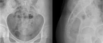
X-ray of the elbow joints (direct projection)
X-ray of the elbow joints (lateral projection)