Understanding the terminology
So, a specialist offers to take a panoramic photo of your teeth, but you don’t understand what it is? Let's understand all the terminology that you may encounter.
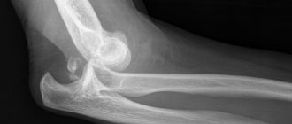
A panoramic dental photograph is an image in which the doctor sees the patient’s entire jaw in cross-section. The image turns out to be two-dimensional (that is, not three-dimensional, but flat); the principle of obtaining the image is based on the action of x-ray radiation. In the resulting image, the specialist will be able to examine the upper and lower jaw, each individual tooth along with the root, the bone tissue of the jaws, the maxillary sinuses, and the temporomandibular joint.
A synonym for a panoramic image is the concept of “OPTG” - this abbreviation stands for orthopantomogram. Scientifically, this term hides one of the methods of x-ray examination and the data obtained as a result of its implementation, i.e. panoramic photographs.
An orthopantomogram should not be confused with an orthopantomograph, since this is the name of the digital device with which the doctor conducts the examination. However, private clinics do not provide exactly this kind of equipment - as a rule, there is a computed tomograph everywhere, which allows you to do OPTG, computed tomography, and teleroentgenogram (read about the differences between these studies below).
On a note! Today, in devices such as orthopantomographs, X-ray films are rarely used - the equipment is considered outdated and produces poor quality images that are quickly erased. The operating principle of modern devices is based on the action of digital sensors, making the procedure fast and safe. Another advantage is that a digital image can subsequently be printed on any medium: photo paper, film. Or you can always store it on disk. But be careful, perhaps in some public dentistry, which provide treatment free of charge under compulsory medical insurance policies, film-type devices may still be used, which give a less accurate image and have higher radiation.
In what cases is an x-ray needed?
During a visual examination, the dentist can see approximately 40% of the oral cavity. However, most problems lie inside: in the canals, roots, interdental space. Therefore, most patient complaints can be resolved only after X-ray diagnosis.
Indications for examination:
- caries in any part of the jaw, including under the crown;
- root destruction: fractures, cracks, bone depletion;
- periodontitis - destruction of bone, while the gums become inflamed and bleed, teeth begin to loosen and hurt;
- broken bite;
- pathology of the dental joint;
- abscesses and tumors, cysts;
- periodontitis is an inflammation of the root, accompanied by the appearance of a cyst that enlarges.
Painful sensations do not always appear, so patients often delay treatment. Advanced periodontitis leads to gumboil and tooth loss.
In addition, canal cleaning, prosthetics and implantation, bone grafting, and orthodontic treatment are not performed without a detailed examination.
How does a panoramic image differ from x-rays and other research methods?
Indeed, many patients are confused when it comes to dental x-rays. It would seem that everything is simple: there is X-ray radiation, with the help of which doctors receive a picture that displays certain data about the patient’s condition. But why today are there so many different research and diagnostic methods, which still cannot be called in one word - x-ray? Because they all have certain differences. See for yourself.
| Research method in dentistry | Peculiarities |
| X-ray |
|
| Orthopantomogram |
|
| Teleradiography |
|
| Computed tomography (CT or tomogram) |
|
So which research method is better? In fact, everything depends on the clinical situation and the goals that the doctor sets for himself. For example, during implantation it is much more useful to trust 3D technologies and undergo a CT scan than OPTG (although the method is also used). When treating one tooth, when the situation is quite simple and clear (for example, caries), a regular x-ray is better.
On a note! It is not uncommon for private centers to ask you to undergo a panoramic scan during your first visit. It will be stored along with your card - so the doctor will be able to track the dynamics and will be able to know which teeth require surgical treatment. Some clinics even offer free images.
Pros and cons of jaw x-rays
The benefits of the study are obvious. In a short period of time, the doctor can clarify the picture and confirm or reject the proposed diagnosis. X-rays will show even those complications that you may not suspect. For example, if osteosynthesis is necessary, an x-ray of the upper or lower jaw is always taken, and if the jaw has pathologies, the doctor will postpone the procedure until a later date. It is impossible to detect this without x-rays. X-rays have become a common procedure in dentistry due to the ease of taking images and obtaining important results.
The disadvantages include certain diagnostic features when it is not possible to establish pathology. However, the fault here is not with the equipment itself, but with the processes that occur in the bone. When certain areas are darkened, the doctor already receives indirect information about the disease. Radiation should not be considered a disadvantage of X-rays, since the doses are small and do not pose a danger.
When you can't do without OPTG
Let's look at in what cases a specialist can refer you to OPTG.
Before starting orthodontic treatment
In this case, an orthodontist will prescribe an examination for you, who, using an image, will determine the condition of the root system of the teeth and, according to the data obtained, will select the most optimal option for correcting the bite. The study will be especially relevant in children; before treatment, the doctor is simply obliged to check and evaluate the anatomical features of the dental system, the presence of unerupted teeth in the bone tissue, the direction of their growth, and the stability of the roots.
For a comprehensive diagnosis of the condition of the maxillofacial apparatus and teeth
In young patients, again, images will be useful for determining the condition of the primary occlusion and identifying possible pathologies. For adults, photographs can be taken during annual dental checkups. The data obtained allows us to confirm the diagnosis, identify and treat even the slightest problems and hidden inflammatory processes, tumors and neoplasms at the initial stage of formation, determine the condition of bone tissue, assess the quality of previously filled root canals, and identify hidden caries.
Before planning surgical interventions
Even when performing manipulations on one tooth, a targeted x-ray is not always enough for the doctor. It is important for him to assess the condition of adjacent teeth and the proximity of their roots, determine the extent of the spread of the inflammatory process, exclude or confirm the presence of root fractures and various jaw injuries.
For example, patients are often referred for OPTG when planning a complex tooth extraction, because... the doctor must assess the condition of the surrounding tissues and the proximity of the nerves.
To identify foci of the inflammatory process
In order to carry out high-quality and adequate treatment, you first need to figure out where the source of infection is hidden (for example, a cyst or granuloma), and it is not always clearly defined. For example, a patient may complain about healthy, adjacent teeth. For this purpose, OPTG data is simply irreplaceable.
For the diagnosis and treatment of periodontitis
OPTG is sometimes also called a “circular photograph of teeth”, because the data obtained allows you to see even the depth of periodontal pockets and determine the severity of periodontitis, determine areas of destruction and resorption of bone tissue.
Before dental implantation
Today, before dental implantation, professional doctors often recommend that patients undergo a CT scan of the jaw, especially when it is necessary to restore teeth in the upper jaw with implants, because the method is much more advanced and informative. It, unlike OPTG, allows you to exclude the presence of an inflammatory process in the nasal sinuses (chronic sinusitis or sinusitis, in which implantation is contraindicated). However, OPTG is also actively used; the data obtained make it possible to assess the general condition of bone tissue and the dental system. For example, at the initial stage (when there is simply an assessment of whether implantation can be carried out) or at the final stage - to control treatment.
For difficult eruption of wisdom teeth
If the teeth are impacted or dystopic, it is important to assess whether they are causing harm to the “neighbors”, how they are affecting them, and whether an infection has occurred during the eruption period, which can affect the patient’s health.
Dental X-ray: types of images, harm, contraindications
One of the traditional and very popular diagnostic methods in the dental industry is x-ray examination. What can be seen on a dental x-ray? A dental photograph allows you to determine the degree of damage to hard tissues, identify hidden caries, assess the quality of previously performed dental treatment, and also timely diagnose foci of inflammation in the periodontium.
X-ray images allow you to clearly demonstrate not only the condition of bone tissue, but also identify many negative changes. This is extremely important for making an accurate diagnosis and determining the appropriate treatment regimen.
If earlier outdated X-ray machines were used, now they have been replaced by more modern radiovisiographs, which have a number of advantages:
- Minimal exposure.
- Fast examination.
- Receiving results on the day of the examination, which is very important for timely diagnosis.
Content
- Are x-rays harmful?
- Indications for the procedure
- Contraindications
- Are dental x-rays harmful during pregnancy?
- What can be seen on a tooth image during an X-ray examination?
- Main types of dental x-rays
- What does a panoramic dental photograph provide?
- Using Contrast
- Preparation for the procedure
- How is a dental x-ray done?
- Decoding the received data
- Why do dental x-rays take place?
- How often can children’s teeth be x-rayed without harming their health?
- Orthopantomogram or CT?
- Where can I do it?
Are x-rays harmful?
Many patients in dental clinics ask the question: is it harmful to their health to take dental x-rays? According to SanPiN standards, the permissible level of gamma ray radiation is 1000 microsieverts (µSv) per year. A feature of modern X-ray examinations is the fact that the exposure time of the device is reduced to a minimum, so the radiation dose is insignificant.
As for specific numbers, one procedure using a digital radiovisiograph is equivalent to 2 microsieverts. As for research using film, the dose is 10 microsieverts.
When answering the question of how harmful dental x-rays are, it should be noted that the frequency of examination is determined by the attending physician. On average, you can take about 100 photos over the course of a calendar year. It is obvious that even in a complex clinical case, such a number of x-ray examinations will not be needed.
Indications for the procedure
X-ray examination of teeth is an extremely important procedure, which allows specialists to determine the exact diagnosis of the patient, as well as determine the sequence of further actions. In the treatment of many diseases, this is the first step towards recovery.
This examination is based on gamma radiation. X-rays pass through organic tissue, resulting in an image. The image is obtained based on the ability of certain body structures to absorb this radiation.
Why are dental x-rays taken? With their help, you can diagnose the following pathologies:
- Periodontitis.
- Abscess.
- Periodontitis.
- Hidden caries.
- Developmental anomalies of the TMJ.
- Cysts and other neoplasms.
Identification of pathologies is not the only indication for this study. X-rays are also required in the following cases:
- Treatment of dental canals.
- Before installing implants.
- After tooth resection.
- Prosthetics.
Contraindications
The radiation dose during an X-ray examination is minimal. But this does not mean that this procedure can be carried out without any restrictions.
The main contraindications include:
- Pregnancy
- Problems with low immunity
- Mental disorders
- Bleeding
Are dental x-rays harmful during pregnancy?
The issue of performing x-rays in pregnant women is quite acute. According to the same SanPiN standards, this procedure is possible even if the woman is “in pregnancy.” The difficulty is that it is impossible to determine the exact effect of radiation on the structure of the fetus, so there are certain limitations.
X-rays are prohibited in the early stages of pregnancy (first trimester), i.e. at a time when the fetus is just beginning to form, so it is susceptible to the negative effects of gamma rays. If we talk about the 2nd and 3rd trimester, then pregnant women can take an X-ray of the tooth, but this procedure should be carried out only in extreme necessity, when other research methods are impossible.
What can be seen on a tooth image during an X-ray examination?
This procedure is considered a fairly informative diagnostic method, allowing to identify up to 90% of all pathological processes. So what does a dental x-ray show? The following changes can be seen in the picture:
- Inflammatory process.
- Atrophy.
- New developments in this area.
- Initial signs indicating the development of caries.
An X-ray also allows you to get a detailed picture and evaluate the quality of the dental canal filling procedure.
Main types of dental x-rays
X-ray is a fairly broad concept that includes a number of diagnostic procedures using various types of equipment. Widely used in dentistry:
- Extraoral radiography.
- Intraoral.
- Electroradiography.
- CT.
- Teleroentgenogram (TRG).
- Orthopantomogram (OPTG).
- X-ray procedure using a contrast agent.
Extraoral radiography
This type of study is carried out if intraoral radiography is impossible for some reason. This may be lockjaw or the patient's gag reflex. The disadvantage of this method is the insufficient accuracy of imaging oral tissues.
Intraoral (bite) examination
This type of examination is carried out using a special film (5x6 or 6x8 cm), which the patient must press between the teeth.
During the procedure you cannot move, and you also need to hold your breath. Otherwise, the image may not be clear. However, this recommendation applies to all types of photographs.
Electroradiography
This study differs from a traditional x-ray in the following features:
- The image is clearer and more informative, so even the slightest nuances are noticeable.
- It is possible to determine the density, condition and volume of periodontal tissues.
- Using these images, you can more accurately diagnose areas of hemorrhage, cysts and other formations.
CT scan
This study is carried out using a special device - a tomograph. This method is based on taking a three-dimensional image (3D visualization effect) in digital format.
The main advantage of this method is its high information content, because in the image it is possible to reproduce the projection of the jaw in a three-dimensional version. A specialist can easily examine the condition of bone tissue and assess the extent of diseases and pathological processes. The exposure to radiation is negligible due to the short duration of the procedure.
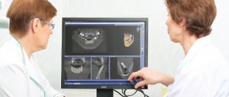
During operation of the tomograph, the image is immediately transmitted to the monitor screen. The result of the study can be easily recorded on digital media for further provision to dental clinics.
Teleroentgenogram (TRG)
This type of diagnosis allows you to examine not only the jaw structure, but also obtain a photograph of the entire skull in front and profile. It is one of the most informative diagnostic procedures in dentistry.
TRG is mandatory before installing braces, and is also widely used before upcoming operations as a preparatory measure.
Orthopantomogram (OPTG)
This is one type of research that is in sufficient demand. Using this technique, it is possible to obtain images of absolutely all jaws and teeth.
What does a panoramic dental photograph provide?
The resulting image gives the specialist the following information:
- The presence of fibroids and other neoplasms.
- Hidden caries.
- Identify unerupted teeth and their exact position.
- Identify bone diseases.
Other indications for OPTG include an upcoming implantation procedure, planned surgery, and other complex procedures.
Using Contrast
The use of X-rays with a contrast agent (sialography) is indicated if it is necessary to assess the condition of the ducts of the salivary glands. Before the procedure, they are filled with iodine-containing compounds. This type of diagnosis allows you to identify a number of pathologies: salivary stone disease, inflammatory foci and the presence of stones.
Preparation for the procedure
No special preparation is required for the upcoming procedure. If your doctor prescribed this examination, it means that you have no contraindications or restrictions. Therefore, preparation only boils down to the fact that you will need to remove all metal objects from yourself, and also put on a special protective vest.
How is a dental x-ray done?
Answering the question of how dental x-rays are taken in dentistry, it should be noted that the procedure depends on its specificity and type. In general, the examination takes no more than 5 minutes, and the radiation itself lasts only a few seconds.
In our diagnostic X-ray center you can take the following types of images:
- 3D (CT) Digital
- 2D (OTPG) Panoramic
- 2D (TRG) Teleradiogram
- 2D (TMJ) Sonogram
Bite shot
This type of research is considered traditional, without losing its relevance to this day. With its help, you can examine several elements of the dentition at once.
The patient heals a special film between the teeth, after which the procedure itself is carried out. Using a bitewing X-ray, you can determine the feasibility of correcting your bite and identify tartar and caries.
Panoramic
If you have been prescribed an x-ray of all teeth at the same time, many may have a question - what is this procedure called? This refers to a panoramic image that is taken using an orthopantomogram.
Before the examination, the patient's head must be securely fixed, because any movement can make the image unclear. After this preparation, the procedure begins: a special apparatus rotates around the head (there is a film on one side of the device, and a tube on the other).
If we talk about how often an orthopantomogram of teeth can be taken, then up to 10 panoramic images per year are permissible, and when using modern equipment, many times more.
Digital (3D)
The resulting digital image is highly detailed. The patient's head is also fixed, after which the tomograph rotates around it. Scanning occurs in three planes, and the image is immediately displayed on the monitor screen.
Decoding the received data
The most important stage of the entire process is the interpretation of the image that was obtained during the study. Healthy tissues of the gums and teeth in the picture are colored gray, fillings and implants will be a characteristic white color. But what does a dental cyst and other neoplasms look like on an X-ray? It is quite easy to see it, because it will appear as a dark area with clear contours (round or oblong shape).
Why do dental x-rays take place?
This question does not sound entirely correct, because this type of research is the main diagnostic tool in many medical fields, including dentistry.
X-ray examination reveals the following complications:
- Cysts and other neoplasms.
- Inflammation characteristic of periodontium.
- Tooth cracks.
- Overhanging fillings.
- Hidden caries.
- Allows you to identify the location of wisdom teeth.
It is also advisable to take an x-ray if dental treatment has already been carried out, but the dentist needs to make sure there are no complications. Bite correction is also necessarily accompanied by x-rays at all stages of this process.
How often can children’s teeth be x-rayed without harming their health?
Exposure to X-rays, one way or another, negatively affects the cells of the human body. The point is only in the dosage of this radiation and the frequency of procedures.
If in the fetus in the womb and a newborn baby the most susceptible organ is the brain, then in children from 3 to 12 years old it is the skeletal system. If the radiation dose is exceeded, there is a risk of slowing bone growth.
Under normal circumstances, any X-ray examination should not be performed on children under 7 years of age. But there are cases when the danger of the disease is much higher than the potential harm from exposure to gamma rays. In this case, an x-ray is a mandatory procedure.
As for frequency, the standard option is only 2 examinations per year. In this case, the radiation dose will be minimal.
Orthopantomogram or CT?
First you need to answer the question - what is the difference between a panoramic dental image and a CT scan? Using OPTG, you can only make 2D images, which are quite accurate, but miss a number of information due to the plane of the resulting image.
Therefore, from the point of view of greater information content, a 3D image using CT will be most preferable. Using a three-dimensional image, you can examine the structure of tissues from different sides, in any section and at any angle. Another advantage is the ability to construct a panoramic orthopantomogram image.
Where can I do it?
If you need to undergo an X-ray examination, then you can use diagnostic services. We have all the necessary equipment to take high quality and detailed images with safety for your health and the health of your children.
To make an appointment, you need to leave an online application on our website, or call our contact phone number.
Sign up for a study by phone
(812) 332-52-54
How the research is carried out
The description of the procedure itself is very simple. All manipulations, from completing the examination to obtaining informative images, will not take you more than 15 minutes. The procedure is performed using an orthopantomograph. This device assumes that the patient is in a standing position, and to take pictures, a special plastic stick is first placed in his mouth, which must be pressed tightly with his lips.
Beforehand, you will definitely be asked to put on a lead protective apron and remove all jewelry (earrings, rings), and you will be warned not to move or tilt your head during the examination. After the specialist checks that you are in the correct position, he will turn on the computer program and start the device. Next, a digital sensor will begin to rotate around your head, producing X-rays. Within one minute after this you can be free, but the doctor will need about 10 more minutes to process the data and save it or transfer it to one of the types of media: photo paper, disk, flash card.
Stages of dental radiography
Before a targeted x-ray is taken, the patient must remove all metal items in the head and neck area (chains, earrings, etc.). To protect the body from radiation, you must wear a special apron with a protective lead membrane. The neck area must be protected with a special collar during the procedure. The healthcare provider places cotton swabs in areas where teeth are missing.
How exactly the radiography will be performed depends on its type. For example, to take an occlusal x-ray of the jaw, the patient must stand near the orthopantomograph. Using this device, images of the lower, upper jaws and jaw joints are obtained. The patient then clamps the plastic tube with his teeth, keeping his lips closed. After this, he must move the device plate tightly to his chest and stand motionless so as not to distort the image. At the final stage, the device begins to move around the head for 20-30 seconds.
If an x-ray of one tooth is needed, the procedure is slightly different. Typically, the patient sits in a chair, pressing photographic film to a certain area of the jaw. After this, the sighting equipment is turned on for a few seconds. An x-ray is produced within 5-10 minutes.
What are the disadvantages of OPTG?
The advantages of this research method are known to everyone: panoramic images make it possible to improve the quality of the doctor’s work and eliminate the risk of errors at any stage of treatment, and to detect a hidden problem in time. But few people talk about the shortcomings, although it’s worth saying a few words about it:
- two-dimensional images: progress does not stand still; research methods have been invented that make it possible to obtain three-dimensional images that are more informative. And the OPTG, for all its advantages, is flat and does not always accurately transmit data,
- the picture does not accurately convey the dimensions: the distortion, depending on the brands of devices used, can range from 15 to 30%,
- the study is not entirely suitable for determining the condition of three-rooted teeth: these include sixes, sevens and eights. The thing is that orthopantomographs reproduce the image in only one section, and the roots of such teeth are in different planes.
On a note! Some doctors call OPTG an approximate research method, because... it shows only a general picture of the dental system, but gives very little information about small processes (for example, it will not be possible to see a perforation or a crack in the root; it will also be difficult to adequately treat curved root canals).
Is it possible to refuse x-rays for a child?
The child's parents have the right to refuse x-rays. However, responsibility for making such a decision rests entirely with them. Such diagnostic methods are not always able to provide a complete picture of the condition of the patient’s oral cavity.
Without X-rays, the doctor cannot arrange prosthetics. If you refuse the procedure, the planned treatment becomes impossible.
Radiography of baby teeth for children is included in the list of the most informative diagnostic methods. This procedure helps to assess the condition of the row elements, nearby soft tissues and identify the rudiments of permanent teeth without harm to the child. For diagnostic purposes, panoramic, targeted and 3D images are created. Digital X-ray is recognized as the most efficient and safe diagnostic tool.
How safe is OPTG?
The need to wear a protective vest during the examination and knowledge of the power of X-ray radiation makes many of us think about the dangers of such procedures and the possible dose of radiation. Regarding OPTG, different researchers today have the following data: one panoramic image is equal to a radiation dose of approximately 17 microsieverts (the maximum permissible recommended rate for one panoramic image, according to the Russian Ministry of Health dated December 21, 201222, should not exceed 24 microsieverts for children under 15 years of age , and 55 microsieverts for adults).
According to research data, it can be concluded that the procedure is safe. Even if it is carried out several times a year.
Baby teeth and molars - what's the difference?
The differences between temporary and permanent are the following characteristics:
- The roots of baby teeth are shorter, widely spaced and angled. This is explained by the need for the formation of molars;
- the enamel of temporary teeth is bluish, it is much thinner, and therefore more susceptible to the development of caries;
- The child’s milk teeth are smaller than the molars;
- The number of some teeth is not equal to the number of others. There are fewer temporary ones, because some elements of the series immediately grow as radicals;
- the tissue of baby teeth is poorly mineralized;
- temporary teeth grow vertically, and the crowns of permanent teeth are directed outward (towards the lips and cheeks).
How often can the procedure be done?
Radiation exposure indicators allow us to judge the safety of orthopantomograms, so if necessary, it is quite acceptable to undergo them as often as necessary, but no more than 80 times a year. It is also worth considering that the share of the total radiation exposure for each person should not exceed 1000 μSv per year. Ideally (if there are no indications), you can limit yourself to a few procedures during the treatment period (before and after treatment) or per year, during preventive examinations at the dentist.
Diagnostic frequency
Many people are interested in the completely natural question of how often dental x-rays can be taken. This information is recorded in a special document. It specifies the maximum number of procedures allowed to diagnose a patient. In this case, the total number of x-rays, both preventive and therapeutic, is taken into account. Naturally, a single diagnosis does not harm the body.
Even using old equipment 2-3 times a month will not have a negative effect on the body. The maximum permissible number of procedures depends on the device on which the examination is performed. The most dangerous class is for film equipment. Digital diagnostics is considered the safest. How many times diagnostics are allowed depends on the modernity of the equipment.
If x-rays are performed using up-to-date equipment, they are not dangerous to the body. You can carry out 8 procedures per day without harm. The permissible quantity per year is 500 photographs. On a film machine, the annual number should not exceed 80 procedures.
There must be indications for the study, otherwise radiation in large quantities can be hazardous to health
What do teeth and jaws look like in a photo?
The result of the examination is an image of a tooth, a separate area or the entire dental system, which can be transferred to film or simply saved on a digital medium. Trying to independently diagnose problems using such an image is almost a useless task, because only an experienced specialist can understand and correctly decipher the image.
Only a specialist can decipher the image
Inability to carry out the procedure
There are times when it is impossible to take an x-ray of a tooth. This occurs due to loss of contact. The main causes of this condition are:
- formation of a cyst in a separate area;
- incorrect canal filling;
- the use of materials that expose the X-ray image;
- initial stage of cementoma.
The development of an abscess leads to deterioration of the image. Children most often suffer from this. If purulent inflammation affects baby teeth, the result will appear as an exceptionally dark spot. There are diagnostic difficulties if you have to examine a wisdom tooth. Dental radiography is a necessary procedure in which radiation exposure is kept to a minimum. Using this method, the doctor will have the most accurate information about the inflammatory process, its location, and the presence of defects.
How much will an X-ray examination cost?
It is quite difficult to give an exact answer as to how much a dental x-ray costs - it all depends on the x-ray method, the size of the area being studied, the type of equipment used and the pricing policy of the medical institution. The table below shows approximate prices for various types of images.
| X-ray method | Approximate cost, rub. |
| Sight shot | 250-500 |
| Orthopantomogram | 1000-1200 |
| CT | 1500-5000 (depending on the scale of the study area) |
A separate fee may require printing the image on film and decoding it in writing by a specialist. As for where you can get an x-ray, it should be noted that most modern dental centers are equipped with the necessary x-ray equipment to obtain targeted and panoramic images.
Another question is if you are facing serious treatment, surgery, and especially implantation using one-stage technology. It is very important to foresee all the nuances in advance and carefully study the condition of the jaw bone tissue in order to select suitable places for implants. You will need to undergo cone beam or multislice tomography, and for this, experts recommend contacting specialized institutions that have high-quality and high-precision equipment.
- Potrakhov NH Low-dose X-ray diagnostics in dentistry and maxillofacial surgery, 2003
Indications for the procedure
An external examination performed by a dentist at each patient’s appointment does not always allow one to accurately determine the cause of the pathological condition. To correctly determine the diagnosis and treatment method, an X-ray machine is used.
Indications for the procedure are the following deviations from the norm in the condition of the teeth:
- abnormal position of the teeth;
- hidden cavity formed as a result of caries;
- periodontal diseases;
- pathological processes occurring under fillings or crowns;
- injury to the internal tissues of the tooth or jaw;
- the presence of tumors or abscesses;
- installation of implants.
The diagnostic results make the specialist’s work easier, giving him the opportunity to accurately determine which method of therapy should be used, or resort to tooth extraction. X-rays may be taken if there are other diseases to determine their course.
Diagnosis without unnecessary blood
Most procedures that involve penetration into the tooth and gums cannot be carried out without prior x-ray diagnostics.
The picture is taken to determine the condition of the bone tissue, roots, as well as the presence of caries under the crown (filling) or in the spaces between the teeth. The device is able to show the condition of the soft tissues inside the gums, identify possible inflammation and cracks in the canals.
Radiography allows you to accurately determine the place where it is necessary to carry out manipulations to eliminate the pathology. The doctor will not need to carry out unnecessary actions that could cause pain to the patient or lead to complications.
X-ray examination is an opportunity to establish the correct action plan for a specialist to treat the disease.
How to prepare for the examination
This diagnostic method does not require any special preparation. Immediately before the procedure, the patient is asked to remove all metal products so as not to disrupt the operation of the device and ultimately not get distorted results. The photo will be ready literally half a minute after turning on the device.
Many patients are also concerned about whether they need to brush their teeth before an x-ray. There are no strict guidelines on this matter. The patient is expected to brush their teeth twice a day, as required. If you had a snack the day before, it’s a good idea to rinse your mouth thoroughly and use dental floss.
X-ray during pregnancy
Dentists should not prescribe diagnostic tests to women in the first trimester of pregnancy.
After this period is over, dental x-rays are taken only in urgent cases, when it is impossible to carry out treatment without it. To reduce radiation exposure, specialists need to use a special film (E-class). It is recommended to use a digital method that will not cause any harm to the woman and her fetus.
It is also possible to take dental x-rays during breastfeeding. Since the radiation dose is small, breast milk does not accumulate any radiation, and accordingly the baby’s body will not be harmed.
In the first trimester, such diagnostics are contraindicated.