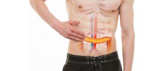
Rectosigmoscopy
– examination of the rectum and sigmoid colon using endoscopic equipment. It is one of the most informative methods for diagnosing diseases of the lower parts of the large intestine. Involves the introduction of a special endoscope (rectosigmoscope) through the anus. Allows for visual inspection of the inner surface of the rectum and sigmoid colon, assessing the condition of the mucous membrane, determining the location, size, shape and prevalence of pathological foci. If necessary, rectosigmoidoscopy is supplemented with a biopsy of the affected areas. It is carried out on an outpatient basis or in a hospital setting. No anesthesia is required. Before rectosigmoidoscopy, special preparation is prescribed, including diet and bowel cleansing measures.
The history of the development of methods for studying the lower parts of the large intestine using special devices inserted through natural openings goes back more than two centuries. The first attempt to create a device for endoscopic examination of the rectum was made by F. Bozzini in 1806. This date is considered the birth of endoscopy. Due to the unpreparedness of the medical community to evaluate the significance of this invention, the technical imperfection of the device and its danger to the human body, the Bozzini apparatus was never used in practice.
The era of rapid development of endoscopy of the large intestine (rectoscopy, rectosigmoidoscopy and colonoscopy) began much later - after the invention of electric lamps, the creation of semi-flexible, and then fiber-optic endoscopes. The first experimental model of a fiber colonoscope was developed in Japan in the early 1960s. In 1966, the creators of the model launched its mass production. Specialists in the field of proctology, gastroenterology and oncology quickly appreciated the advantages of the new technique. Rectoscopy, rectosigmoidoscopy and colonoscopy began to be used in clinical practice.
In the mid-80s, electronic video endoscopes appeared, providing a significant increase in the information content of endoscopic studies. Over time, rectosigmoidoscopy has become a routine procedure used in the diagnosis of lesions in the lower colon. Currently, rectosigmoidoscopy is included in the list of studies carried out for proctitis and sigmoiditis of various origins, benign and malignant neoplasia of the rectum and sigmoid colon and some other diseases.
Description of the procedure
What is rectosigmoidoscopy, and for what purposes is it used? The method is widely used in the diagnosis of diseases of the large intestine. The advantage of this examination is its relative painlessness, minimal traumatic effects on the intestinal walls and the high informativeness of the research result.
The procedure is performed using a rectosigmoidoscope, which is an instrument in the form of a hollow flexible tube up to 60 cm long, equipped with an optical system, that is, a light bulb and a video camera that transmits an enlarged image several times to the monitor screen.
The procedure is carried out by a specialist who, moving the rectosigmoscope along the intestine, visually examines it, noting various pathological points on the intestinal mucosa and taking material for biopsy from problem areas.
Rectosigmoscopy makes it possible to identify pathologies of the lower intestine in the early stages of development, especially of a malignant nature, as well as to prevent various complications. Rectosigmocolonoscopy is used to examine the entire large intestine. This method makes it possible to assess the condition of the uppermost parts of the organ.
Rectosigmoscopy: what is it?
When conducting this endoscopic examination, the doctor is able to visually examine the condition of the walls of the distal intestine. The procedure is performed using a sigmoidoscope - a long tube that is placed into the intestines at a distance of up to 60 cm. The movement of the flexible tube is completely controlled by the doctor, focusing on the image displayed on the monitor. A small camera is pre-attached to the sigmoidoscope, as well as a lighting device.
Using rectosigmoidoscopy, you can identify or confirm the presence of any neoplasms on the intestinal walls, in particular polyps. Also, during the examination, fragments of narrowing of the intestinal walls and the appearance of dilated hemorrhoidal veins are detected. If internal bleeding is suspected, a sigmoidoscope can be used to identify the area that is bleeding intermittently. Rectosigmoscopy is performed only if indicated. The need for this procedure is determined by a coloproctologist.
Sigmoscope
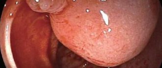
Attention! Thanks to the presence of high-precision eyepieces, the doctor is able to identify tumors that are minimal in size. If necessary, a small section of tissue is taken for further histological examination. As a result, it is possible to identify malignant neoplasms in the early stages, which improves the prognosis of the disease and saves the patient’s life.
When performing rectosigmoidoscopy, it becomes possible to identify many diseases and pathologies in the structure of the mucous membrane at an early stage, since the following parameters are visualized on the monitor:
- Shade, tone of the mucous membrane.
- General condition, relief of the intestinal walls.
- Elasticity of blood vessels.
- Any irregularities, including ulcers, areas of erosion, and the emergence of hemorrhoids.
- Cracking of the surface, scars from previously healed injuries, the presence of foreign bodies.
What is rectosigmoidoscopy
Attention! If rectosigmoidoscopy is performed by a professional specialist, he will be able to identify inflammatory processes and determine the stage of development of the disease. Also, with the help of a visual examination of the mucous membrane, you can determine the performance of the intestines in order to confirm a preliminary diagnosis.
Possibilities of rectosigmoidoscopy:
- Taking a piece of tissue for a biopsy if pathological changes are noticed in a specific area of the rectum.
- Removal of polyps without surgery.
- Elimination of tumors whose size does not exceed a few millimeters.
Video - Review of the rectoscope
Indications for rectosigmoidoscopy
Diseases of the large intestine are a common pathology, so videorectosigmoscopy is widely used in the following pathological conditions:
- persistent disturbances in bowel function in the form of alternating constipation and diarrhea;
- the presence of mucous or purulent impurities in the stool;
- discharge of blood during defecation or during pushing;
- suspicion of hemorrhoids or already diagnosed pathology to determine the stage of the disease;
- causeless loss of body weight;
- discomfort in the intestines in the form of incomplete emptying after defecation;
- the presence of anemia, low hemoglobin and weakness without obvious reasons;
- determination of a tumor formation along the intestine upon palpation of the abdomen;
- frequent and persistent constipation that cannot be corrected with diet and medications;
- positive stool occult blood test;
- the presence of polyps that must be removed.
The appointment for examination of the large intestine is based on the patient’s complaints, objective and laboratory data. The procedure for examining the rectum and sigmoid colon does not present any particular difficulties compared to colonoscopy, that is, a complete examination of the large intestine.
What preparation is required before the diagnosis?
Rectosigmoscopy requires proper preparation so that the procedure is painless, without subsequent complications and provides the most informative result. Preparatory measures begin two days before the examination. Its first stage is following a special slag-free diet, which is prescribed two days in advance. Its important rules are the following:
- exclude bakery products;
- give preference to low-calorie foods;
- do not eat porridge other than oatmeal and rice;
- stick to fractional meals;
- replace sugary drinks with water, herbal infusions, green tea;
- avoid eating all those foods that increase gas formation in the stomach;
- It is advisable to steam or boil dishes.
A day before the examination, a fasting diet is prescribed. You are allowed to drink only ordinary purified water.
After this comes the stage of cleaning the rectum and sigmoid colon (the day before), for which several methods are used. This is an enema that must be given in the evening and immediately before the procedure (several hours before). As an alternative, laxatives are used, the time and method of taking which must be found in the instructions. In addition, doctors recommend drinking a tablespoon of castor oil three hours before.
Read: what should be the prevention of dysentery.
Find out which foods cause heartburn.
We recommend that you find out what alcoholic gastritis is.
Contraindications
What is this sigmoidoscopy and when is it not performed? This is an examination of the rectum and sigmoid colon using endoscopic equipment, which, despite being well tolerated by patients, has a number of contraindications, namely:
- the presence of chronic cardiac and respiratory pathology in the stage of decompensation;
- vascular disorders due to arterial hypertension;
- inflammatory processes in the acute phase (hemorrhoids accompanied by bleeding, paraproctitis, rectal fissures);
- the presence of a narrowing of the intestine in a certain area;
- pronounced emotional stress on the conduct of the study;
- severe patient conditions with severe fever.
These contraindications to rectosigmoidoscopy may be delayed. After relief of inflammatory phenomena and removal of symptoms of decompensation of diseases, the procedure can be performed.
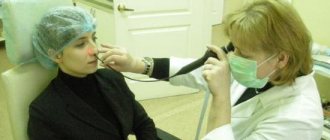
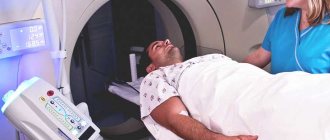
How does the examination take place?
Rectosigmoscopy is a special type of endoscopic examination. Many potential patients are interested in the question of how it differs from a colonoscopy. These are practically two similar procedures. Rectosigmoidoscopy allows you to assess the condition of the rectum and sigmoid colon. Colonoscopy examines similar parts of the intestine, but it allows a more detailed examination of the large intestine. This is due to the functional features of the colonoscope; by its nature, it has a longer and more flexible structure.
How is the procedure performed? This type of examination is absolutely safe and does not require the use of anesthesia. If during the examination the patient feels acute pain, anesthesia is used. The presence of pain indicates a low sensitivity threshold or serious illness.
| Aspects | Characteristic |
| Patient position. | During the examination, the patient must take a knee-elbow position. In this position, maximum muscle relaxation is achieved, and insertion of the endoscope becomes more convenient. |
| Time spending. | The inspection takes only a few minutes. The inspection must be carried out by an experienced specialist. The procedure does not accept sudden movements; it requires an increased level of delicacy. |
| Doctor's actions. | As the endoscope is inserted deep into the intestine, the specialist assesses the condition of the mucous membrane and its surface. If tumors are present, biopsy tests are taken using the device. During the study, all data from the endoscope is displayed on the screen. |
The manipulation is informative and fast. Many patients do not dare to undergo this type of study, but in some cases confirmation of the diagnosis is impossible without rectosigmoidoscopy. If the patient is categorically against it, a different type of study is selected.
Preparation for rectosigmoidoscopy
The main goal of preparation is high-quality cleansing of the intestines from the accumulation of gases and feces. Both the reliability of the research result and the patient’s comfort during the examination will depend on this.
Preparation for rectosigmoidoscopy begins 3 to 4 days before the scheduled procedure with dietary adjustments. A diet is prescribed that excludes the consumption of foods that cause gas formation in the intestines and also contain large amounts of fiber.
Legumes, cabbage, fatty protein foods, confectionery products, and rye bread are excluded from the diet. It is mandatory to maintain a water regime with the exclusion of alcohol and carbonated drinks. During the day, food intake is completely excluded.
Colon cleansing is carried out using an enema, on the eve of the procedure and on the day of it, 2 - 3 hours before the diagnosis. An addition to a more thorough cleansing of the intestines is taking the medications Duphalac, Fortrans according to the regimen.
What to take with you
To ensure that fibrorectosigmoidoscopy does not cause increased discomfort due to the nature of the procedure, it is recommended to have the following items during the examination:
- a sheet, a second shoe;
- special underwear that leaves only the field open for manipulation;
- wet wipes;
- toilet paper;
- socks;
- documents (medical policy, laboratory tests, passport, referral).
Procedure
When performing rectosigmoidoscopy, the following measures are sequentially carried out:
| Actions | Execution Features |
| Consultation with a doctor | The patient should warn if he is bothered by any unpleasant sensations or if there are suspicions of complications of chronic diseases |
| Digital examination of the rectum | Carried out by a specialist to identify pathologies in the structure of the rectum |
| Preparation for the procedure | The patient should remove clothing below the waist. If a person feels embarrassed, you can purchase special underwear designed for endoscopic examinations at the pharmacy. There is a hole in the panties for trouble-free examination |
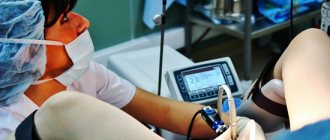
| The patient lies down on the couch | The person can move to their side or take a knee-elbow position. The exact position is chosen by the doctor. Usually the patient lies down with emphasis on bent knees and elbows, since it is in this case that the minimum discomfort appears during the examination |
| Conducting a survey | The sterile tip is lubricated with Vaseline, then inserted into the intestine at a distance of up to 60 cm. Based on the data displayed on the monitor, the doctor diagnoses the condition of the mucous membrane, the presence of tumors, and foci of pathologically changed tissues |
To avoid complications, for example, bleeding, exacerbation of chronic diseases, you must notify your doctor in advance if you have the following risk factors:
- Early pregnancy.
- Diabetes mellitus has been diagnosed or there is suspicion of its presence.
- Diseases of the cardiovascular system.
- Chronic bronchitis or asthma.
- Taking medications that the doctor conducting the examination does not know about.
Carrying out rectosigmoidoscopy
Procedure and features of rectosigmoidoscopy
The technique of this examination is minimally invasive and painless. Some patients may experience minor pain during the test. But they are usually relieved with painkillers. This diagnosis is carried out without anesthesia and, for the most part, is well tolerated by patients.
The examination is carried out in one of 3 positions, which the patient occupies before the manipulation:
- on a special chair or examination table, lying on your back with your legs bent at the hip joints;
- standing in a knee-elbow position on the couch;
- lying on your side with your legs bent and pulled to your chest.
Before inserting the endoscope into the anus, a mandatory digital examination of the rectum is performed to determine its patency. After a preliminary examination, an optical device is inserted into the rectum. By gently pumping air into the intestine, the walls of the organ are straightened, which facilitates visual examination of the intestine.
In this case, according to indications, a biopsy is taken from the pathological area of the rectum or sigmoid colon for histology. With rectosigmoidoscopy, it is possible to remove polyps or stop bleeding from small vessels.
How the research is carried out
If, apart from research, no action is expected, then the procedure itself takes from 10 to 30 minutes. The patient is changed into a clean robe and placed on a specially prepared couch on his left side. In this case, the legs should be bent at the knees.
The doctor lubricates the endoscope with Vaseline and very slowly, carefully inserts it into the anus. Air is then pumped into the intestines to ensure the most effective examination. Afterwards, the intestinal sections are carefully examined sequentially.
Important! During the procedure itself, it is strictly forbidden to twitch, change the position of the legs, or make other sudden movements, as these actions can damage the intestinal walls. The main thing is to remain calm and breathe deeply. If these measures are followed, the procedure will be completed more quickly, calmly and efficiently.
After completing the study, the patient lies on the couch for some time to allow all the pumped air to come out.
Sometimes, if necessary, material is taken for a biopsy. Such an analysis is necessary if Crohn's disease, a tumor process, and some other pathologies are suspected.
Possible complications after the examination
As a rule, diagnosis proceeds without complications and is well tolerated by patients. But sometimes complications occur and they are associated not only with the mistakes of the endoscopist, but also with the individual characteristics of the patient.
The most serious complications are:
- bleeding due to damage to the intestinal mucosa by careless insertion of a diagnostic instrument or when removing a polyp;
- perforation of the intestinal wall, leading to the development of peritonitis.
These complications are emergencies that require immediate medical attention.
Recovery after rectosigmoidoscopy
After the examination, patients may experience abdominal discomfort in the form of bloating, cramping, accompanied by minor pain for several hours. As a rule, these unpleasant sensations disappear after a short time.
The diet is limited only to heavy fatty foods and alcoholic beverages for 2 to 3 days. After diagnosis, patients almost immediately switch to their usual daily routine.
Diseases for which the procedure is effective
Examination of the large intestine, that is, proctoscopy, allows not only to identify various pathologies, but also to successfully combat them. These include:
- polyps;
- malignant and benign formations;
- ulcerative colitis;
- rectal diverticulosis;
- Crohn's disease;
- various inflammatory processes of the large intestine and the degree of their development.
Timely diagnosis will help establish not only the correct diagnosis, but also make a differential diagnosis with other pathologies.
Indications
Rectosigmoscopy is performed when the following disorders are detected:
- Frequent constipation, in which the condition of the digestive system gradually worsens. It is important to identify disorders in a timely manner if constipation cannot be eliminated by changing the diet or taking herbal medicines.
- Frequent stool pathologies, constipation followed by diarrhea.
- A feeling of incomplete bowel movement after bowel movements, occurring several times in a row.
- Pain, burning, and other unpleasant sensations in the lower abdomen, manifested in acute or chronic form.
- The appearance of pus or mucus in the stool.
- Signs of hemorrhoids.
- Presumptive diagnosis of the presence of malignant neoplasms or benign tumors in the intestine. Significant weight loss without obvious predisposing factors.
- The need for a preventive examination in the presence of a congenital predisposition to cancer.
- Discharge of blood from the anus.
- Symptoms of anemia, decreased hemoglobin levels, accompanied by constant weakness and loss of ability to work.
- The presence of occult blood in the stool.
Indications for rectosigmoidoscopy
Advantages of rectosigmoidoscopy when compared with colonoscopy:
- Minimal trauma, reduced risk of complications such as bleeding and discomfort. Immediately after the procedure, patients have the opportunity to get up and do their usual activities.
- Usually performed without local anesthesia. The exception is when the intestinal walls are severely inflamed or damaged.
- The duration of the procedure is from 5 minutes.
- Easy preparation for the procedure.
- Allows a thorough examination of the lower intestines.
Scheme of the procedure
Diseases that can be detected during rectosigmoidoscopy:
- The presence of malignant neoplasms. You can not only make or confirm a diagnosis, but also take a tissue sample for histological examination.
- Crohn's disease.
- Ulcerative colitis.
- Proctitis.
- Pathologies in the structure of the mucosa, the presence of inflammation.
- Polyposis, formation of cracks, areas of erosion.
- Congenital or acquired disorders in the structure of the intestine.
- Haemorrhoids.
Diseases that can be detected during rectosigmoidoscopy
Advantages of the method
The rectosigmoidoscopy method is minimally invasive, which allows one to examine the large intestine at a fairly large distance, about 60 cm. Since the procedure is painless and does not last long (15 minutes), it does not involve the use of general anesthesia. The technique does not require complex preparation and a long recovery period after it.
In addition, the method does not affect the patient’s ability to work and his quality of life, but, being highly informative, contributes to the early detection of organ pathologies, especially with a malignant course.
What should you take with you?
To avoid any discomfort during the procedure, you must take the following items with you:
- Disposable slippers, bed sheet.
- Warm socks if you are worried that your feet will get cold.
- Medical documentation: insurance policy, results of tests you did previously. It is advisable to discuss the complete list of required papers with your doctor in advance.
- Passport.
- Wet wipes, toilet paper.
- Disposable underwear and shoe covers can be purchased at the pharmacy located next to the medical facility.
Disposable underwear for rectosigmoidoscopy